


Courtesy of Dr. Ronald Green.

Courtesy of Dr. Ronald Green.
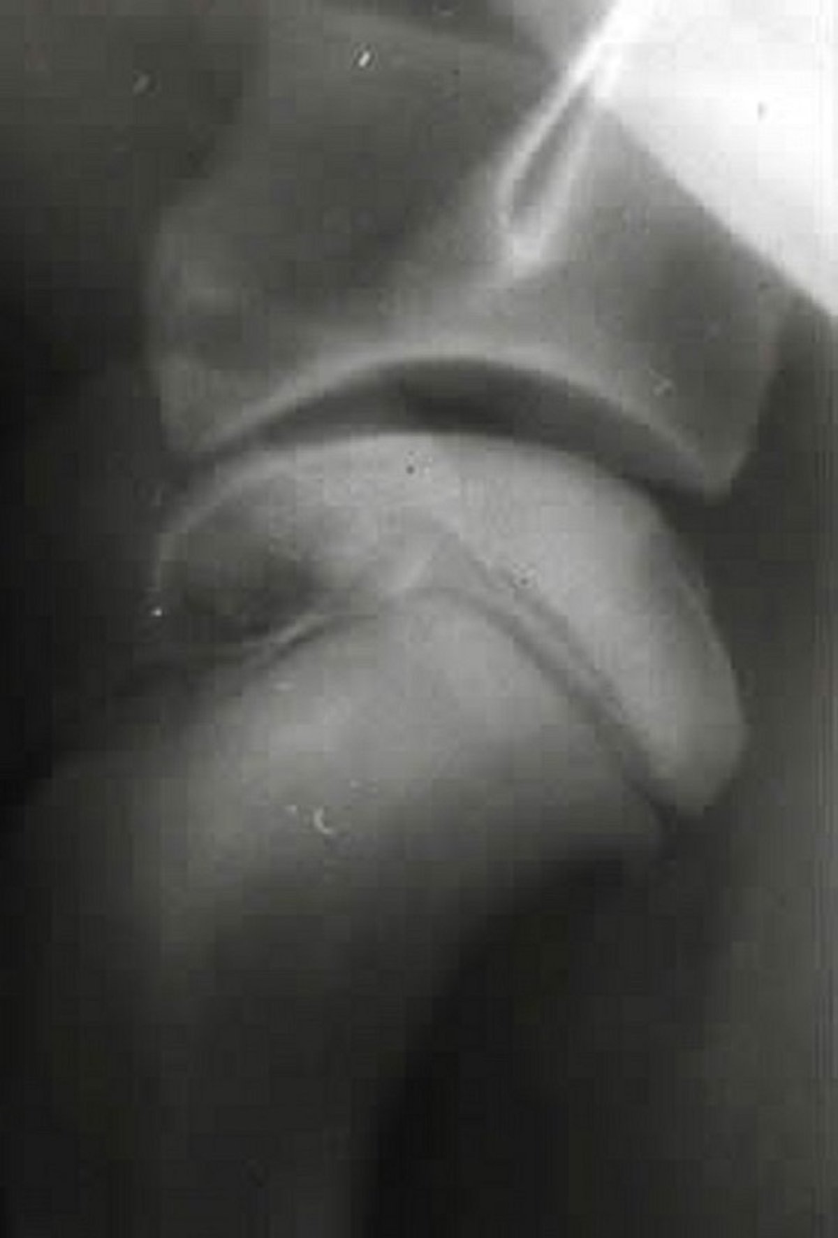
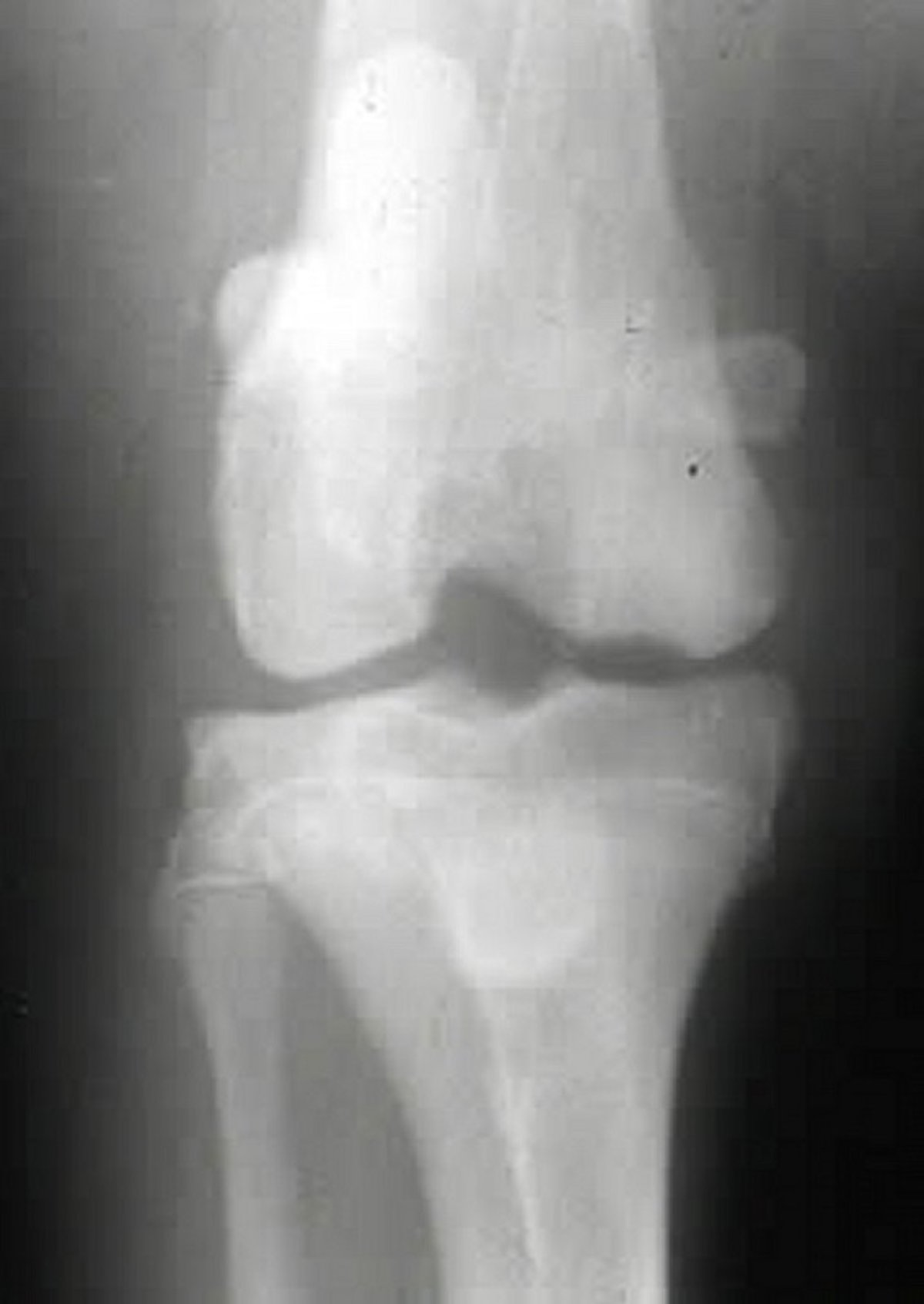
Osteochondrosis is a developmental disorder of medium and large rapidly growing dogs that is characterized by abnormal endochondral ossification of epiphyseal cartilage in the shoulder, elbow, stifle, and hock joints. Although the exact cause is unknown, excessive nutrition, rapid growth, trauma, and a hereditary component are suspected to be contributing factors. As a result of abnormal maturation and vascularity, basal cartilage cells thicken and weaken, thus leading to cartilage cracks, fissures, and flap formation (osteochondritis dissecans) after minor trauma or normal pressure to the joint. Abnormal cartilage congruency and joint debris lead to a synovitis and subsequent arthritis and continued cartilage breakdown. Cartilage flaps can break loose and attach to the joint capsule or migrate and deleteriously affect joint motion.
Clinical signs are lameness, joint effusion, and reduced range of motion in affected joints or limbs. Locations of the lesions include the head of the humerus (shoulder joint), the medial aspect of the humeral condyle (elbow joint), the femoral condyles (stifle joint), and the trochlear ridges of the talus (hock joint). Additionally, fragmented medial coronoid process and ununited anconeal process in the elbow joint may be related conditions. Radiography is useful in identifying joint lesions; changes may include flattening of joint surfaces, subchondral bone lucency or sclerosis, osteophytosis, joint effusion, and “joint mice.” Arthrography can be used to delineate cartilage flaps, and arthroscopy can also be performed to identify and treat cartilage or joint lesions. CT imaging also helps identify subchondral bone changes.
Treatment involves surgical excision of cartilage flaps or free-floating fragments and curettage of subchondral bone to stimulate fibrocartilage formation. Animals with degenerative joint disease may benefit from NSAIDs—eg, carprofen (2.2 mg/kg, PO, every 12 hours), deracoxib (1–2 mg/kg, PO, every 24 hours), firocoxib (5 mg/kg, PO, every 24 hours), or meloxicam (0.1 mg/kg, PO, every 24 hours). Joint fluid modifiers such as polysulfated glycosaminoglycan (4.4 mg/kg, IM, twice a week for 4 weeks) may also help prevent cartilage degeneration. Prognosis for recovery is excellent for the shoulders, good for the stifle joint, and fair for the elbow and tarsal joints. Concomitant signs of degenerative joint disease, other joint conditions, or instability (hock joint) deleteriously affect recovery.
For More Information
Also see pet health content regarding osteochondrosis in dogs.


