Search
SectionsIndex
  • Behavior
  • Circulatory System
  • Clinical Pathology and Procedures
  • Digestive System
  • Emergency Medicine and Critical Care
  • Endocrine System
  • Exotic and Laboratory Animals
  • Eye and Ear
  • Generalized Conditions
  • Immune System
  • Integumentary System
  • Management and Nutrition
  • Metabolic Disorders
  • Musculoskeletal System
  • Nervous System
  • Pharmacology
  • Poultry
  • Reproductive System
  • Respiratory System
  • Toxicology
  • Urinary System
  • Zoonoses
ABCDEFGHI
JKLMNOPQR
STUVWXYZ
In This Topic
Musculoskeletal System
Lameness in Horses
Overview of Lameness in Horses
Back to Top
Resources
  • About The Merck Veterinary Manual
  • Reference Guides
  • Multimedia
Manuals available online
'/home/index.html' + bookPageLink
 
'/vet/index.html'
These and other Manuals available
in print, online, and as mobile applications.

See more at MerckManuals.com
Sections in Veterinary Professionals
  • Behavior
  • Circulatory System
  • Clinical Pathology and Procedures
  • Digestive System
  • Emergency Medicine and Critical Care
  • Endocrine System
  • Exotic and Laboratory Animals
  • Eye and Ear
  • Generalized Conditions
  • Immune System
  • Integumentary System
  • Management and Nutrition
  • Metabolic Disorders
  • Musculoskeletal System
  • Nervous System
  • Pharmacology
  • Poultry
  • Reproductive System
  • Respiratory System
  • Toxicology
  • Urinary System
  • Zoonoses
Chapters in Musculoskeletal System
  • Musculoskeletal System Introduction
  • Congenital and Inherited Anomalies of the Musculoskeletal System
  • Dystrophies Associated with Calcium, Phosphorus, and Vitamin D
  • Arthropathies in Large Animals
  • Lameness in Cattle
  • Lameness in Goats
  • Lameness in Horses
  • Lameness in Pigs
  • Lameness in Sheep
  • Myopathies in Ruminants and Pigs
  • Myopathies in Horses
  • Bovine Secondary Recumbency
  • Lameness in Small Animals
  • Arthropathies and Related Disorders in Small Animals
  • Myopathies in Small Animals
  • Osteopathies in Small Animals
  • Sarcocystosis
Topics in Lameness in Horses
  • Overview of Lameness in Horses
  • The Lameness Examination in Horses
  • Imaging Techniques in Equine Lameness
  • Arthroscopy in Equine Lameness
  • Regional Anesthesia in Equine Lameness
  • Osseous Cyst-Like Lesions in the Distal Phalanx in Horses
  • Bruised Sole and Corns in Horses
  • Canker in Horses
  • Fracture of Navicular Bone in Horses
  • Fracture of Distal Phalanx in Horses
  • Keratoma in Horses
  • Laminitis in Horses
  • Navicular Disease in Horses
  • Pedal Osteitis in Horses
  • Puncture Wounds of the Foot in Horses
  • Pyramidal Disease in Horses
  • Quittor in Horses
  • Quarter Crack in Horses
  • Scratches in Horses
  • White Line Disease in Horses
  • Sheared Heels in Horses
  • Sidebone in Horses
  • Thrush in Horses
  • Fracture of Phalanges and Proximal Sesamoids in Horses
  • Osteoarthritis in Horses (Fetlock and Pastern)
  • Palmar/Plantar Osteochondral Disease in Horses
  • Sesamoiditis in Horses
  • Chronic Proliferative Synovitis in Horses
  • Digital Sheath Tenosynovitis in Horses
  • Disorders of the Carpus and Metacarpus in Horses
  • Bucked Shins in Horses
  • Subchondral Bone Disease of the Carpal Bones in Horses
  • Desmitis or Sprain of the Inferior Check Ligament in Horses
  • Fracture of the Carpal Bones in Horses
  • Fractures of the Small Metacarpal and Metatarsal (Splint) Bones in Horses
  • Fracture of the Third Metacarpal (Cannon) Bone in Horses
  • Hygroma in Horses
  • Osteoarthritis in Horses (Carpus and Metacarpus)
  • Osteochondritis Dissecans in Horses
  • Osteochondroma of the Distal Radius in Horses
  • Rupture of the Common Digital Extensor Tendon in Horses
  • Splints in Horses
  • Subchondral Bone Cysts and Septic Arthritis in Horses
  • Suspensory Desmitis in Horses
  • Synovial Hernia and Ganglion and Synovial Fistulae in Horses
  • Tearing of the Medial Palmar Intercarpal Ligament in Horses
  • Tenosynovitis of the Tendon Sheaths Associated with the Carpus in Horses
  • Traumatic Synovitis and Capsulitis in Horses
  • Shoulder Disorders in Horses
  • Elbow Disorders in Horses
  • Disorders of the Tarsus and Metatarsus in Horses
  • Bog Spavin in Horses
  • Bone Spavin in Horses
  • Curb in Horses
  • Displacement of the Superficial Flexor Tendon from the Point of the Hock in Horses
  • Fracture of the Tarsus in Horses
  • Luxation of the Hock in Horses
  • Hindlimb Tendon Ruptures in Horses
  • Rupture of the Peroneus Tertius Muscle in Horses
  • Stringhalt in Horses
  • Thoroughpin in Horses
  • Fractures of the Third Metatarsal Bone in Horses
  • Large Wounds in the Proximal Metatarsal Region in Horses
  • Osteochondrosis of the Stifle in Horses
  • Subchondral Cystic Lesions in Horses
  • Meniscus and Meniscal Ligament Injuries in Horses
  • Cranial and Caudal Cruciate Ligament Injuries in Horses
  • Collateral Ligament Injuries in Horses
  • Intermittent Upward Fixation of Patella and Delayed Patella Release in Horses
  • Fragmentation of the Patella in Horses
  • Patellar Luxation in Horses
  • Patellar Ligament Injuries in Horses
  • Gonitis and Osteoarthritis in Horses
  • Fractures in Horses
  • Disorders of the Hip in Horses
  • Luxation of the Coxofemoral Joint in Horses
  • Pelvic Fracture in Horses
  • Osteoarthritis and other Coxofemoral Joint Diseases in Horses
  • Disorders of the Back and Pelvis in Horses
  • Spinal Processes and Associated Ligaments in Horses
  • Articular Process-Synovial Intervertebral Articulation Complexes in Horses
  • Vertebral Bodies and Disks in Horses
  • Muscle Strain and Soreness in Horses
  • Lumbosacral Junction Abnormalities in Horses
  • Sacroiliac Joint Abnormalities in Horses
  • Tendinitis in Horses
  • Developmental Orthopedic Disease in Horses
  • Osteochondrosis in Horses
  • Physitis in Horses
  • Flexion Deformities in Horses
 
  • Merck Manual
  • >
  • Veterinary Professionals
  • >
  • Musculoskeletal System
  • >
  • Lameness in Horses
  • 4
 
Overview of Lameness in Horses

Share This

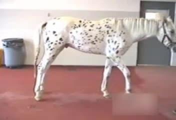
Lameness is defined as an abnormal stance or gait caused by either a structural or a functional disorder of the locomotor system. The horse is either unwilling or unable to stand or move normally. Lameness is the most common cause of loss of use in horses. It can be caused by trauma, congenital or acquired disorders, infection, metabolic disorders, and nervous and circulatory system disease.

Lameness is not a disease per se but a clinical sign. It is a manifestation of pain, mechanical restrictions causing alteration of stance or gait, or neuromuscular disease. Pain is the most common cause of lameness in all horses. Mechanical lameness is best typified by complete upward fixation of the patella with its characteristic gait abnormality, but can also be the result of fibrotic myopathy of the semitendinosus muscle or of restrictions caused by annular ligaments, adhesions, or severe fibrosis.

It is critical to correctly determine the cause of the lameness, because treatment varies greatly depending on the cause. For example, the mechanical lameness of complete upward fixation of the patella will not respond to analgesics, whereas lameness caused by pain often responds to systemic or local analgesics and anti-inflammatory drugs. Some causes of lameness produce very characteristic and classically described gaits. In fibrotic myopathy, a mechanical lameness, the affected limb is pulled back and down quickly before the end of the protraction phase, giving the impression that the foot “slaps down” on the ground. The signs are most obvious at the walk. In stringhalt, a neuromuscular disorder, the affected limb is hyperflexed during the cranial or swing phase, while the stepwise caudal jerking movement before foot contact does not occur. Unfortunately, many causes of lameness do not produce a characteristic gait abnormality, making diagnosis a challenge.
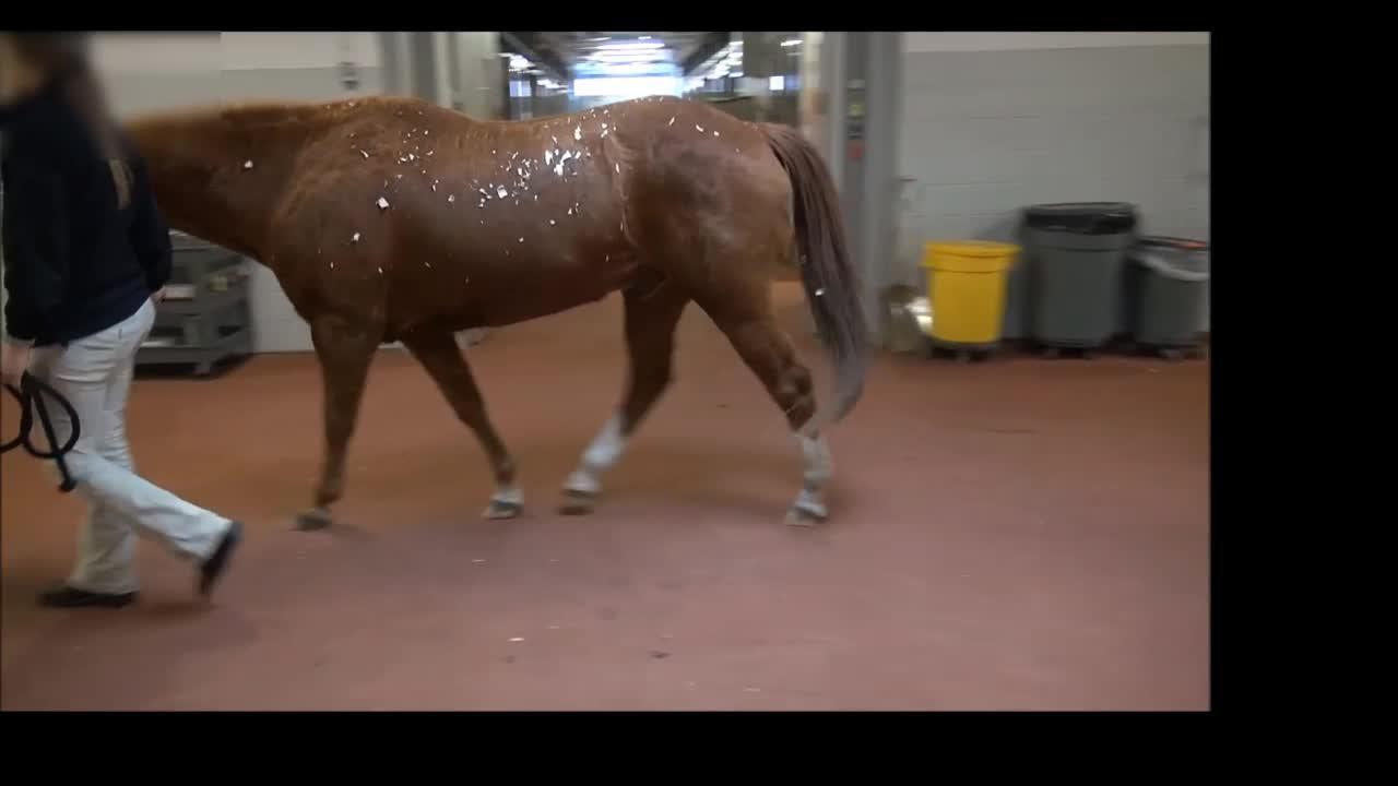
Pain-related lameness can be classified as weight bearing (supporting leg) or nonweight bearing (swinging leg) lameness. Although lameness is most often observed as a weight-bearing deficit, it may be composed of both. A supporting leg lameness is seen when the horse reduces the amount of time or reduces the amount of force applied to the weight-bearing limb. The most consistent and easily recognized clinical signs of lameness are the head nod associated with forelimb lameness and the sacral rise, also called a pelvic rise or hip hike, associated with hindlimb lameness. Hindlimb lameness should be assessed from the side as well as from behind, because this provides an opportunity to assess arc of foot flight, duration of protraction and retraction phases, length of weight-bearing phase, and the presence or absence of a sacral rise. Hindlimb and forelimb lameness in many horses will be accentuated when the horse is worked in a circle with the affected limb on the inside.

Factors that predispose horses to lameness include physical immaturity, which may occur in premature or dysmature foals, and training older foals prior to maturity. Other factors include preexisting developmental orthopedic disease (eg, osteochondrosis, flexural limb and angular limb deformities); poor conformation; improper hoof balance or shoeing; failure to adequately condition performance horses; monotonous repetitive stresses on bones, tendons, ligaments, and joints in performance horses; hard, slippery, or rocky surfaces upon which horses work; and extremely athletic activities. Inciting factors in lameness include direct or indirect trauma, fatigue resulting in incoordination of muscles (which often occurs in racehorses at the end of races), inflammation, infection, and failure to recognize early disease before it creates significant pain.

Lameness in one part of a limb often results in secondary soreness in another area of the same limb and may result in lameness of the contralateral forelimb or hindlimb from overuse due to compensation. The entire horse should be evaluated for secondary lameness even when the cause of the primary problem is obvious. Secondary lamenesses are very common in performance horses but may occur in all types of horses. A dramatic example of a secondary lameness occurs when biomechanical laminitis develops in the normal contralateral limb of a horse with a severe orthopedic problem due to a shifting of weight from the injured limb to the normal limb.

Last full review/revision March 2012 by Stephen B. Adams

Buy the Book

Back to Top

Previous: Overview of Lameness in Goats

Next: The Lameness Examination in Horses

Audio
Figures
Photographs
Sidebars
Tables
Videos

Copyright     © 2010-2013 Merck Sharp & Dohme Corp., a subsidiary of Merck & Co., Inc., Whitehouse Station, N.J., U.S.A.    Privacy    Terms of Use