


Fowl cholera is a contagious bacterial disease of birds caused by Pasteurella multocida. Acutely, it causes elevated mortality rate. Chronically, it may cause lameness, swollen wattles (in chickens), pneumonia (in turkeys), and/or torticollis; however, birds can also be subclinically affected carriers. Both live, attenuated vaccines and adjuvanted bacterins are available to aid in prevention, and the organism is susceptible to some antimicrobials.
Fowl cholera affects domestic and wild birds worldwide. It usually occurs as a septicemia of sudden onset with high morbidity and mortality rates, but chronic infections and subclinical carrier states may also occur.
Etiology and Transmission of Fowl Cholera
Pasteurella multocida, the causative agent of fowl cholera, is a small, gram-negative, nonmotile rod with a capsule that may exhibit pleomorphism after repeated subculture. P multocida is considered a single species although it includes three subspecies: multocida, septica, and gallicida. Subspecies multocida is the most common cause of disease, but septica and gallicida may also cause cholera-like disease.
In freshly isolated cultures or in tissues, the bacteria have a bipolar appearance when stained with Wright stain. Although P multocida may infect a wide variety of animals, strains isolated from most nonavian hosts generally do not produce fowl cholera.
Strains that cause fowl cholera represent a number of immunotypes (or serotypes). P multocida can be subgrouped by capsule serogroup antigens into five capsular types (A, B, C, D, and F) and into 16 somatic serotypes. Turkeys and waterfowl are more susceptible than chickens, older chickens are more susceptible than young ones, and some breeds of chickens are more susceptible than others.
Chronically infected birds and subclinical carriers are considered to be major sources of infection. Wild birds may introduce the organism into a poultry flock; however, mammals (including rodents, pigs, dogs, and cats) may also carry the infection. However, the role of these as a reservoir has not been thoroughly investigated.
Dissemination of P multocida within a flock and between houses is primarily by excretions from the mouth, nose, and conjunctiva of diseased birds that contaminate their environment. Pecking and cannibalism of infected carcasses contributes to spread of the organism through affected flocks. In addition, P multocida survives long enough in the environment to be spread by contaminated crates, feed bags, shoes, and other equipment. The infection does not seem to be egg-transmitted.
Clinical Findings of Fowl Cholera
Clinical findings from fowl cholera vary greatly, depending on the species and age affected and the course of disease. In acute fowl cholera, finding a large number of dead birds without previous clinical signs is usually the first indication of disease. Mortality rate often increases rapidly. In more protracted cases, listlessness, anorexia, mucoid discharge from the mouth, ruffled feathers, diarrhea, and increased respiratory rate are usually observed. Pneumonia is particularly common in turkeys.
In chronic fowl cholera, clinical signs and lesions are generally related to localized infections of the sternal bursae, wattles, joints, tendon sheaths, and footpads, which often are swollen because of accumulated caseous or fibrinous exudate (see swollen wattles image). There may be lameness, as well as exudative conjunctivitis and pharyngitis. Torticollis may result when the meninges, middle ear, or cranial bones are infected.

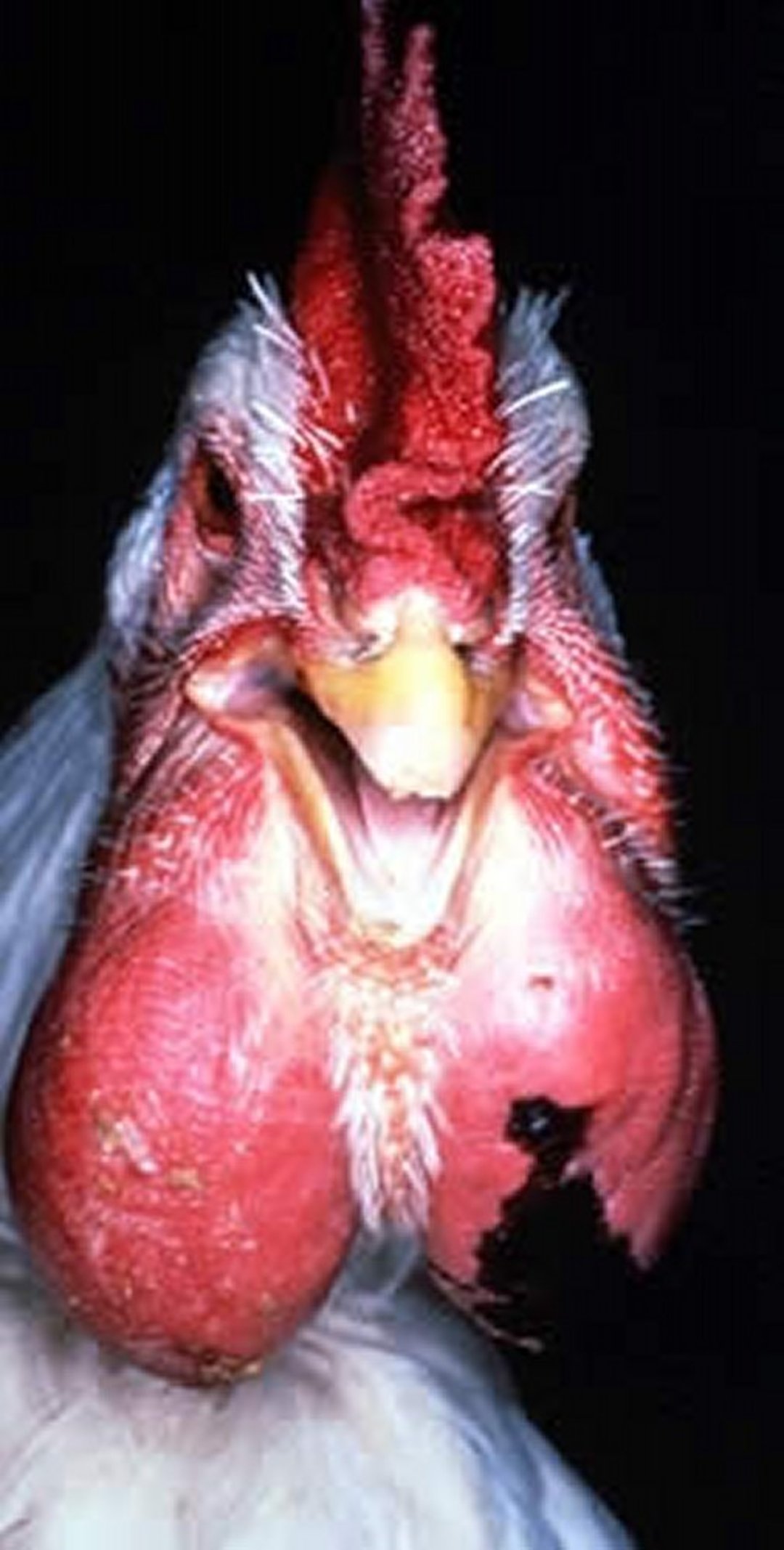
Courtesy of Dr. Jean Sander.
Lesions
Lesions observed in peracute and acute forms of the disease are primarily vascular disturbances. These include general passive hyperemia and congestion throughout the carcass, accompanied by enlargement of the liver and spleen. Petechial and ecchymotic hemorrhages are common, particularly in subepicardial and subserosal locations.
Increased amounts of peritoneal and pericardial fluids are frequently present. In addition, acute oophoritis with hyperemic follicles may be observed.
In subacute cases, multiple small necrotic foci may be disseminated throughout the liver and spleen (see bacterial septicemia lesions image). Severe, often unilateral, fibrinous pneumonia is observed in turkeys with fowl cholera.

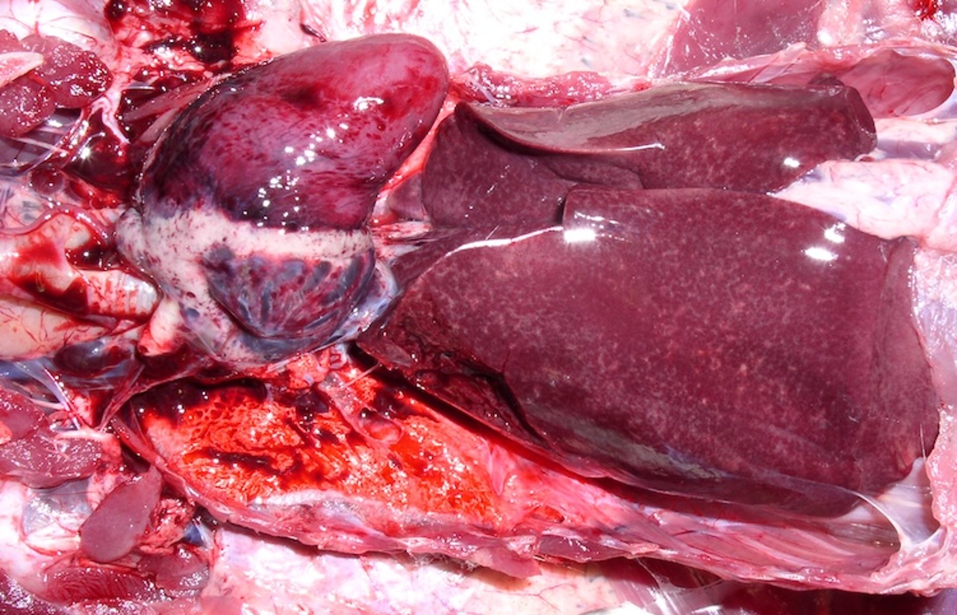
Courtesy of Dr. Megan Lighty.
In chronic forms of fowl cholera, lesions may be widely distributed, often involving the respiratory tract, the conjunctiva, and adjacent tissues of the head. Caseous arthritis and productive inflammation of the peritoneal cavity and the oviduct are common in chronic infections.
A fibrinonecrotic dermatitis that includes caudal parts of the dorsum, abdomen, and breast and involves the cutis, subcutis, and underlying muscle has been observed in turkeys and broilers. Localized dermatitis, cellulitis, and myositis may be observed in cases of fowl cholera after transmission via predator bite.
Sequestered necrotic lung lesions in poultry should always raise suspicion of cholera (see consolidated lung image).

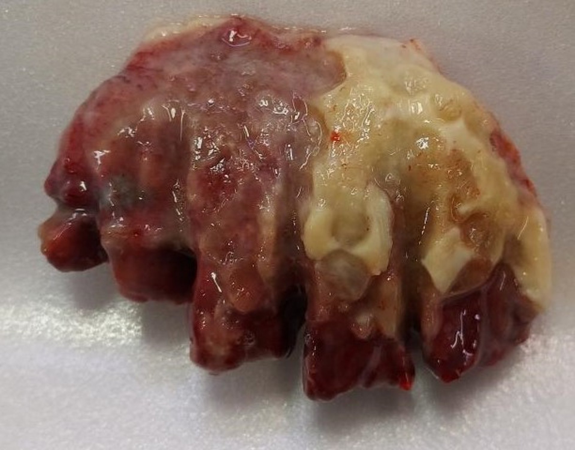
Courtesy of Dr. Megan Lighty.
Diagnosis of Fowl Cholera
Bacterial culture
PCR assays
Although the history, clinical signs, and lesions may aid field diagnosis of fowl cholera, isolation of P multocida is the standard for confirmation. Primary isolation can be accomplished using media such as blood agar, dextrose starch agar, or trypticase soy agar. Isolation may be improved by the addition of 5% heat-inactivated serum.
P multocida can be readily isolated from viscera of birds dying from peracute or acute fowl cholera, whereas isolation from suppurative lesions of chronic cholera may be more difficult. At necropsy, bipolar microorganisms may be demonstrated by the use of Wright or Giemsa stain of impression smears obtained from the liver in the case of acute cholera. In addition, immunofluorescent microscopy and in situ hybridization have been used to identify P multocida in infected tissues and exudates.
PCR assay has been used for the detection of P multocida in pure and mixed cultures and clinical samples. This method may help identify carrier animals within flocks. However, the specificity and sensitivity of the test must be improved. Conventional serotyping suffers from problems with reproducibility and reliability, and the methods are quite laborious. A multiplex PCR assay has been developed that can differentiate between different somatic serotypes and may enable more efficient vaccine development.
Serological testing can be done by rapid whole blood agglutination, serum plate agglutination, agar diffusion tests, and ELISA. Such testing may be used to evaluate vaccine responses but has very limited value for diagnostic purposes; therefore, serological testing is not readily available at most diagnostic laboratories in the US.
Several bacterial infections may be confused with fowl cholera based solely on the gross lesions. Escherichia coli, Salmonella enterica, Ornithobacterium rhinotracheale, gram-positive cocci, and Erysipelothrix rhusiopathiae (erysipelas) may all produce lesions indistinguishable from those caused by P multocida.
Prevention of Fowl Cholera
Good management practices, including a high level of biosecurity
Vaccines
For prevention of fowl cholera, rodents, wild birds, pets, and other animals that may be carriers of P multocida must be excluded from poultry houses. The organism is susceptible to ordinary disinfectants, sunlight, drying, and heat.
Adjuvant bacterins are widely used and generally effective. Because bacterins are only effective in preventing disease caused by the same serotypes included in the vaccine, somatic serotyping is important. Thus, it is important to know the most prevalent serotypes within an area. Autogenous bacterins are recommended when polyvalent bacterins are found to be ineffective.
Live, attenuated vaccines are available for administration in drinking water to turkeys and by wing-web inoculation to chickens. These live vaccines can effectively induce immunity against different serotypes of P multocida. They are recommended for use in healthy flocks only.
Treatment and Control of Fowl Cholera
Depopulation, thorough cleaning, and disinfection
Antimicrobials
Prompt removal of carcasses
A number of antibacterial drugs will decrease deaths from fowl cholera; however, deaths may resume when treatment is discontinued, showing that treatment does not eliminate P multocida from a flock. Pecking and cannibalism contribute to spread of disease through a flock; therefore, prompt removal and disposal of carcasses is critical to decrease further losses.
Eradication of infection requires depopulation and cleaning and disinfection of buildings and equipment. The premise should then be kept free of poultry for a few weeks.
When antimicrobials are used, early treatment and adequate dosages are important. Antimicrobial resistance testing should be used to guide drug selection because of the emergence of multiresistant strains.
Sulfamethazine or sulfadimethoxine in feed or water usually controls mortality rates. Sulfa drugs should be used with caution in breeders because of potential toxicity and cannot be used in hens laying eggs for human consumption. Oxytetracycline and chlortetracycline are labeled for control of P multocida and may be useful. Penicillin is often effective in turkeys for sulfa-resistant infections but is not labeled for this indication.
Key Points
Fowl cholera is a bacterial disease of chickens, turkeys, waterfowl, and other birds, caused by P multocida.
Lesions are typically found in the conjunctiva and other structures of the head, lungs, and liver.
Fowl cholera causes acute death and chronic caseous to fibrinous inflammation and necrosis.
It is controlled through good biosecurity, vaccination, and antimicrobials.
For More Information
Blackall PJ, Hofacre CL. Fowl cholera. In: Swayne DE, Boulianne M, Logue CM, et al, eds. Diseases of Poultry. 14th ed. Wiley; 2019:831-846.
Abdul-Aziz T, Barnes HJ. Pasteurella multocida infection (fowl cholera). In: Gross Pathology of Avian Diseases. American Association of Avian Pathologists; 2018:18-23.


