


Gout is caused by the precipitation of urates in the viscera (acute urate deposition) or the joints (chronic urate deposition). Acute and chronic urate deposition differ in etiology, morphology, and pathogenesis. Acute urate deposition (ie, visceral gout) is the counterpart to uremia in mammals. Chronic urate deposition (ie, articular gout) is the equivalent of gout in mammals.

Courtesy of Dr. Rocio Crespo.
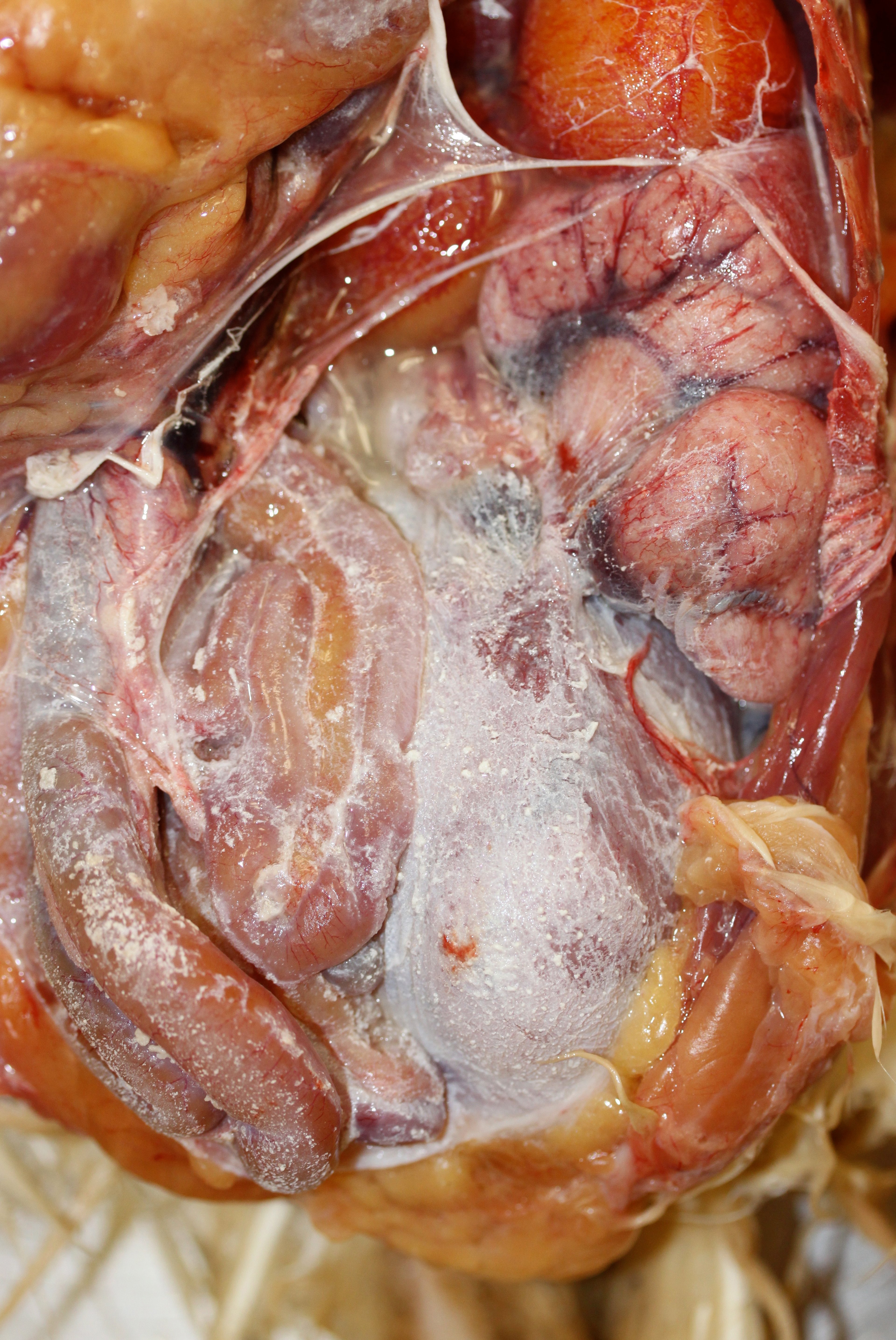
Birds excrete nitrogenous wastes as urates bound in colloidal form with mucus in their urine. Renal dysfunction decreases the clearance of uric acid from the blood, which results in hyperuricemia with precipitation of insoluble products within the kidney itself or other organs, leading to urate deposition or urolithiasis. Urate deposits are white and semisolid and must be differentiated from yellow fibrinous or purulent inflammatory exudates that are secondary to infectious causes, such as synovitis, peritonitis, perihepatitis, and pericarditis.
Visceral urate deposition occurs after rapidly progressing renal failure or as a terminal event with acute decompensation of chronic renal disease. Deposits develop most commonly on the pericardium, peritoneum, and liver capsule and rarely on synovial surfaces of joints and tendons. Microscopically, urate deposits are often seen as feathery crystals or basophilic spherical masses, usually with little inflammation due to the rapid course. Visceral urate deposition may be secondary to urolithiasis, which is common in older laying chickens. Progressive obstruction of the ureters by uroliths causes kidney atrophy “upstream” of the site of ureteral obstruction and compensatory hypertrophy by the undamaged portions of the kidney. Distended ureters often contain brittle, white, staghorn calcium urate calculi or uroliths.

Courtesy of Dr. Rocio Crespo.
Predisposing factors for visceral urate deposition and urolithiasis in poultry include infectious bronchitis virus, avian nephritis virus, and cryptosporidiosis. Noninfectious causes include dehydration, ingestion of feed containing > 3% calcium by nonlaying chickens, vitamin A deficiency, and exposure to myotoxins (eg, oosporein). Other avian species commonly develop visceral deposits secondary to nephrotoxin exposure, most commonly aminoglycoside antibiotics or heavy metals.
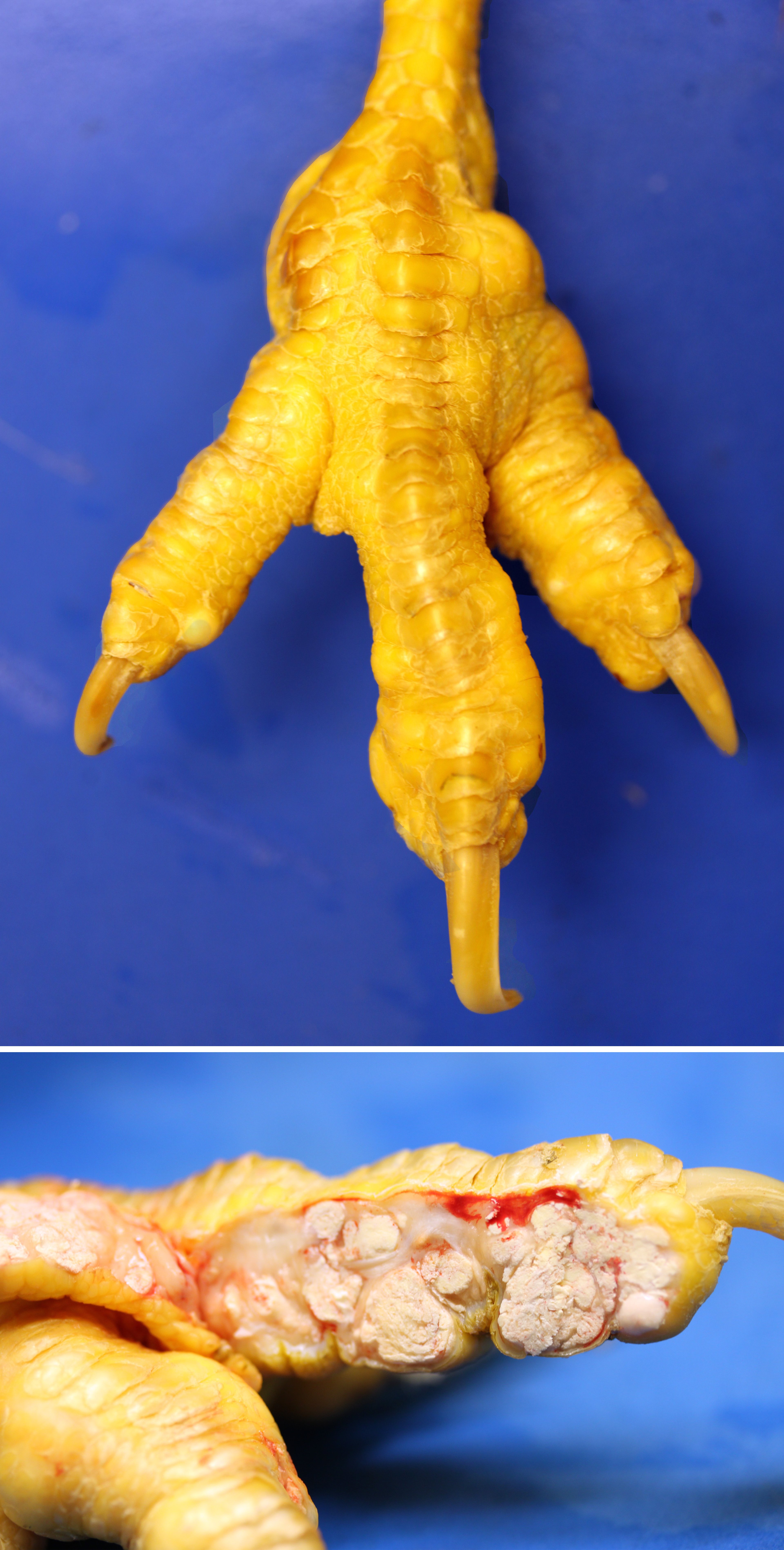
Articular urate deposition is less common and occurs after longterm increases in serum levels of uric acid. Deposits develop on synovial membranes in the toes and wing joints and incite a chronic granulomatous reaction to urate crystals (tophi). Joints are enlarged, and the feet appear deformed. Unlike visceral urate deposition, the kidneys are usually grossly normal. Articular urate deposition may be seen in birds that have hereditary defects in uric acid metabolism or that are fed excessive protein.


