


Necrotic enteritis is caused by intestinal overgrowth of Clostridium perfringens Type A and C in young broilers and sometimes laying hens. Predisposing factors that disturb the intestinal microflora create a permissive environment for this overgrowth; Eimeria infection or certain dietary substrates are the main causes. The disease is rapidly fatal in up to 50% of affected animals. Treatment is by antimicrobial-medicated drinking water.
Necrotic enteritis is an acute enterotoxemia. The clinical signs are usually very short-lived, and often the only sign is a severe depression followed quickly by a sudden increase in flock mortality. The disease affects primarily broiler chickens (2–5 weeks old) and turkeys (7–12 weeks old) raised on litter; however, it can also affect commercial layer pullets raised in cages.
Early mortality is often related to concurrent coccidiosis, with Eimeria cycling in these flocks. In individual chickens, the disease appears to be peracute, leading to death or resolution within 3–4 days. Because of the asynchronous challenge of individual birds with predisposing Eimeria, the incidence may rise and decline over ≥ 2 weeks in commercial flocks. Postmortem examination of the mucosal epithelium (see below) is usually diagnostic.
Etiology and Pathogenesis of Necrotic Enteritis in Poultry
The agent that causes necrotic enteritis is the gram-positive, obligate, anaerobic bacterium Clostridium perfringens. It is usually isolated on blood agar, incubated anaerobically at 37°C (98.6°F), on which it produces a double zone of hemolysis. Two primary C perfringens types, A and C, are associated with necrotic enteritis in poultry. Toxins produced by the bacteria damage the small intestine and liver.
C perfringens is a nearly ubiquitous bacterium readily found in soil, dust, feces, feed, and used poultry litter. It is also a normal inhabitant of the intestines of healthy chickens and turkeys. The enterotoxemia that leads to clinical signs most often occurs either after a change in the intestinal microflora or as an effect of a condition that results in damage to the intestinal mucosa (eg, coccidiosis, mycotoxicosis, salmonellosis, ascaridiasis).
High dietary amounts of animal by-products (eg, fish meal), wheat, barley, oats, or rye predispose birds to necrotic enteritis. Anything that promotes excessive bacterial growth and toxin production or slows the passage rate of feed in the small intestine could promote necrotic enteritis.
In many cases, concurrent coccidiosis (especially when due to Eimeria maxima or, to a lesser extent, Eimeria acervulina) is associated with outbreaks in commercial broilers. Clostridial isolates positive for the cytotoxic pore-forming toxin necrotic enteritis B (NetB) can cause disease without predisposition from Eimeria infections, but the majority of necrotic enteritis breaks are associated with predisposing Eimeria infections.
Clinical Findings and Lesions of Necrotic Enteritis in Poultry
Usually, the only clinical sign of necrotic enteritis in a flock is a sudden increase in mortality. However, birds with depression, ruffled feathers, and diarrhea may also be observed. The gross lesions are found primarily in the small intestine (jejunum/ileum), which may be ballooned and friable, and may contain a foul-smelling, brown fluid.

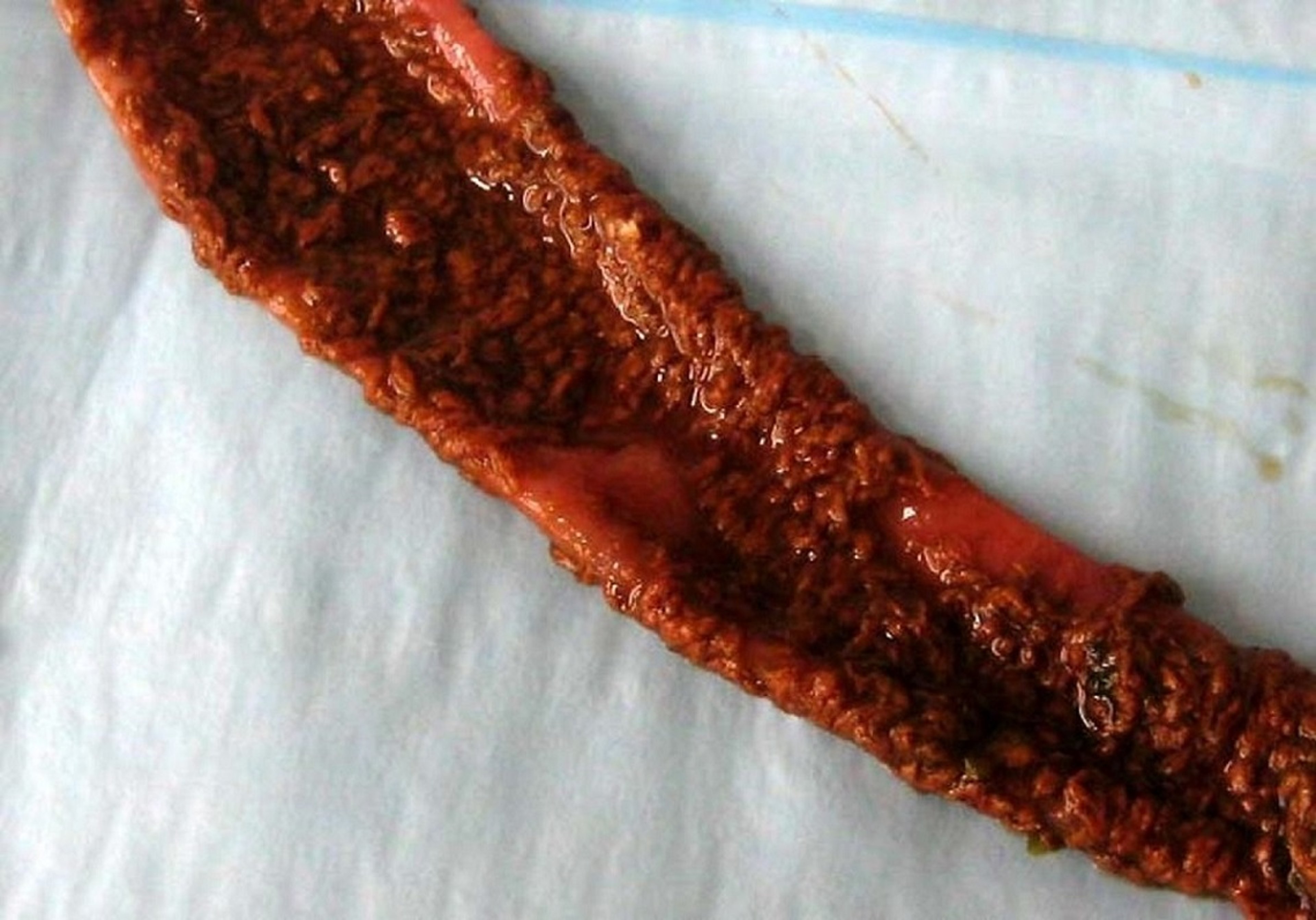
Courtesy of Dr. Billy Hargis.
The mucosa is usually covered with a tan to yellow pseudomembrane, progressing to a diphtheritic-like appearance. The color, which may range from a slight orange to a deep brown, is assumed to be related to fresh or oxidized blood combined with bile staining. This pseudomembrane may extend throughout the small intestine or be localized, and severe cases may be marked by sloughing of the epithelium in some regions. The disease usually persists in a flock for 5–14 days, and mortality is 2%–50%.
Diagnosis of Necrotic Enteritis in Poultry
Presumptive diagnosis: clinical signs of diarrhea, depression
Confirmation: based on gross lesions in small intestine and microscopic observation of gram-positive rods
A presumptive diagnosis of necrotic enteritis is based on gross lesions and a Gram-stained smear of a mucosal scraping that exhibits large, gram-positive rods. Histologic findings consist of coagulative necrosis of one-third to one-half the thickness of the intestinal mucosa, as well as masses of short, thick bacterial rods in the fibrinonecrotic debris.
Isolation of large numbers of C perfringens from intestinal contents that produce the double zone of hemolysis as described above can confirm the diagnosis. Double-zone hemolysis should not be the sole criterion for identification of C perfringens, however, because some strains do not produce both toxins responsible for the hemolysis characteristics. Differential media specifically designed for isolation of C perfringens are available and may be useful for diagnosis.
Unsporulated Eimeria oocysts may or may not be present, because the coccidiosis in affected chickens precedes clostridial overgrowth by 3–7 days. Moreover, intensive epithelial necrosis often precludes further active oocyst shedding.
Necrotic enteritis must be differentiated from lesions produced by Eimeria brunetti and also from ulcerative enteritis. Uncomplicated coccidiosis rarely produces lesions as acute or severe as those that occur with necrotic enteritis. Ulcerative enteritis due to Clostridium colinum usually produces focal lesions from the distal portion of the small intestine (ileum) to the ceca and is almost always accompanied by hepatic necrosis.
Prevention, Control, and Treatment of Necrotic Enteritis in Poultry
Prevention of coccidiosis
Avoidance of animal by-products, rye, fish meal, wheat, barley in the diet
Administration of probiotics or competitive cultures
Treatment with antimicrobial-medicated drinking water
Because C perfringens is nearly ubiquitous, the key to controlling necrotic enteritisis to prevent coccidiosis, which is the main predisposing factor for C perfringens overgrowth and infection. E acervulina and E maxima are particularly problematic species of coccidia. In addition, dietary management of poultry should avoid any changes that could alter the intestinal microflora and promote the growth of C perfringens.
Historically, coccidiosis was prevented by the addition of antimicrobials in the feed, such as virginiamycin (22 g/metric tonne of feed), bacitracin (55 g/metric tonne of feed), and lincomycin (2.2 g/metric tonne of feed), as well as ionophore-class anticoccidial drugs. The move to antimicrobial-free feeds in the late 1990s and early 2000s was initially associated with a marked increase in the use of live coccidiosis vaccines, and in some cases Eimeria infections occurred after vaccination, along with subsequent necrotic enteritis. Since that time, coccidial control strategies have continued to evolve: modified live vaccines have been improved, recombinant antigen-only vaccines have been developed, and the occurrence of necrotic enteritis is generally reduced by prevention of coccidiosis.
Avoiding drastic changes in feed and minimizing the amounts of fish meal, wheat, barley, and rye in the diet can also help prevent necrotic enteritis. When higher amounts of wheat, barley, or rye are necessary, adding enzymes for non-starch polysaccharides to the feed has decreased the incidence of necrotic enteritis in flocks fed these cereals. Adding selected probiotics or competitive exclusion cultures to feed has been successful in both preventing and treating clinical necrotic enteritis (presumably by preventing the proliferation of C perfringens).
Antimicrobials to treat necrotic enteritis are usually administered in drinking water. The most commonly used antimicrobials are bacitracin (52.8 mg/L for 5–7 days), penicillin (400,000 U/L for 5 days), and lincomycin (17 mg/L for 7 days). In each case, the medicated drinking water should be the sole source of water. Moribund birds should be removed promptly because they can be a source of toxicosis or infection as a result of cannibalism.
Necrotic enteritis is an acute to peracute disease in individual chickens, and outbreaks are usually short. The response to treatment may be overestimated because the disease is likely to be self-limiting in a flock regardless of treatment.
Key Points
Necrotic enteritis in poultry results from overgrowth of C perfringens.
Disease outbreaks are short-lived within a flock; mortality is as high as 50%.
Affected flocks are treated with antimicrobials in drinking water.
Prevention of coccidiosis and avoidance of certain feedstuffs are keys to preventing necrotic enteritis.


