


Cows
In cows, metritis is a common polymicrobial disease, especially within the first 2 weeks after parturition. Acute puerperal metritis refers to a severe postpartum uterine infection that results in systemic signs of toxemia. Clinical metritis is used as a general term for postpartum uterine infections, which may not be associated with systemic signs. Infectious reproductive system disease (eg, brucellosis, leptospirosis, trichomoniasis, and campylobacteriosis) may also cause metritis.
Etiology and Pathogenesis of Metritis in Cows
As a result of advancements in culture-independent techniques and sequencing, it is now understood that the uterine microbiome of cows developing postpartum metritis deviates in favor of a higher relative abundance of Bacteroidetes and Fusobacteria and lesser relative abundance of Proteobacteria and Tenericutes. The shift in the uterine microbiome has been suggested to be a dysbiosis characterized by a loss of heterogeneity and a decrease in bacterial richness, with Bacteroides, Porphyromonas, and Fusobacterium having the strongest association with development of metritis.
Epidemiology of Metritis in Cows
Risk factors associated with the occurrence of postpartum metritis include stillbirth; twins; dystocia; retention of fetal membranes; male calf; primiparity; reduced feed intake 2–3 weeks before calving; subclinical hypocalcemia; high nonesterified fatty acids, beta-hydroxybutyrate, and haptoglobin in the first week after parturition; and poor perineal area hygiene. The cumulative incidence of postpartum metritis ranges from 10%–25% of cows in the lactation period on most dairy farms.1 Affected cows have reduced milk production, impaired reproductive performance, and higher likelihood of endometritis, culling, and death.
References
Zwald NR, Weigel KA, Chang YM, Welper RD, Clay JS. Genetic selection for health traits using producer-recorded data. I. Incidence rates, heritability estimates, and sire breeding values. J Dairy Sci 2004;87:4287–94.
Clinical Findings of Metritis in Cows
The distinction between acute puerperal metritis and clinical metritis is pivotal because acute puerperal metritis requires treatment, whereas clinical metritis does not.
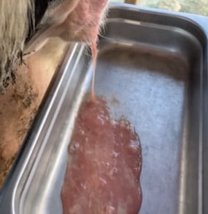
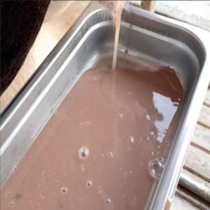
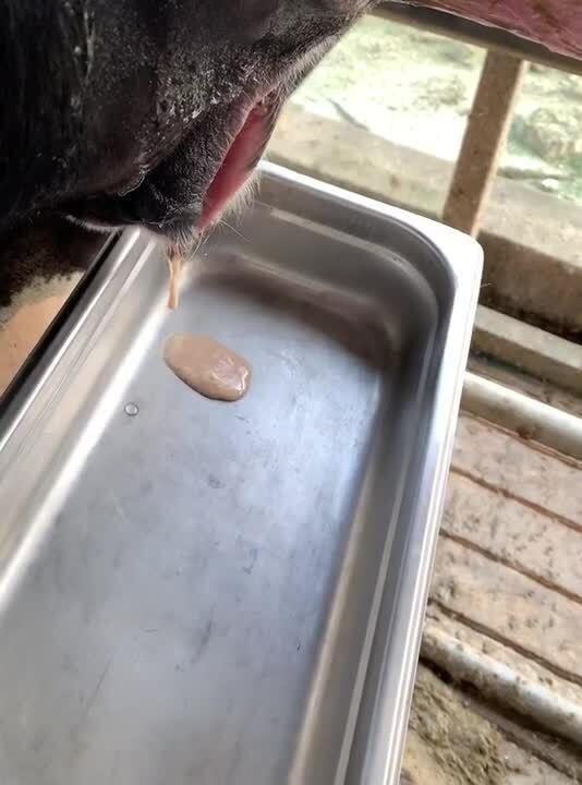
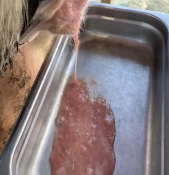
Acute puerperal metritis is characterized by an abnormally enlarged uterus and the presence of fetid, watery, reddish-brownish uterine discharge generally associated with signs of systemic illness such as decreased milk production, inappetence, depression, and fever >39.5°C.
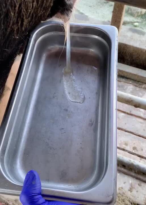
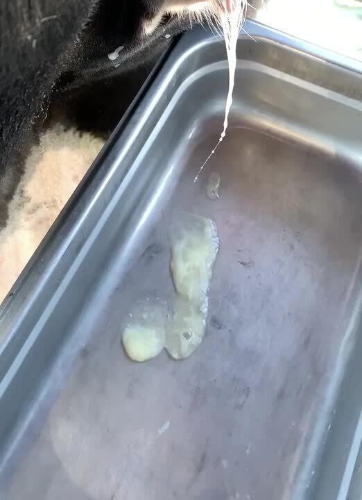
Clinical metritis is characterized by an abnormally enlarged uterus and purulent uterine discharge detectable in the vagina within 21 days after parturition.
Diagnosis of Metritis in Cows
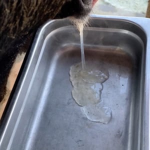
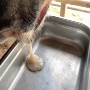
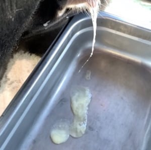
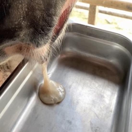
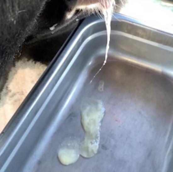
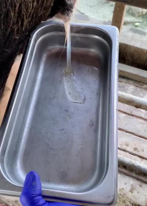
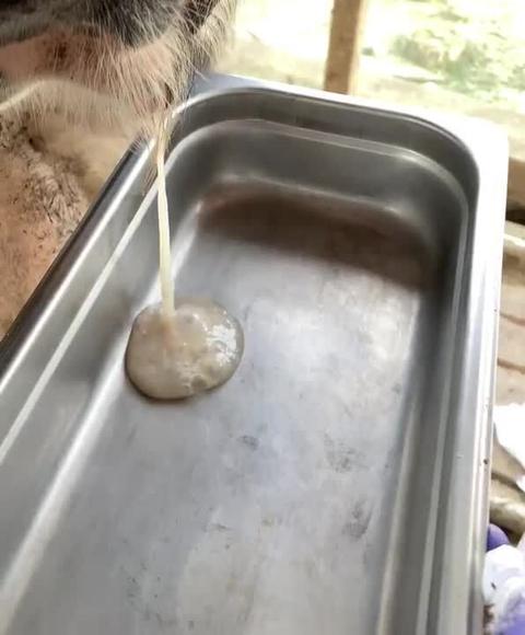
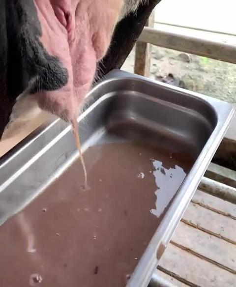
Metritis can be diagnosed based on visual observation of the characteristic discharge at the perineal area. Vaginal discharge collection devices (consisting of a rubber collection cup on a stainless steel rod)1 to collect the discharge accumulated in the cranial vagina are commercially available. A vaginal discharge scoring system for cows in the early postpartum period has been described.

Courtesy of Jessica Prim, Segundo Casaro, and Klibs Galvao.
Courtesy of Jessica Prim, Segundo Casaro, and Klibs Galvao.
Courtesy of Jessica Prim, Segundo Casaro, and Klibs Galvao.
Courtesy of Jessica Prim, Segundo Casaro, and Klibs Galvao.
Courtesy of Jessica Prim, Segundo Casaro, and Klibs Galvao.
Courtesy of Jessica Prim, Segundo Casaro, and Klibs Galvao.

Courtesy of Jessica Prim, Segundo Casaro, and Klibs Galvao.
Courtesy of Jessica Prim, Segundo Casaro, and Klibs Galvao.
Courtesy of Jessica Prim, Segundo Casaro, and Klibs Galvao.

Courtesy of Jessica Prim, Segundo Casaro, and Klibs Galvao.


Courtesy of Jessica Prim, Segundo Casaro, and Klibs Galvao.
Courtesy of Jessica Prim, Segundo Casaro, and Klibs Galvao.

Courtesy of Jessica Prim, Segundo Casaro, and Klibs Galvao.

Courtesy of Jessica Prim, Segundo Casaro, and Klibs Galvao.

Courtesy of Jessica Prim, Segundo Casaro, and Klibs Galvao.

Courtesy of Jessica Prim, Segundo Casaro, and Klibs Galvao.
Courtesy of Jessica Prim, Segundo Casaro, and Klibs Galvao.

Courtesy of Jessica Prim, Segundo Casaro, and Klibs Galvao.

Courtesy of Jessica Prim, Segundo Casaro, and Klibs Galvao.
Courtesy of Jessica Prim, Segundo Casaro, and Klibs Galvao.
Notes
Metricheck, Simcro Ltd, Hamilton, New Zealand
Treatment of Metritis in Cows
Antimicrobial treatment
Fluid therapy
Anti-inflammatory treatment
The primary antimicrobial used to treat metritis in cows is ceftiofur. Two formulations are available on the market: ceftiofur hydrochloride (2.2 mg/kg, IM, every 24 hours for 5 days) and ceftiofur crystalline free acid (6.6 mg/kg, SC in the fat pad behind the ear twice, 72 hours apart). Other antimicrobials that have been used to treat metritis with an efficacy similar to that of ceftiofur include procaine G penicillin (22,000 U/kg, IM, every 24 hours for 5 days), oxytetracycline (11 mg/kg, IV or SC, once at diagnosis, or 6 g, intrauterine, twice a week), and ampicillin trihydrate (11 mg/kg, IM, every 24 hours for 5 days). Only the two ceftiofur formulations and oxytetracycline are labeled to be used in the US.
Cows with more severe clinical signs such as lethargy, anorexia, and fever might benefit from additional supportive care. Fluid therapy should be administered, including 50% dextrose (500 mL, IV slowly; may be repeated in several hours or on successive days as needed), as most metritic cows are ketotic, and hypertonic saline (7.2%) solution (500 mL, IV, as needed judged on clinical condition of patient; provide adequate access to drinking water during administration), as most metritic cows are dehydrated. Anti-inflammatory treatment may be provided with flunixin meglumine (2.2 mg/kg, IV, every 24 hours for 3 days), ketoprofen (3 mg/kg, IM or IV, every 24 hours for up to 3 days), or aspirin (5–300 mg/kg, PO, every 24 hours for 5 days).
On organic dairy farms, alternative therapies used include povidone-iodine (200 mL diluted in 2 L of distilled water) and essential oil based on carvacrol (3.75 mL diluted in 117 mL of distilled water).
Prevention of Metritis in Cows
Several new preventives under development have been shown to reduce the incidence of metritis. These include a vaccine based on strains of Escherichia coli, Fusobacterium necrophorum, and Trueperella pyogenes and virulence factors from these bacteria; a recombinant bovine interleukin-8 formulation; and probiotics based on a combination of Lactobacillus sakei and strains of Pediococcus acidilactici. Other measures that might help reduce the incidence of metritis include keeping the maternity area clean, using bulls that produce small calves, and feeding antioxidants. The antioxidants vitamin E, selenium, and beta-carotene have been shown to reduce the incidence of retained fetal membranes, which ultimately might help reduce the incidence of metritis.
Horses
In mares, two classifications of metritis are relevant: postpartum metritis (acute metritis) and contagious equine metritis (CEM). Postpartum metritis is generally associated with the trauma at foaling and retention of fetal membranes occurring within 10 days after parturition (more commonly 2–4 days after parturition). In contrast, CEM is an acute venereal disease caused by the bacteria Taylorella equigenitalis, which establishes subclinical infections in stallions and chronically infected mares. The disease is more commonly diagnosed in Europe, but technical challenges in the propagation of the causative organism prevent accurate determination of the precise distribution of the disease.
Etiology and Pathogenesis of Metritis in Horses
The specific pathogenesis of postpartum metritis remains unclear. It has been hypothesized that disruption of the endometrial mucosal barrier either is caused during dystocia or gradually progresses after retention of fetal membranes leading to excessive bacterial proliferation and, in some cases, toxemia. The major pathogens, virulence factors, and specific mechanisms are not well elucidated, but more commonly isolated bacteria include Escherichia coli, Streptococcus spp, and sometimes Bacteroides fragilis and clostridial species.
The other type of metritis, CEM, is caused by the gram-negative, microaerophilic coccobacillus Taylorella equigenitalis. It is fastidious and slow growing. Notable strain differences exist. Although transmitted primarily at mating, infected fomites also play a role. Undetected infected mares and stallions are the sources of new outbreaks. Infected stallions show no signs and shelter the organism in the smegma of the prepuce and the surface of the penis, especially in the urethral fossa.
Epidemiology of Metritis in Horses
The transmission rate of CEM is exceptionally high; virtually every mare mated by an infected stallion becomes infected.
Contagious equine metritis is an OIE-listed disease. The US is considered CEM free, and CEM is included on the US National List of Reportable Animal Diseases. It is immediately reportable to the USDA and State Animal Health Officials in all 50 states and territories. Strict import regulations exist in many countries to avoid the introduction of CEM, and the prevalence of the disease appears to be low.
Clinical Signs of Metritis in Horses
Postpartum metritis in horses often is associated with a fetid copious vulvar discharge that may have remnants of fetal membranes, particularly in mares with trauma and necrosis of the birth canal. In some cases, septic shock might develop without evident vulvar copious discharge (likely a result of uterine atony and inertia), and the cases might also lead to the development of laminitis. Repeated abdominal straining occasionally arises when vaginal necrosis, lacerations, or placental remnants are present. The first noticeable clinical signs are inappetence, lethargy, fever, and in some cases, lameness. If the mare develops toxemia, dehydration will follow. The foal frequently suckles, but because of the decreased milk production, it does not seem to be satisfied. The heart and respiratory rates are often increased in mares that have a fever. Mares with postpartum septic metritis seem to be more predisposed to develop laminitis.
In mares with CEM, a copious, mucopurulent vaginal discharge is seen 10–14 days after infected matings. Early return to estrus after a shortened cycle occurs in most affected mares. Although the discharge subsides after a few days, mares may remain infected for several months. Chronically infected mares show no signs. Most mares do not conceive at the time of infected mating. If they do, they may infect the foal at or shortly after birth. Foals so infected may become carriers when they reach sexual maturity.
Diagnosis of Metritis in Horses
Physical examination
Ultrasonography
Bacterial culture
Molecular diagnostic testing
To diagnose metritis in mares, an aseptic manual assessment of the birth canal should be performed to identify associated swelling, hemorrhage, or necrotic tissue. Rectal palpation or transrectal ultrasonographic examination can identify an enlarged flaccid uterus with an excessive amount of variably echogenic contents. A digital inspection of the uterine lumen may reveal placental remnants, including a retained tip of the fetal membranes. Swabbing the uterine lining and lochia for bacterial culture is recommended to identify the organisms present and to guide antimicrobial selection. If the mare is septic, a CBC often reveals absolute neutropenia (ie, often <1,500 neutrophils/mcL) with a degenerative left shift. When concurrent foot soreness is present, radiography is recommended to assess potential problems such as laminitis with rotation and sinking of the distal phalanx.
For CEM, the specific diagnosis depends on isolation of the causative organism. In mares, swabs for culture should be taken from the endometrium (preferably during estrus) and the clitoral fossa and sinuses. Swabs from suspected stallions should be taken from the urethral fossa, urethra, preputial cavity, shaft of the penis, and, if possible, pre-ejaculatory fluid or ejaculate. Stallions should be sampled at least three times before being declared free of the disease. Test-mating suspect stallions to susceptible mares that are then screened bacteriologically constitutes a satisfactory way to determine CEM status. Swabs should be placed in Amies transport medium and kept on ice or at 4°C for delivery to a qualified laboratory within 24 hours (or frozen, if transport takes longer). Some strains are sensitive to streptomycin, while others are resistant, which helps isolate this fastidious, slow-growing organism from contaminants. It is best cultured on chocolate Eugon agar at 37°C in an atmosphere of 5%–10% CO2 in air. Taylorella equigenitalis is asaccharolytic but positive for catalase, cytochrome oxidase, and phosphatase and is unreactive to other conventional biochemical tests. Various serologic tests have been developed, but none is capable of reliably detecting the carrier status. Molecular testing methods such as PCR assay are now commonly used to detect Taylorella from swabs and culture plates and can differentiate between T equigenitalis and T asinigenitalis.
Lesions in mares with CEM include edema and hyperemia of the endometrium, the endocervix, and the vaginal mucosa. The microscopic lesions include invasion of the affected tissues by neutrophils during the acute stage, and by lymphocytes, macrophages, and plasma cells later in the course of the infection.
Treatment of Metritis in Horses
To treat postpartum metritis, broad-spectrum antimicrobials effective against both gram-positive and gram-negative bacteria are recommended. Some of the common antimicrobials used include gentamicin (6.6 mg/kg, IV, every 24 hours) and potassium penicillin (22,000–44,000 U/kg, IV or IM, every 6 hours until 2 days after clinical signs resolve). When endotoxemia is suspected, polymyxin B (1,000–6,000 U/kg, IV slowly, every 6–8 hours) and pentoxifylline (7.5–10 mg/kg, PO or IV, every 8–12 hours) may be added. The latter may improve the malleability of RBCs and improve circulation to the vasculature of the foot. Administration of flunixin meglumine (0.25 mg/kg, IV, every 8 hours) is also recommended in cases where endotoxemia is suspected. Specific treatment of the genital tract includes oxytocin (10–20 U per mare, IM, every 6 hours as needed), stimulating uterine contractions (lasting 20–50 minutes) that aid in the expulsion of uterine contents. Whether there is an additional benefit in administering local antimicrobials by uterine infusion is controversial. There may be some benefit from controlled exercise if foot soreness is not present.
For CEM, in most mares, the uterine infection is eliminated after a few weeks. Those that become chronically infected harbor the T equigenitalis in the clitoral fossa or sinuses. These mares can be treated by thoroughly cleaning the clitoral area with chlorhexidine surgical scrub and then applying nitrofurazone ointment (as for the stallion). In some mares, surgical excision of the clitoral sinuses may be required to rid them of infection. Stallions can be treated by thoroughly cleaning the extended penis with chlorhexidine surgical scrub and then applying nitrofurazone ointment. This treatment should be repeated daily for 5 days and the stallion retested at least 10 days after treatment.
Prevention and Control of Metritis in Mares
Postpartum metritis is highly associated with trauma at foaling and retention of fetal membranes. Thus, the prevention of these conditions may reduce indirectly the odds of postpartum development of metritis.
Control of CEM depends on the identification of infected carrier animals and their treatment or elimination from breeding programs.
Pigs
Metritis in sows and gilts has been associated with prolonged or manual-assisted farrowing. It has also been linked to mastitis and agalactia syndrome. The urinary tract has been suggested as a source of endotoxins and bacteria that can infect the genital tract, leading to metritis.
Etiology and Pathogenesis of Metritis in Pigs
The pathogenesis of metritis in sows and gilts remains unclear. It has been speculated that the factors predisposing sows to urinary tract infection also increase their susceptibility to metritis. The uterine microbiome of sows shares a large number of microbes with the swine fecal microbiome. Some of these microbes commonly cultured in cases of metritis (ie, Escherichia coli, Staphylococcus aureus, and Trueperella pyogenes) have been identified as potential pathogens acquired at parturition. Environmental contamination with feces during farrowing and increased bacterial proliferation and infection of the uterus (in the setting of immunosuppression by progesterone and pregnancy) can lead to the development of metritis. Infusion practices and unclean intervention during parturition also seem to increase the risk of metritis development.
Clinical Signs of Metritis in Pigs
Metritis in sows is characterized by a white to brown, necrotic, malodorous discharge that often has remnants of fetal membranes within 48 hours after arrowing. Metritis may be associated with fever, decreased appetite, anorexia, sepsis, mastitis, and agalactia that can lead to an increased mortality rate for piglets.
Diagnosis of Metritis in Pigs
The diagnosis of metritis in sows is based on the appearance of vaginal discharge and the malodorous nature of this discharge.
Treatment of Metritis in Pigs
The treatment of metritis is based on the use of systemic antimicrobials selected on the basis of in vitro susceptibility. Intrauterine infusions seem to result in an uncertain outcome, with narrow-spectrum antimicrobials reducing some pathogens but increasing others.
Prevention of Metritis in Pigs
Proper hygiene, especially the removal of fecal material during and after parturition, is critical to minimize the risks of metritis development. When manual intervention is needed at farrowing, assistants must wash their hands and arms, wear well-lubricated arm-length sleeves, and wash the perineal region of the sow before manual entry into the birth canal. The facilities used for farrowing and nursing must be cleaned with hot water (>95°C), disinfected, and dried before sows are moved in. It may be helpful to wash sows with soap and water, with particular attention given to the mammary area, before entry into the farrowing facility. All sows requiring manual assistance during parturition should be identified and treated with an effective antimicrobial. Rectal temperatures should be monitored daily, and sows with a rectal temperature >40°C should be treated for 3–5 days.
Goats and sheep
Metritis is often associated with retention of fetal membranes or trauma to the uterus during dystocia, and it may lead to infertility in some goats. In ewes, metritis is often associated with a dead fetus, assisted delivery of multiple lambs without proper hygiene, and uterine prolapse.
Etiology and Pathogenesis of Metritis in Goats and Sheep
The pathogenesis of metritis in ewes and goats is not well understood, but in goats, clostridial organisms, frequently Clostridium tetani and C perfringens, may colonize the uterus and cause toxemia and death of affected does.
Clinical Signs of Metritis in Goats and Sheep
Acute, postpartum metritis in goats and sheep is characterized by a malodorous, dark red uterine discharge. Does are febrile (40°C–41.5°C) and anorectic. In goats, severe metritis may be complicated by peritonitis. Ewes with metritis have a red to brown discharge on the wool of the tail and perineum, and also may have a swollen vulva. The ewe may be lethargic, with a reduced appetite, and a reluctance to nurse that is associated with persistent suckling attempts of the hungry lambs. The ewe may spend extended periods in sternal recumbency.
Treatment of Metritis in Goats and Sheep
Therapy for metritis includes systemic antimicrobials and fluid therapy, if necessary. It has been suggested that repeated oxytocin or prostaglandin injections may facilitate uterine evacuation. Ancillary therapy for tetanus and endotoxemia may be helpful.


