


Vaginal hyperplasia (also known as vaginal fold prolapse, estrus hypertrophy, vaginal eversion, or vaginal protrusion) is the protrusion of edematous vaginal tissue into the vaginal lumen and often through the vulvar lips of the female dog.
Etiology of Vaginal Hyperplasia in Small Animals
Although the exact pathophysiology of vaginal hyperplasia is unknown, during proestrus and estrus, the normal vaginal hyperemia and edema resulting from estrogen stimulus is increased.
Hyperestrogenism is not present when this occurs.
Barring other disorders of the reproductive tract, dogs with vaginal hyperplasia remain fertile. It occurs most commonly during the time of peak estrogen concentration in sexually intact female dogs.
It may occur near parturition due to the decrease in progesterone and increase of estrogen at this time.
Estrogenic stimulation could be confirmed by the following:
Cornification of the vaginal epithelial cells
Characteristic serosanguineous estrous discharge
Estrous behavior
There are 3 degrees of vaginal hyperplasia and prolapse:
Type I is slight to moderate eversion of the vagina floor with no protrusion of the vaginal tissue through the vulva lips.
Type II is a prolapse of the floor and lateral walls of the vaginal through the vulvar lips, forming a tongue or pear-shaped mass.
Type III is a prolapse of the entire vagina circumference as a “doughnut”-shaped mass with a lumen.
Vaginal hyperplasia originates from the floor of the vagina, cranial to the urethral papilla, so that the external urethral orifice is on the ventral surface of the prolapse tissue; however, dysuria rarely occurs. True vaginal prolapse is rare but has been associated with dystocia and precipitous separation of the male and female during coitus.
Epidemiology of Vaginal Hyperplasia in Small Animals
Vaginal hyperplasia has been reported to occur in as many as 8%–10% of bitches. There is no documented breed predisposition, but brachycephalic breeds are overrepresented.

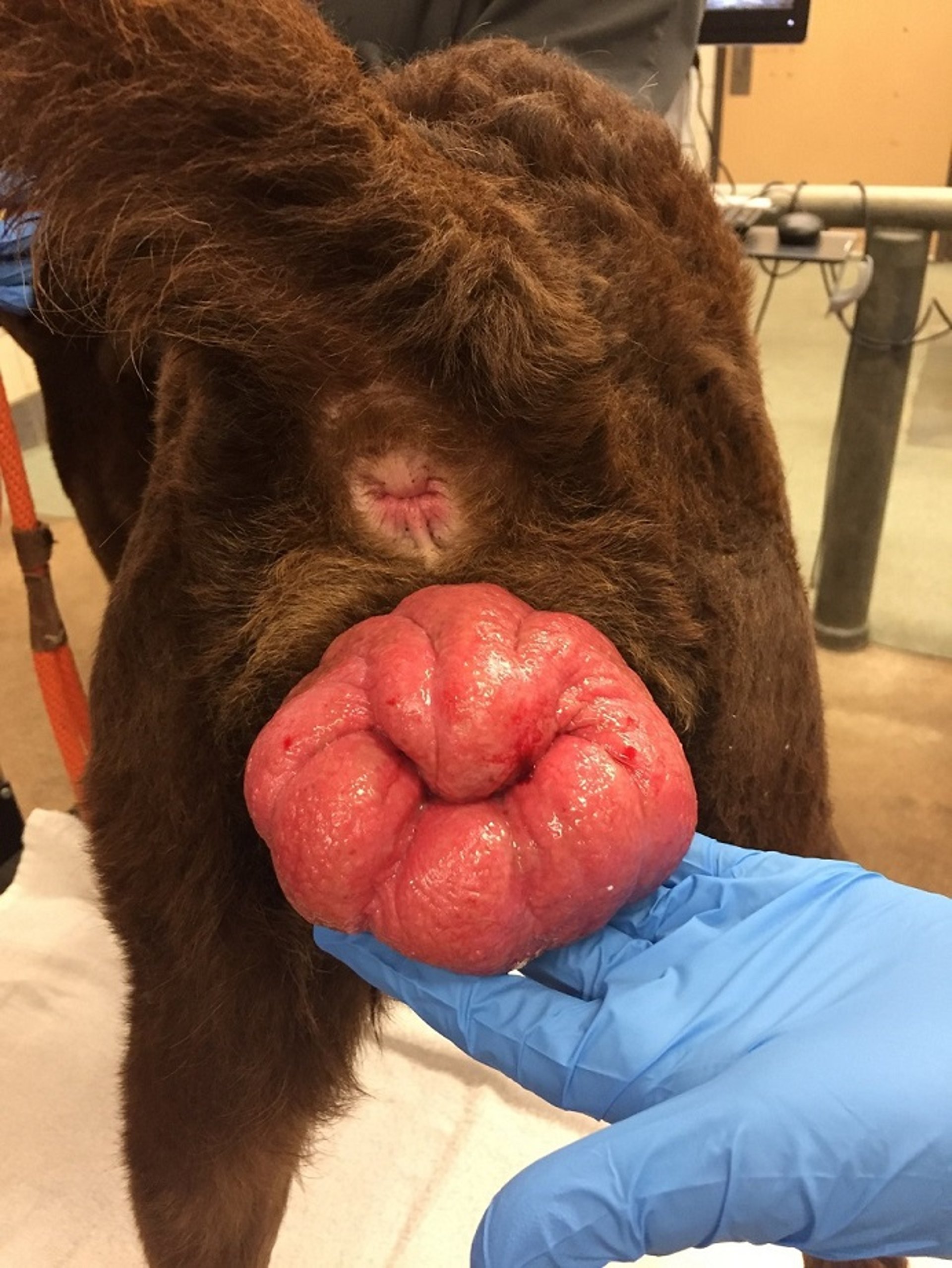
Courtesy of Dr. Jenny Sones.
Clinical Signs of Vaginal Hyperplasia in Small Animals
Clinical signs of vaginal hyperplasia include:
a visible mass protruding from the vulvar lips
reluctance to breed
The primary differential diagnosis is vaginal neoplasia.
Treatment of Vaginal Hyperplasia in Small Animals
Treatment goals are to keep exposed vaginal tissue clean and free from trauma. An E-collar is needed to prevent self-mutilation.
Vaginal prolapses may resolve and regress spontaneously as the estrous cycle progresses and estrogen no longer is the dominate hormone.
Affected dogs that have not ovulated could be induced to ovulate with administration of gonadotropin-releasing hormone (GnRH) or human chorionic gonadotropin (hCG).
Surgical treatment for canine vaginal prolapse include purse-string sutures, hysteropexy, circumferential excision of prolapsed tissue, and ovariohysterectomy. Manual reduction of the prolapse with the placement of purse-string sutures have been reported to be a successful treatment, but this procedure is not recommended because of the discomfort and perivulvar trauma. Recurrence is common even after surgical resection.
Vaginal hyperplasia usually does not recur after complete ovariohysterectomy.


