


Diseases of the Upper Respiratory Tract of Sheep and Goats
Diseases of the upper respiratory tract of sheep and goats include sinusitis, nasal foreign bodies, nasal tumors, and pharyngeal-laryngeal disorders. Clinical signs associated with sinusitis may include unilateral or bilateral, serous to mucopurulent nasal discharge; decreased or lack of airflow through one or both nostrils; coughing; sneezing; and mild to severe respiratory distress. The types of nasal neoplasms reported include adenopapillomas (nasal polyps), adenomas, adenocarcinomas, lymphosarcomas (goats), and squamous cell carcinomas (sheep).
Enzootic nasal tumors are due to an exogenous retrovirus referred to as enzootic nasal tumor virus (ENTV)—specifically, ovine nasal adenocarcinoma virus (ONAV) or caprine nasal adenocarcinoma virus (CNAV). The virus is related to the jaagsiekte sheep retrovirus (JSRV), which causes ovine pulmonary adenocarcinoma. ENTV can be transmitted experimentally by tumor homogenates, thus explaining the widespread occurrence of this condition within some flocks. Enzootic nasal tumors generally affect mature animals (2–4 years old); however, they have been reported in animals as young as 4 months old. The lesions may be unilateral or bilateral, resulting in serous, mucoid, or mucopurulent nasal discharge. Advanced unilateral tumors may cause deviation of the nasal septum, resulting in bilateral nasal discharge.
Affected animals develop progressive signs of inspiratory dyspnea, including open-mouth breathing, decreased airflow at the nares, dullness on percussion over the turbinates, sneezing, and head shaking. In addition, compression of the larynx by enlarged retropharyngeal lymph nodes associated with abscesses of the head may result in stridor. With advancing tumor growth, exophthalmos and facial deformity may occur. Metastatic spread is uncommon. The outcome depends on the tumor type, condition of the animal, and extent of the lesion. In most commercial situations the animal is culled for animal welfare and economic reasons; surgical removal of a noninvasive tumor is rarely undertaken.
The most common problems associated with the pharynx are trauma and abscess formation. Pharyngeal trauma usually results from overly aggressive use of equipment to administer boluses or drenches, or to deworm. Injuries may result in discrete abscesses or extensive and diffuse cellulitis, both of which can interfere with swallowing and lead to respiratory difficulty or distress. Bacteria commonly isolated after an incident of pharyngeal trauma include Trueperella pyogenes, Pasteurella multocida, Mannheimia haemolytica, and Fusobacterium necrophorum.

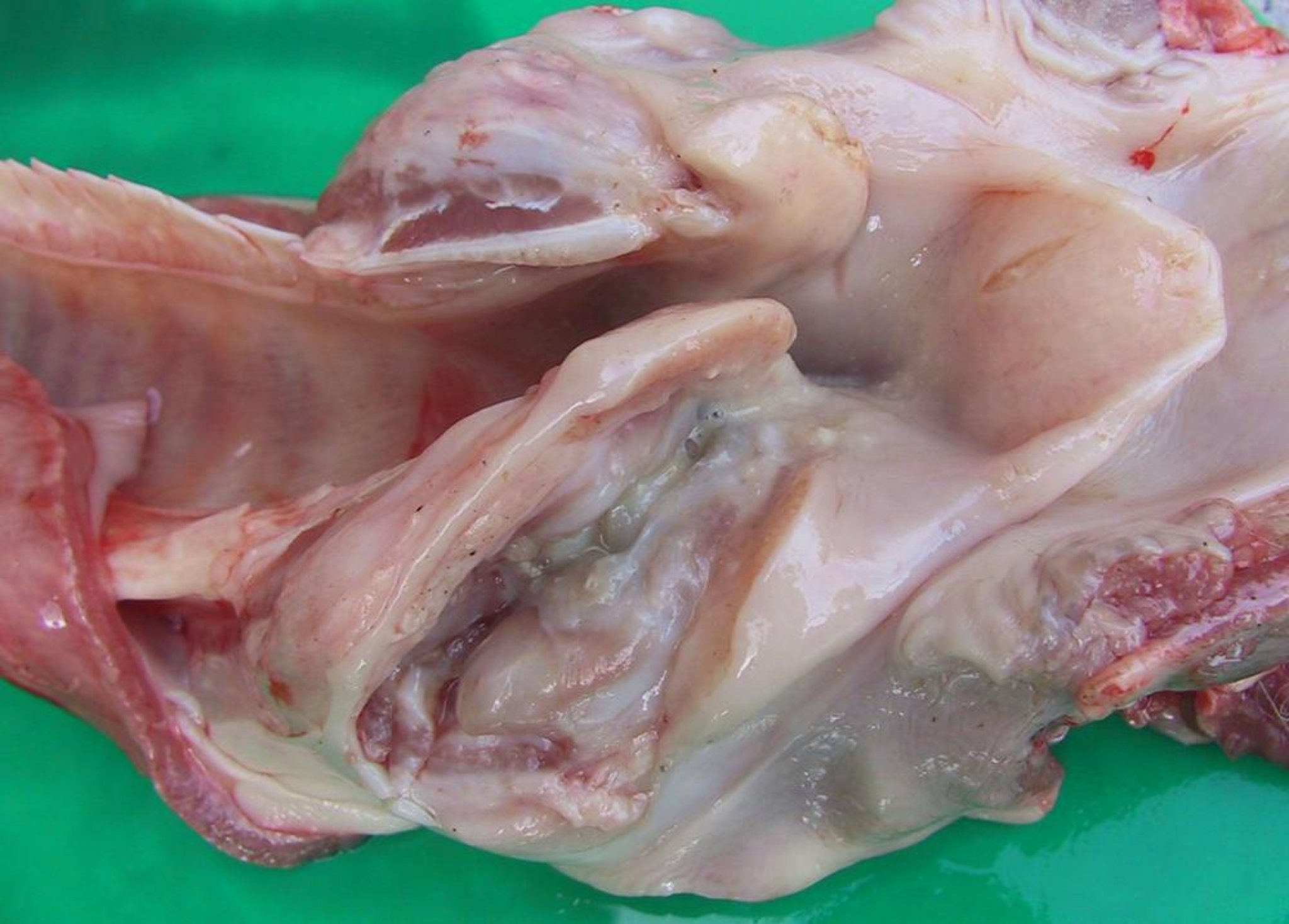
Courtesy of Dr. Philip Scott.

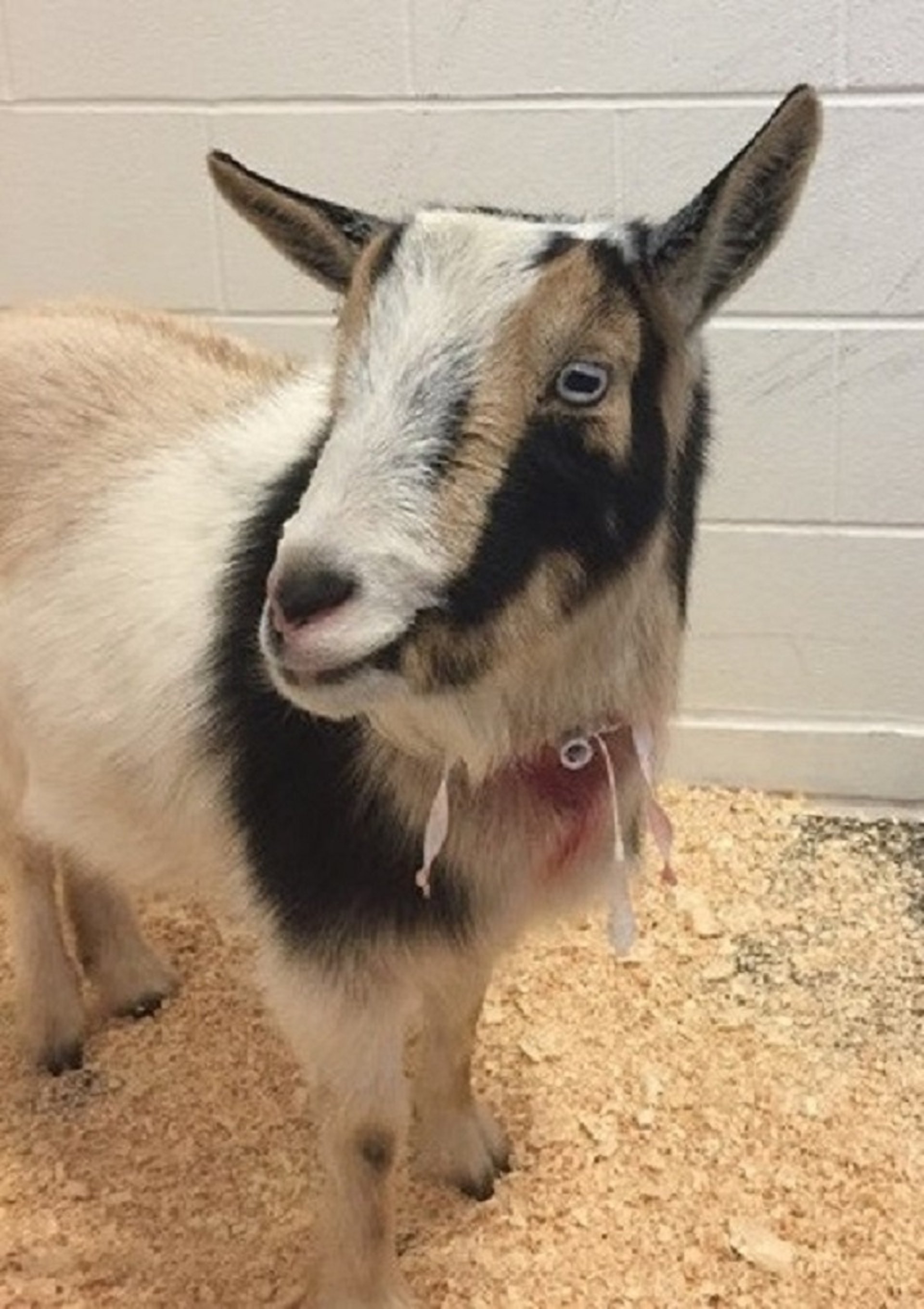
Courtesy of Dr. Jennifer Schleining.
Laryngeal chondritis is an obstructive upper respiratory tract disease characterized by severe dyspnea. It is most common in meat-breed rams and bucks 18–24 months old. Acute onset of severe respiratory distress with marked inspiratory effort and stertor is due to edema of the arytenoid cartilages of the larynx, resulting in narrowing of the lumen. Affected animals stand with the neck extended, head lowered, nostrils flared, and mouth open; they are reluctant to move because of dyspnea.
Delayed identification or inadequate duration of antimicrobial treatment may result in abscess formation within the arytenoid cartilages. Diagnosis can be made with endoscopic evaluation; however, radiography and ultrasonography may reveal abscesses or calcification of affected cartilages. Dyspneic animals can be treated with a temporary tracheostomy, anti-inflammatories, and prolonged antimicrobial treatment. Permanent tracheostomy and resection of affected cartilages have been described; however, response to treatment varies, and the prognosis is guarded to fair.
Diseases of the Lower Respiratory Tract of Sheep and Goats
The most common problem associated with the lower respiratory tract in small ruminants is pneumonia. Pneumonia can be viral, bacteria, or parasitic, and the clinical course may be acute, chronic, or progressive.
Viruses associated with acute pneumonia include parainfluenza type 3 (PI-3), adenovirus, and respiratory syncytial virus. These viral pneumonias most often affect lambs and kids.
PI-3 is an enveloped RNA virus (family Paramyxoviridae) that induces mild interstitial pneumonia. Clinical signs may include coughing, serous nasal or ocular discharge, fever (40°–41°C [104°–106°F]), and an increased respiratory rate. The single ovine PI-3 serotype that has been identified is distinct from the bovine PI-3 serotype. Infection with this virus can be confirmed by its isolation from nasal swabs from affected animals, or by comparison of acute and convalescent serum antibody concentrations. Treatment is usually not warranted in mildly affected animals. In severely affected animals in which secondary pathogens are suspected, antimicrobial treatment is recommended, using drugs with efficacy against the most likely organisms, such as P multocida, M haemolytica, and Mycoplasmaspp. No PI-3 vaccines are designed specifically for use in small ruminants.
Chronic, progressive viral pneumonia is most common in adults. One form is progressive interstitial retroviral pneumonia: in sheep, ovine progressive pneumonia, or maedi; in goats, pneumonia induced by caprine arthritis encephalitis virus (see Caprine Arthritis and Encephalitis). Another form is sheep pulmonary adenomatosis, or ovine pulmonary adenocarcinoma, also known as jaagsiekte, or the infectious lung tumor of sheep and, infrequently, goats.
Chronic, progressive, proliferative changes in the lungs are usually associated with the lentiviruses (family Retroviridae), or so-called slow-virus infections. In both progressive pneumonia and pulmonary adenocarcinoma, the entire lung can change in a gradual process of abnormal cell proliferation. In affected animals, the loss of functional lung tissue results in progressive dyspnea, anorexia, and weight loss.
M haemolytica, P multocida, Bibersteinia trehalosi, Mycoplasma spp, Chlamydia pneumoniae, and Salmonella spp are associated with either primary or secondary bronchopneumonia in sheep and goats. Both P multocida and M haemolytica can be cultured from the upper respiratory tract of normal sheep and goats.
Not all predisposing factors for acute respiratory diseases are known; however, acute viral infections in a susceptible population can alter the protective mechanisms in the respiratory tract so that bacteria may invade lung tissue, multiply, and cause serious disease. An initial infection with the PI-3 virus may predispose an animal to infection with pathogenic Mannheimia haemolytica. Mycoplasma ovipneumoniae alone can cause a mild bronchopneumonia; however, it is often isolated along with M haemolytica from sheep and goats with severe pneumonia, suggesting that the Mycoplasma may predispose the lung to invasion by M haemolytica. In addition, the introduction of new animals, high-density stocking, poor ventilation, and any sudden change to a high plane of nutrition can be stress factors that predispose the animal to developing pneumonia.
Caseous lymphadenitis due to Corynebacterium pseudotuberculosis may result in abscesses of the lungs and mediastinal lymph nodes. The result may be a progressive debilitation in sheep and goats, with or without obvious clinical signs of respiratory disease.
Parasitic or verminous pneumonias of sheep and goats are most commonly due to infection with Dictyocaulus filaria, Muellerius capillaris, or Protostrongylus rufescens. (Also see Lungworm Infection.) In contrast to the acute viral and bacterial pneumonias, which result in a cranioventral bronchopneumonia, verminous pneumonia affects the margins of the caudal lung lobes. Dictyocaulus has a direct life cycle, whereas Protostrongylus and Muellerius have indirect life cycles and rely on a variety of snails and slugs to serve as intermediate hosts. Adult forms of Dictyocaulus and Protostrongylus live in the bronchi, but they rarely cause clinical signs. Adult Muellerius live in alveoli and lung parenchymal tissue and are considered the least pathogenic of the three lungworms. Muellerius appears to cause more problems for goats than for sheep.
Diagnosis of lungworm infection requires Baermann examination of fecal material (also see Internal Parasite Diagnosis in Small Animals). Treatment for lungworm infection is rarely indicated; however, sheep with such infections frequently also carry other nematodes that cause parasitic gastroenteritis and limit production.


