


Potentially dangerous to a vast array of mammals and birds, anticoagulant rodenticides are a relatively common cause of poisoning in pets and wildlife. Intoxications in nontarget domestic animals and wildlife have resulted from direct ingestion of bait product intended for other target species; contamination of feed with anticoagulant concentrate; ingestion of prey or scavenged carrion, resulting in relay toxicity; and, rarely, malicious intent.

Courtesy of Dr. Frederick W. Oehme.

Courtesy of Dr. Frederick W. Oehme.
Anticoagulant rodenticides come in a variety of formulations and active ingredients. First-generation anticoagulant rodenticides (eg, warfarin, pindone, chlorphacinone, and diphacinone) generally require multiple feedings by the target species before causing death. Second-generation anticoagulant rodenticides have median lethal doses (LD50) that are 2.5–200 times lower than those of first-generation anticoagulant rodenticides and generally require only a single feeding to result in the death of the target species. Examples include brodifacoum, bromadiolone, difethiolone, and difenacoum. The toxic-dose calculations in companion animals are widely accepted as based on one-tenth of the lowest reported LD50 for a particular active ingredient in a particular species.
Anticoagulant rodenticides mechanistically inhibit the enzyme vitamin K epoxide reductase, which is crucial in the recycling and production of vitamin K1, a necessary component for clotting factors II, VII, IX, and X. When the production of these clotting factors in the liver is inhibited, prothrombin cannot be adequately converted to thrombin, and coagulopathy results. The serum half-life of the affected coagulation factors ranges from 6.2 to 16.5 hours, and circulating supplies are generally exhausted 24–64 hours after ingestion of a toxic dose of the rodenticide. Elevation of coagulation parameters is thus delayed for 2–5 days after ingestion, with clinical evidence of bleeding typically noted 3–7 days after ingestion of a toxic dose.
Clinical Signs of Anticoagulant Rodenticide Poisoning in Animals
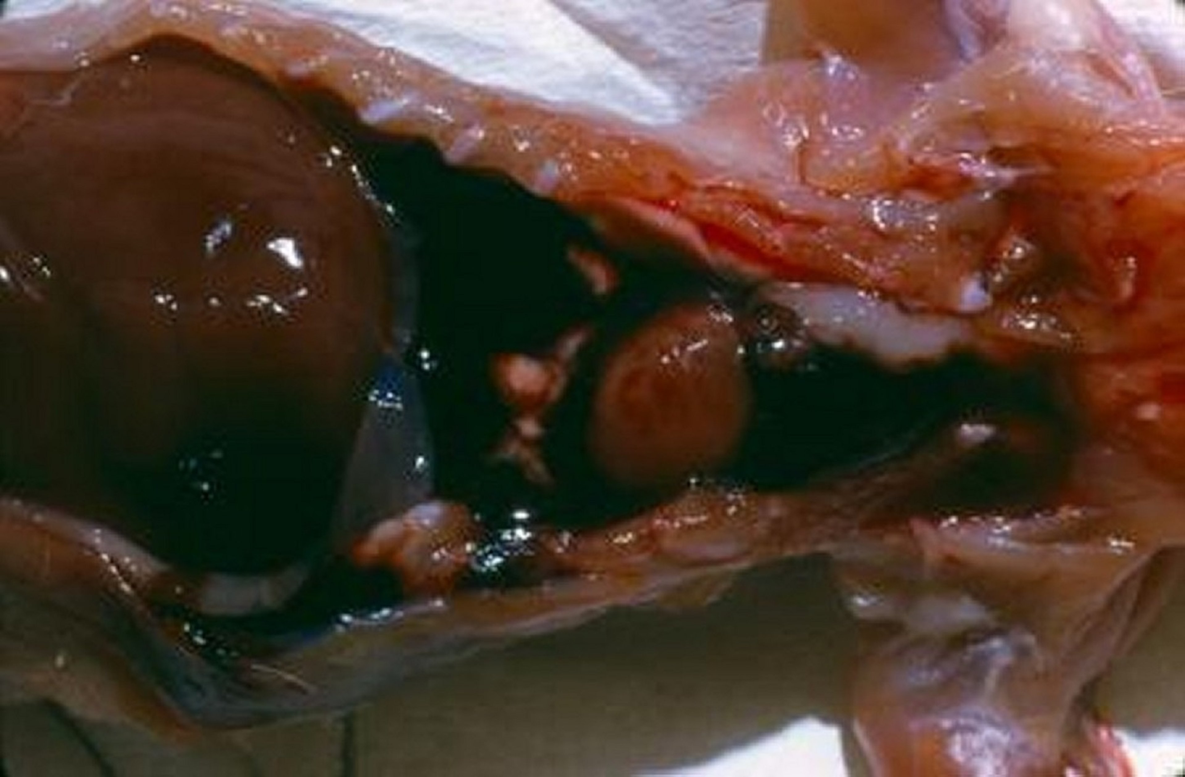
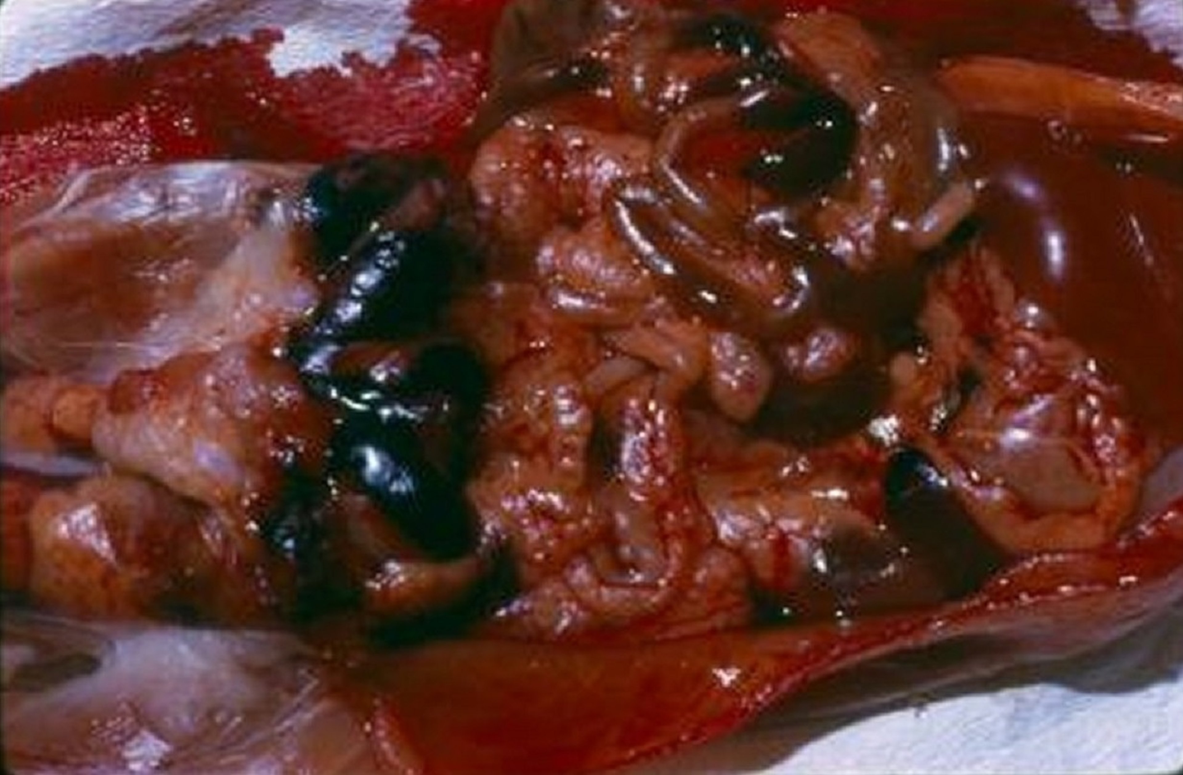
Clinical signs of poisoning by anticoagulant rodenticides are a manifestation of coagulopathy and bleeding. They may include inappetence, lethargy, weakness, epistaxis, respiratory distress (secondary to pleural hemorrhage and resulting hemothorax or pulmonary hemorrhage and resulting hemoptysis), hematoma or petechiation with pallor, cavitary bleeding, hematemesis, melena, hematochezia, hematuria, tachycardia, and hypovolemia. Atypical signs depend on the location of bleeding and may include joint pain, ataxia, seizures, pharyngeal swelling, tracheal compression, or collapse with consequent respiratory distress. Sudden death, though rare, is also possible.
Decontamination of Anticoagulant Rodenticide Poisoning in Animals
For decontamination in non-clinically affected patients:
Induction of emesis, if ingestion occurred within 4 hours, using the following:
In dogs: apomorphine or hydrogen peroxide
In cats: dexmedetomidine, hydromorphone, or xylazine
Activated charcoal (1–3 g/kg, PO as aqueous slurry, repeated as needed at 4- to 6-hour intervals) with a cathartic
Not pursued by all clinicians, given the availability of vitamin K1 antidote
Diagnosis of Anticoagulant Rodenticide Poisoning in Animals
Diagnostic tests in non-clinically affected patients include:
Baseline PCV and total protein concentration (PCV/TP) or CBC; prothrombin time (PT), partial thromboplastin time (PTT), or both PT and PTT
If exposure is questionable and vitamin K1 is withheld: PT 48 hours after ingestion. If PT is elevated, treatment should proceed.
If vitamin K1 is administered: PT 2–3 days after treatment
Diagnostic tests in clinically affected patients include:
Baseline PCV/TP or CBC, coagulation profile, cross-match in preparation for transfusion
PT 2–3 days after vitamin K1 treatment
Serial monitoring of PCV/TP and coagulation profiles every 6–12 hours until normalized
Thoracic or abdominal radiographs in the stable patient if clinical signs dictate need:
For pleural hemorrhage with hemothorax: interlobar fissure lines, extraluminal tracheal deviation or compression
For pulmonary hemorrhage: air bronchogram
For hemoabdomen: loss of serosal detail
Anemia and thrombocytopenia are generally mild to moderate at the time clinical signs of anticoagulant rodenticide poisoning are noted by the animal's owner; however, the extent of anemia may be severe in some cases. The PT will elevate first in dogs and cats; PTT will elevate shortly thereafter. By the time there is clinical evidence of a bleeding event, both PT and PTT are typically elevated.
A prolonged PT, PTT, or thrombin time in the presence of relatively normal fibrinogen, fibrin degradation products, and platelet counts strongly suggests anticoagulant rodenticide toxicosis, as does a positive therapeutic response to vitamin K1. Gastric contents, serum, or plasma can be analyzed for the presence of anticoagulant to confirm a diagnosis. Some veterinary diagnostic laboratories offer an anticoagulant screen to detect most of the anticoagulant rodenticides available in the market in the serum, plasma, liver, or kidney; however, such screens are rarely performed.
Differential diagnoses when hemorrhage is encountered may include disseminated intravascular coagulation, congenital factor deficiencies, von Willebrand disease, platelet deficiencies, and canine ehrlichiosis.
Treatment of Anticoagulant Rodenticide Poisoning in Animals
Treatment for non-clinically affected patients consists of:
Vitamin K1 (2.5 mg/kg, PO, every 12 hours for 28 days, or 5 mg/kg, PO, every 24 hours for 28 days)
The short half-life of warfarin (15 days) indicates that a 2-week course of vitamin K1 is typically sufficient in this less common exposure.
Reassessment of PT 2–3 days after treatment, and if treatment is prolonged, reimplementation of vitamin K1 for an additional 1–2 weeks, until posttreatment PT has normalized
Treatment for clinically affected patients consists of:
Fluid resuscitation of the hypovolemic patient, followed by IV fluid therapy at 1.5–2 times maintenance rates until the patient is noncoagulopathic and stable
Vitamin K1 (2.5 mg/kg, PO, every 12 hours for 28 days, or 5 mg/kg, PO, every 24 hours for 28 days)
Subcutaneous dosing may be considered until the patient can tolerate oral medications.
Oral dosing with food to enhance bioavailability is preferred so as to decrease the risk of hematoma formation with needle sticks.
Coagulation factors are produced within 6–12 hours of implementing treatment; PT and PTT improve within 12–24 hours.
Transfusion of fresh frozen plasma or whole blood in the coagulopathic patient, depending on availability and patient needs, with a focus on providing necessary coagulation factors
Monitoring of PCV/TP and PT every 6–12 hours until values normalize and the patient is stable. Typical duration of inpatient care ranges from 1 to 3 days.
Supportive measures such as gastrointestinal support, antiemetic agents, and oxygen therapy as needed
Restriction of activity until the patient is noncoagulopathic


