Elbow fractures may involve the upper arm bone (humerus) near the elbow—called lower (distal) humeral fractures—or one of the upper forearm bones (radius or ulna)—called radial head fractures or olecranon fractures.
(See also Overview of Fractures.)
Lower Humeral Fractures
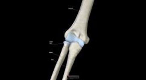
Lower humeral fractures occur in the lower part of the upper arm bone (humerus), which is part of the elbow joint.
Lower humeral fractures usually result from a fall on an outstretched arm or direct force.
Fractures of the lower humerus may damage an artery in the arm, cause bleeding in the joint, or damage nerves that run through the elbow, causing numbness and problems moving the hand and fingers.
Doctors base the diagnosis on results of the physical examination and x-rays.
Treatment involves consultation with an orthopedic surgeon and splinting (when the bones have not moved out of alignment) or surgery (when the broken pieces are separated and/or out of alignment).
Lower humeral fractures often occur in children aged 3 to 11 years old. They usually result from a fall on an outstretched arm or direct force.
The fracture usually extends into the joint and may cause bleeding in the joint.
The main artery of the upper arm (brachial artery) may be damaged, particularly if the broken bones are separated (displaced) or out of alignment. When this artery is damaged, compartment syndrome may develop. As a result, the elbow and wrist may become a permanently stiff (called a contracture).
Sometimes nerves that run through the elbow (radial or median nerve) are damaged. When the radial nerve is damaged, people cannot cock their hand up. When the median nerve (the nerve that is compressed in carpal tunnel syndrome) is damaged, people may have difficulty pinching their thumb and little finger together.
Symptoms of Lower Humeral Fractures
The elbow area is painful and swollen in people with lower humeral fractures. The ability to bend the elbow may be limited.
People may have bruises on their lower forearm. The bruises suggest that a blood vessel is injured.
The forearm and hand may be numb, and people may not be able to move their hand and fingers normally. These symptoms suggest that a nerve is injured.
Diagnosis of Lower Humeral Fractures
X-rays
(See also Diagnosis of Fractures.)
If people think they or a child may have fractured their elbow, the injured person should be seen by a doctor immediately.
Did You Know...
|
Doctors ask people to describe what happened and what their symptoms are. Doctors also examine the elbow.
To determine whether there is a fracture, doctors take x-rays of the elbow joint from different angles.
If doctors suspect a lower humeral fracture but x-rays do not show one, they splint the elbow and have the person come back for additional x-rays, usually in 7 to 10 days.
If doctors suspect a fracture, they also check for damage to blood vessels and nerves in the arm. For example, they check the pulse at the wrist to determine whether blood flow to the hand is normal. To check whether a nerve is damaged, they ask the person to move the fingers and hand and ask whether the person can feel things with the fingers.
Treatment of Lower Humeral Fractures
Consultation with an orthopedic surgeon
Usually surgery to realign the broken bones
Rarely only a splint
An orthopedic surgeon is usually consulted because lower humeral fractures that are close to the elbow often involve nerves or blood vessels and can cause long-term problems.
If the bones have not moved out of alignment, a splint can be used to immobilize the broken bone. Most people are admitted to the hospital so that doctors can determine whether blood vessels or nerves have been damaged. However, if people agree to return for another examination the next day, they may be allowed to go home.
Typically, if the broken pieces are separated and/or out of alignment, surgery (open reduction with internal fixation, or ORIF) is done to realign and immobilize the broken pieces. Because aligning the broken pieces (reduction) can damage nearby nerves and blood vessels, surgery is usually done by a specialist.
Fractures of Part of the Elbow
The pointy bony area of the ulna that projects behind the elbow is called the olecranon (or the olecranon process). Fractures of the olecranon are usually due to a fall on an outstretched hand or a direct blow to the elbow. Avulsion fractures of the olecranon (which occurs when a small chunk of bone attached to a tendon or ligament gets pulled away from the main part of the bone) rarely occur when the triceps tendon pulls off a piece of the olecranon.
Symptoms of Olecranon Fractures
A fractured olecranon is painful, swollen, and tender. It may also be deformed. Elbow extension (straightening the elbow joint) may be limited or impossible. Because the ulnar nerve is sometimes damaged when the olecranon is fractured, the doctor checks for such damage, which may cause numbness and/or tingling in the little finger and part of the ring finger.
Diagnosis of Olecranon Fractures
X-rays
Typically, the doctor x-rays the fracture from two different angles to determine the exact location of the fracture.

ANTONIA REEVE/SCIENCE PHOTO LIBRARY
Treatment of Olecranon Fractures
Splinting
Sometimes surgery
Sometimes antibiotics (for open fractures)
Most olecranon fractures are treated with a long arm posterior splint. Often, the bone fragments can be put back in place (reduced) by manipulation, then held in place by a splint. However, sometimes surgery is required.
Severely displaced fractures (those with a wide gap between the pieces of bone) and those that are comminuted (fractured in at least two places) usually require surgery to align the pieces of bone, which helps them heal in the correct position.
People with open fractures (where the bone pieces break through the skin) are immediately given antibiotics and need to be seen by an orthopedist.
Fractures of the Upper Forearm
Upper forearm fractures can occur in the top (head) of the larger forearm bone (radius), which is part of the elbow joint.
Upper forearm (radial head) fractures usually result from a fall on an outstretched arm.
The elbow is swollen and painful.
Doctors take x-rays from different angles, but these fractures are often hard to see, so diagnosis relies heavily on results of the physical examination.
Most of these fractures can be treated with a sling, but some require surgery.
Range-of-motion exercises are started as soon as possible.
Radial head fractures usually result from a fall on an outstretched arm. They often occur in active adults. They are more common among adults than children.
Fractures in the lower end of the forearm bones are considered wrist fractures.
Symptoms of Upper Forearm Fractures
Moving the elbow is painful, and one side of the elbow is tender when touched.
Blood may leak into the elbow joint, causing swelling. Often, people cannot fully straighten their arm.
Diagnosis of Upper Forearm Fractures
A doctor's evaluation
X-rays
(See also Diagnosis of Fractures.)
Doctors ask people to describe what happened and what their symptoms are. Doctors also examine the elbow.
To check for radial head fractures, doctors take x-rays from different angles. But radial head fractures may be hard to see, so diagnosis relies heavily on results of the physical examination. However, x-rays usually show fluid inside the elbow joint if a fracture is present.
Doctors may insert a needle into the space around the elbow joint and remove fluid (called joint aspiration or arthrocentesis). With the fluid removed, doctors can sometimes determine whether the elbow's movement is limited because of a fracture or because of pain and muscle spasms.
Doctors also gently try to move the elbow to determine whether ligaments are affected.
Treatment of Upper Forearm Fractures
Usually a sling
Range-of-motion exercises
For severe fractures, surgery
Most radial head fractures can be treated with a sling. If the fracture is severe, surgery is done.
Exercises to move the elbow through its full range of motion are started as soon as people can tolerate them (often after a few days). These exercises can prevent permanent stiffness.