


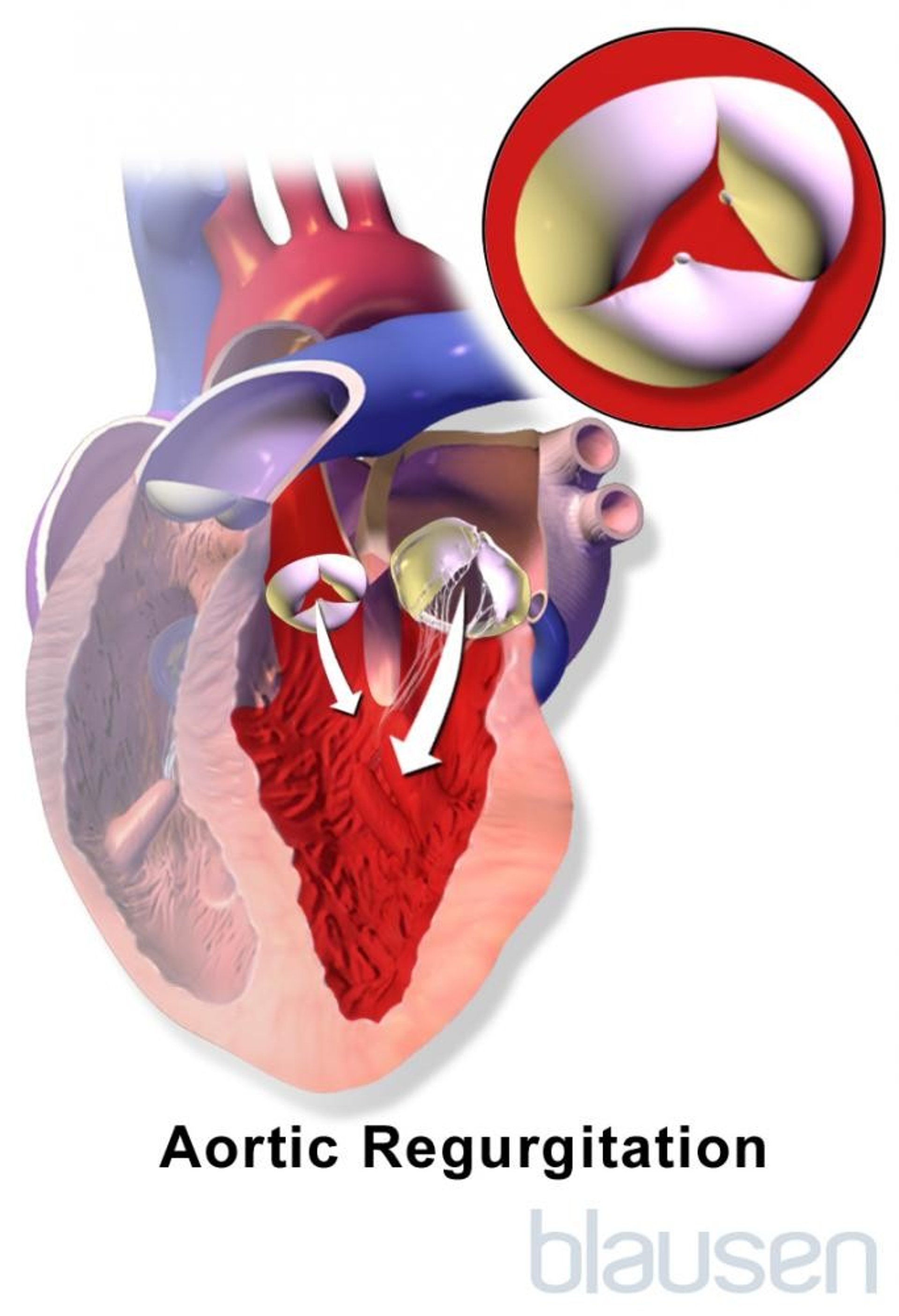
Aortic regurgitation (AR) is incompetency of the aortic valve causing backflow from the aorta into the left ventricle during diastole. Causes include valvular degeneration and aortic root dilation (with or without a bicuspid valve), rheumatic fever, endocarditis, myxomatous degeneration, aortic root dissection, and connective tissue (eg, Marfan syndrome) or rheumatologic disorders. Symptoms include exertional dyspnea, orthopnea, paroxysmal nocturnal dyspnea, palpitations, and chest pain. Signs include widened pulse pressure and an early diastolic murmur. Diagnosis is by physical examination and echocardiography. Treatment is surgical aortic valve replacement or repair.
(See also Overview of Cardiac Valvular Disorders.)
Etiology of Aortic Regurgitation
Aortic regurgitation may be acute (very uncommonly) or chronic.
The primary causes of acute aortic regurgitation are
The primary causes of chronic aortic regurgitation in adults are
Degeneration of the aortic valve and root (with or without a bicuspid valve)
Myxomatous degeneration
Trauma
The most common cause of chronic aortic regurgitation in children is
Ventricular septal defect with aortic valve prolapse
Aortic regurgitation due to myxomatous degeneration may develop in patients with Marfan syndrome or Ehlers-Danlos syndrome.
Rarely, aortic regurgitation is caused by seronegative spondyloarthropathies (eg, ankylosing spondylitis, reactive arthritis, psoriatic arthritis), rheumatoid arthritis, systemic lupus erythematosus, arthritis associated with ulcerative colitis, luetic (syphilitic) aortitis, osteogenesis imperfecta, supravalvular or discrete membranous subaortic stenosis, Takayasu arteritis, rupture of a sinus of Valsalva, acromegaly, and giant cell arteritis.
Pathophysiology of Aortic Regurgitation
In aortic regurgitation, volume overload of the left ventricle (LV) occurs because the LV receives blood regurgitated from the aorta during diastole in addition to blood from the left atrium.
In acute aortic regurgitation, the LV does not have time to dilate to accommodate the increased volume, which then causes a rapid increase in left ventricular pressure and subsequently pulmonary edema and decreased cardiac output.

In chronic aortic regurgitation, LV hypertrophy and dilation can gradually occur, so normal left ventricular pressures and cardiac output are maintained. But decompensation eventually develops, ultimately causing arrhythmias, LV impairment, and heart failure (HF).
Symptoms and Signs of Aortic Regurgitation
Acute aortic regurgitation causes symptoms of heart failure (dyspnea, fatigue, weakness, edema) and cardiogenic shock (hypotension with resultant multisystem organ damage).
Chronic aortic regurgitation is typically asymptomatic for years; progressive exertional dyspnea, orthopnea, paroxysmal nocturnal dyspnea, and palpitations develop insidiously.
Symptoms of heart failure correlate poorly with objective measures of left ventricular function. Chest pain (angina pectoris) affects only about 5% of patients who do not have coexisting coronary artery disease (CAD) and, when it occurs, is especially common at night. Patients may present with endocarditis (eg, fever, anemia, weight loss, embolic phenomena) because the abnormal aortic valve is predisposed to bacterial seeding.
Signs vary by severity and acuity. Signs in acute aortic regurgitation reflect heart failure and cardiogenic shock and typically include tachycardia, cool extremities, lung crackles, and low blood pressure (BP). The first heart sound (S1) is usually absent (because aortic and LV diastolic pressures equalize), and a third heart sound (S3) is common. An AR murmur may be absent even if AR is severe, although an Austin Flint murmur is common.
As chronic disease progresses, systolic blood pressure increases while diastolic blood pressure decreases, creating a widened pulse pressure. With time, the LV impulse may become enlarged, increased in amplitude, and displaced downward and laterally, with systolic depression of the entire left parasternal area, giving a rocking motion to the left chest.
A systolic apical or carotid thrill may become palpable in later stages of AR; it is caused by large forward stroke volumes and low aortic diastolic pressure.
Auscultatory findings include a normal S1 and a nonsplit, loud, sharp or slapping second heart sound (S2) caused by increased elastic aortic recoil. The murmur of AR is often unimpressive. The murmur is blowing, high-pitched, diastolic, and decrescendo, beginning soon after the aortic component of S2 (A2); it is loudest at the third or fourth left parasternal intercostal space. The murmur is heard best with the diaphragm of the stethoscope when the patient is leaning forward, with breath held at end-expiration. It increases in volume in response to maneuvers that increase afterload (eg, squatting, isometric handgrip). If AR is slight, the murmur may occur only in early diastole. If LV diastolic pressure is very high, the murmur is short because aortic and LV diastolic pressures equalize earlier in diastole.
Other abnormal sounds include a forward ejection and backward regurgitant flow (to-and-fro) murmur, an ejection click soon after the S1, and an aortic ejection flow murmur. A diastolic murmur heard near the axilla or mid left thorax (Cole-Cecil murmur) is caused by fusion of the aortic murmur with the S3, which is due to simultaneous filling of LV from the left atrium and AR. A mid-to-late diastolic rumble heard at the apex (Austin Flint murmur) may result from rapid regurgitant flow into the LV, causing mitral valve leaflet vibration at the peak of atrial flow; this murmur mimics the diastolic murmur of mitral stenosis.
Other signs are unusual; sensitivity and specificity are low or unknown. Visible signs include
Head bobbing (de Musset sign)
Pulsation of the fingernail capillaries (Quincke sign, best seen while applying slight pressure)
Pulsation of the uvula (Müller sign)
Palpable signs include
Large-volume pulse with rapid rise and fall (slapping, water-hammer, or collapsing pulse)
Pulsation of the carotid arteries (Corrigan sign)
Pulsation of the retinal arteries (Becker sign)
Pulsation of the liver (Rosenbach sign)
Pulsation of the spleen (Gerhard sign)
BP findings may include
Popliteal systolic pressure ≥ 60 mm Hg higher than brachial pressure (Hill sign)
A fall in diastolic BP of > 15 mm Hg with arm elevation (Mayne sign)
Auscultatory signs include a
Sharp sound heard over the femoral pulse (pistol-shot sound, or Traube sign)
Femoral systolic bruit distal and a diastolic bruit proximal to arterial compression (Duroziez sign)
Diagnosis of Aortic Regurgitation
Echocardiography
Diagnosis of aortic regurgitation is suspected based on history and physical examination findings and confirmed by echocardiography. Doppler echocardiography is the test of choice to detect and quantify the magnitude of regurgitant blood flow and grade overall severity of the AR. Two-dimensional echocardiography can quantify aortic root size and anatomy and LV function.
Severe chronic aortic regurgitation is suggested by any of the following:
Color Doppler jet width ≥ 65% of the LV outflow tract diameter
Holodiastolic flow reversal in the abdominal aorta (specific for severe AR)
Regurgitant volume ≥ 60 mL/beat
Regurgitation fraction ≥ 50%
Vena contracta > 6 mm (the narrowest diameter of the fluid stream downstream of the abnormal valve orifice)
Echocardiography can also assess severity of pulmonary hypertension secondary to LV failure, detect vegetations or pericardial effusions (eg, in aortic dissection), and provide information about prognosis. Coarctation is associated with bicuspid valve and is detected by placing the ultrasound transducer at the suprasternal notch. Transesophageal echocardiography provides additional delineation of aortic dilatation and valve anatomy, which is especially useful when surgical repair is being considered. If the aorta is enlarged, gated CT or MRI is recommended to evaluate the entire thoracic aorta. MRI also can help assess LV function and degree of AR when echocardiographic images are suboptimal.
ECG and chest x-ray should be done.
ECG may show repolarization abnormalities with or without QRS voltage criteria of LV hypertrophy, left atrial enlargement, and T-wave inversion with ST-segment depression in precordial leads.
Chest x-ray may show cardiomegaly and a prominent aortic root in patients with chronic progressive AR. If AR is severe, signs of pulmonary edema and HF may also be present. Exercise testing may help assess functional capacity and symptoms in patients with documented AR and equivocal symptoms.
Coronary angiography should be done before surgery, even if no angina is present because about 20% of patients with severe AR have significant coronary artery disease, which may need concomitant coronary artery bypass graft surgery.
First-degree relatives of patients with a bicuspid valve should be screened using echocardiography because 20 to 30% will be similarly affected.
Treatment of Aortic Regurgitation
Aortic valve replacement or repair
Sometimes vasodilators, diuretics, and nitrates
When aortic root dilatation is part of the mechanism of aortic regurgitation, angiotensin-receptor blockers may slow progression of root dilation, making them favored medications for patients with concomitant hypertension. These medications do not reduce the severity of aortic regurgitation or alter disease progression.
Intervention is either surgical aortic valve replacement or (less commonly) valve repair. Percutaneous options are being developed (1Anticoagulation for patients with a prosthetic cardiac valve).
Transcatheter aortic valve implantation (TAVI) is challenging because of the dilated aortic annulus and lack of sufficient leaflet calcification, leading to prosthetic valve migration and/or paravalvular leak.
Patients who are not candidates for surgery benefit from treatment of heart failure (eg, with diuretics, vasodilators, nitrates). Beta-blockers should be used with caution because they block compensatory tachycardia and worsen AR by prolonging diastole. Intra-aortic balloon pump insertion is contraindicated because the diastolic balloon inflation worsens AR.
Patients with severe AR who do not meet the criteria for intervention should be reevaluated by physical examination and echocardiography every 6 to 12 months.
Antibiotic prophylaxis against endocarditis is no longer recommended for aortic regurgitation except for patients who have had valve replacement (see table Recommended Endocarditis Prophylaxis During Oral-Dental or Respiratory Tract Procedures).
Indications for intervention
Intervention is indicated when
AR is severe and is causing symptoms
AR is severe and is causing LV dysfunction (EF ≤ 55%, LV end-systolic dimension > 50 mm, or indexed to body surface area > 25 mm/m2)
AR is severe and at least 3 serial studies show a progressive decline in EF to 55 to 60%, or progressive increase in LV end-diastolic dimension to > 65 mm
Patients with an enlarged ascending aorta frequently have AR as well as an increased risk of aortic dissection. A high risk of aortic dissection may be the first indication for cardiac surgery, which should be undertaken when
Ascending aortic diameter is> 55 mm
Bicuspid valve is present with ascending aortic diameter 50 to 55 mm and aortic growth rate > 5 mm/year, aortic coarctation, or a family history of aortic dissection
Marfan syndrome is present with ascending aortic diameter > 50 mm (or less if the aortic growth rate is > 5 mm/year) or a family history of aortic dissection at < 50 mm diameter
When cardiac surgery is being done for other reasons, concomitant aortic surgery is indicated if the ascending aortic diameter is ≥ 45 mm.
Treatment reference
1. Otto CM, Nishimura RA, Bonow RO, et al: 2020 ACC/AHA Guideline for the Management of Patients With Valvular Heart Disease: Executive Summary: A Report of the American College of Cardiology/American Heart Association Joint Committee on Clinical Practice Guidelines. Circulation 143(5):e35–e71, 2021. doi: 10.1161/CIR.0000000000000932
Prognosis for Aortic Regurgitation
With treatment, the 10-year survival for patients with mild to moderate aortic regurgitation is 80 to 95%. With appropriately timed valve replacement (ie, before heart failure and using accepted criteria for intervention), long-term prognosis for patients with moderate to severe AR is good. However, the prognosis for patients with severe AR and heart failure is considerably poorer.
Key Points
The primary causes of acute aortic regurgitation (AR) are infective endocarditis and dissection of the ascending aorta; chronic AR in adults is most often caused by degeneration of the aortic valve or root.
Acute AR causes symptoms of heart failure and cardiogenic shock, but signs of AR may be absent.
Chronic AR is typically asymptomatic for years followed by progressive exertional dyspnea, orthopnea, and paroxysmal nocturnal dyspnea.
Typical heart sounds with chronic AR include a normal first heart sound (S1) followed by a sharp or slapping second heart sound (S2) and a blowing, high-pitched, decrescendo diastolic murmur.
Acute AR requires prompt aortic valve replacement or repair.
Chronic AR requires aortic valve replacement or repair when symptoms or left ventricular dysfunction develops; patients who meet criteria but are not candidates for surgery benefit from treatment of heart failure.
AR is sometimes accompanied by a dilated ascending aorta. Indications for surgery on the ascending aorta may occur before surgery is indicated for AR.


