


Diagnostic procedures may be needed to confirm a diagnosis suggested by the medical history and neurologic examination.
Imaging Tests
Imaging tests commonly used to diagnose nervous system (neurologic) disorders include the following:
Computed tomography (CT)
Spinal Tap
Cerebrospinal fluid flows through a channel (the subarachnoid space) between the layers of tissue (meninges) that cover the brain and spinal cord. This fluid, which surrounds the brain and spinal cord, helps cushion them against sudden jarring and minor injury.
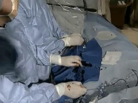
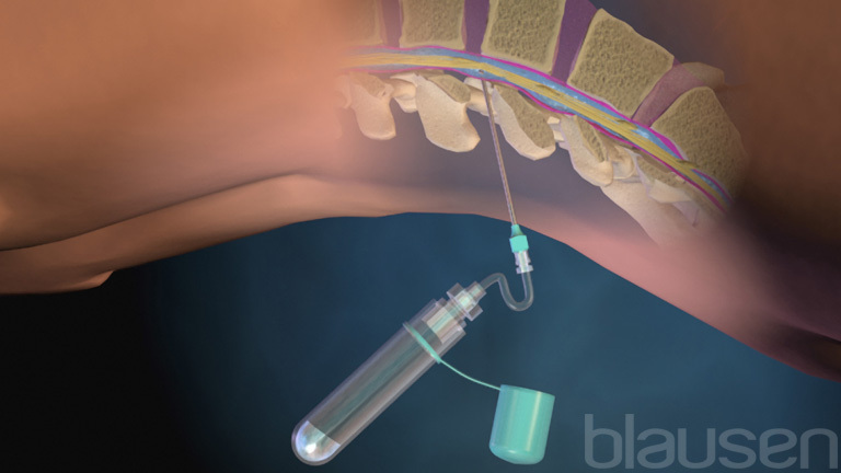
For a spinal tap (lumbar puncture), a sample of cerebrospinal fluid is withdrawn with a needle and sent to a laboratory for examination.
The cerebrospinal fluid is checked for evidence of infections, tumors, and bleeding in the brain and spinal cord. These disorders may change the content and appearance of the cerebrospinal fluid, which normally contains few red and white blood cells and is clear and colorless. For example, the following findings suggest certain disorders:
An increase in the number of white blood cells in the cerebrospinal fluid suggests an infection or inflammation of the brain and spinal cord.
Cloudy fluid, due to the presence of many white blood cells, suggests meningitis (infection and inflammation of the tissues covering the brain and spinal cord) or sometimes encephalitis (infection and inflammation of the brain).
High protein levels in the fluid may result from any injury of the brain, the spinal cord, or a spinal nerve root (the part of a spinal nerve next to the spinal cord).
Abnormal antibodies in the fluid suggest multiple sclerosis or an infection.
Low sugar (glucose) levels suggest meningitis or cancer.
Blood in the fluid may indicate a brain hemorrhage—for example, when a bulge in a weakened artery in the brain (aneurysm) bursts (ruptures).
An increase in the fluid’s pressure can result from many disorders, including brain tumors and meningitis.
Doctors do not do a spinal tap when the pressure within the skull is increased, for example, when there is a mass (such as a tumor or abscess) in the brain. In such cases, a spinal tap may suddenly reduce pressure below the brain. As a result, the brain may shift and be pressed through one of the small openings in the relatively rigid tissues that separate the brain into compartments (called herniation). Herniation puts pressure on the brain and may be fatal. The medical history and neurologic examination help doctors determine whether herniation is a risk. For example, doctors use an ophthalmoscope to examine the optic nerve, which bulges when the pressure within the skull is increased. As another precaution before a spinal tap is done, CT or MRI of the head is often done to check for masses.
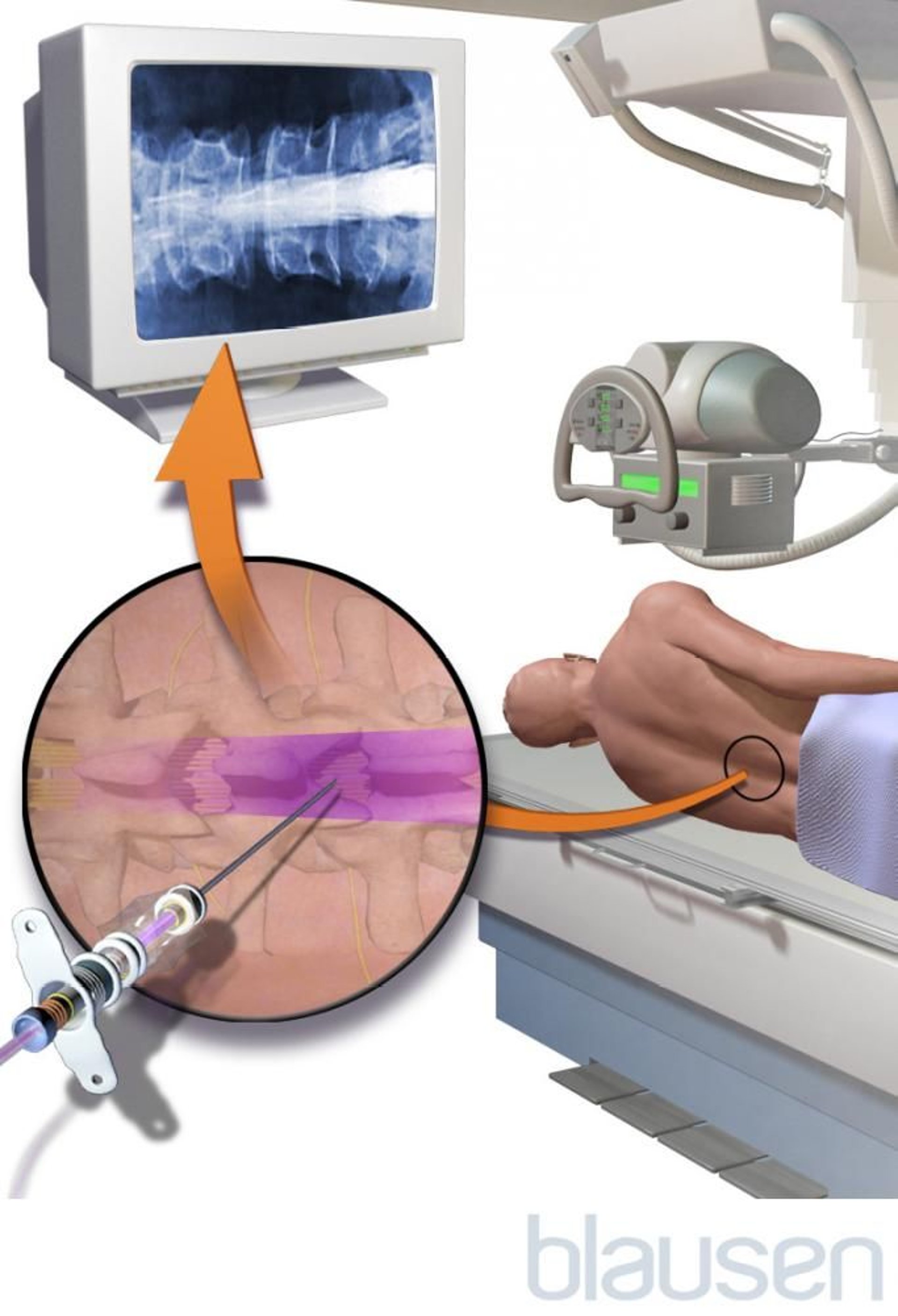
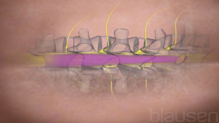
How a Spinal Tap Is Done
Cerebrospinal fluid flows through a channel (called the subarachnoid space) between the middle and inner layers of tissue (meninges) that cover the brain and spinal cord. To remove a sample of this fluid, a doctor inserts a small, hollow needle between two bones (vertebrae) in the lower spine, usually the 3rd and 4th or the 4th and 5th lumbar vertebrae, below the point where the spinal cord ends, and then into the subarachnoid space—the space between the layers of tissue (meninges) that cover the spinal cord (and brain). Usually, people lie on their side with their knees curled to their chest. This position widens the space between the vertebrae, so that the doctor can avoid hitting the bones when the needle is inserted. Cerebrospinal fluid is then allowed to drip into test tubes, and the samples are sent to a laboratory for examination. |

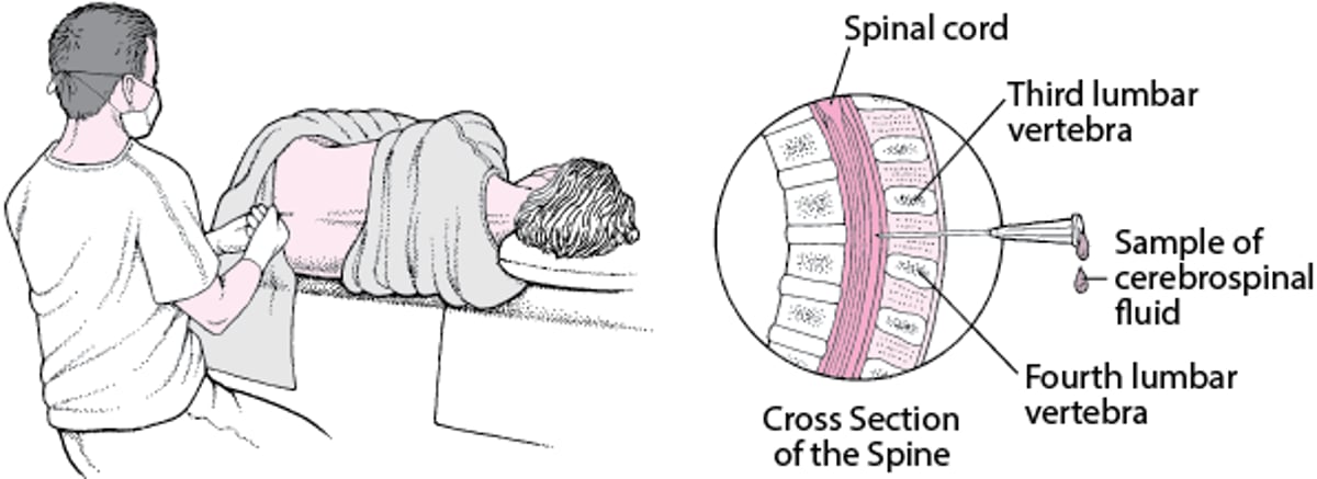
For a spinal tap, people typically lie on their side in a bed and draw their knees to their chest. A local anesthetic is used to numb the insertion site. Then, a needle is inserted between two vertebrae in the lower spine below the end of the spinal cord.
During a spinal tap, doctors can measure the pressure within the skull. Pressure can be higher than normal in people with idiopathic intracranial hypertension and certain other disorders of the brain and surrounding structures. Pressure is measured by attaching a gauge (manometer) to the needle used for the spinal tap and noting the height of the cerebrospinal fluid in the gauge.
A spinal tap may be done for other reasons:
To reduce pressure within the skull (intracranial pressure) in people with idiopathic intracranial hypertension
To give a radiopaque contrast agent before myelography
To give drugs when they are needed to work quickly or to target a specific area of the brain, spinal cord, or meninges—for example, to treat infections or cancer affecting these structures
A spinal tap usually takes no more than 15 minutes.
About 1 of 10 people develops a headache when standing up after a spinal tap (called a low-pressure headache). The headache usually disappears after a few days to weeks. However, if the headache is still troublesome after a few days, doctors may inject a small amount of the person's blood into the area around where the spinal tap was done. This procedure, called a blood patch, slows the leakage of cerebrospinal fluid and may relieve the headache. Other problems are very rare.
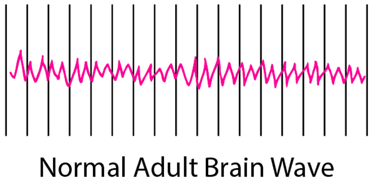
Electroencephalography
Electroencephalography (EEG) is a simple, painless procedure in which the brain’s electrical activity is recorded as wave patterns, printed on paper, and/or recorded in a computer. EEG can help identify the following:
Certain metabolic or structural disorders of the brain
Rare conditions such as Creutzfeld-Jacob disease
For example, EEG can help identify where a seizure originates and show changes in electrical activity associated with confusion, which may result from disorders such as liver failure (liver encephalopathy) or certain drugs.
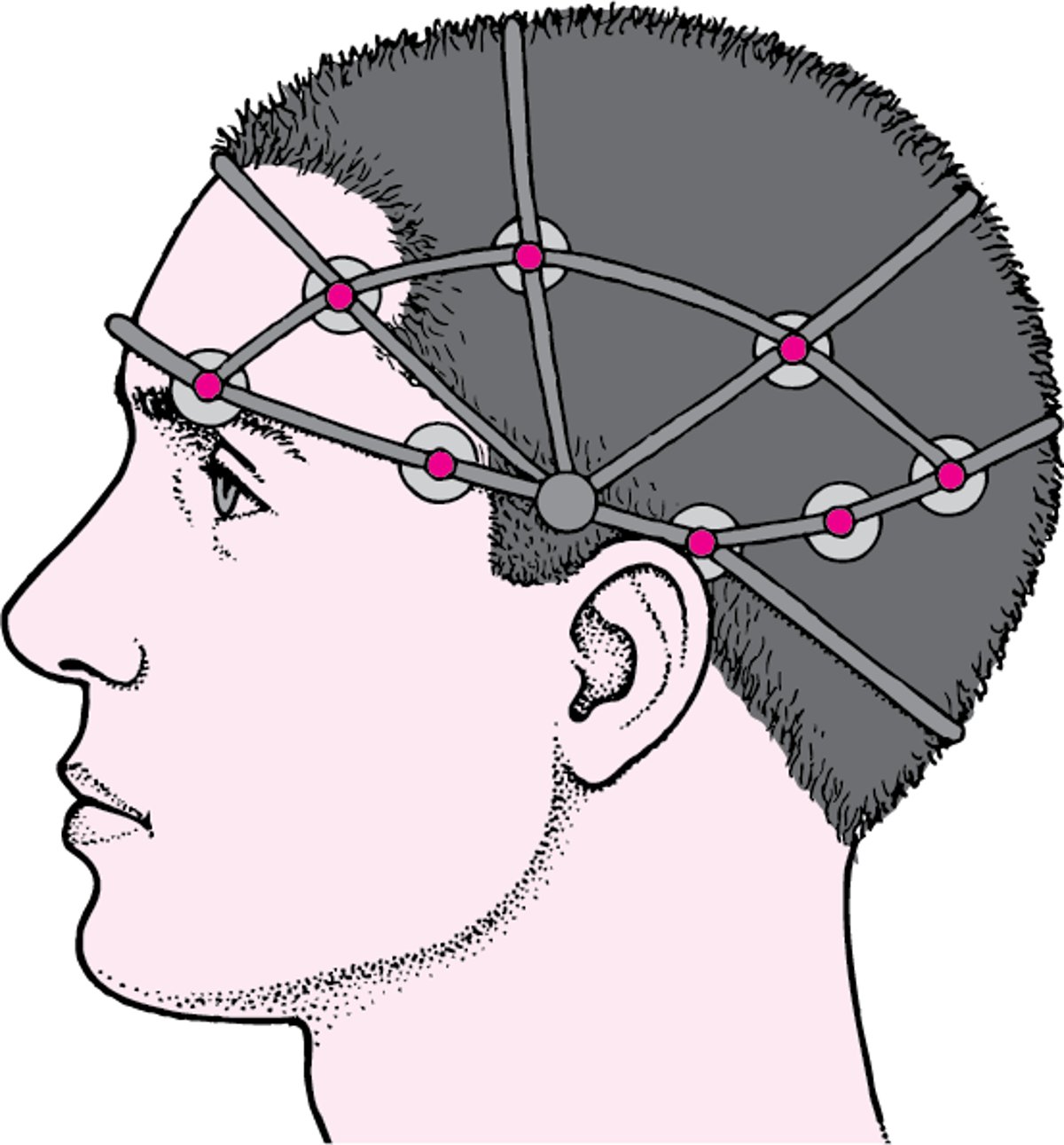
For the procedure, an examiner places small, round adhesive sensors (electrodes) on the person’s scalp. The electrodes are connected by wires to a machine, which produces a record (tracing) of small changes in voltage detected by each electrode. These tracings constitute the electroencephalogram (the EEG).
If a seizure disorder is suspected but the initial EEG is normal, another EEG is done after using a tactic that makes seizure activity more likely. For example, the person may be deprived of sleep, be asked to breathe deeply and rapidly (hyperventilate), or be exposed to a flashing light (stroboscope).
Sometimes (for example, when behavior that resembles a seizure is difficult to distinguish from a psychiatric disorder), the brain’s electrical activity is recorded for 24 hours or longer while people are monitored in the hospital by a video camera. This procedure is called video EEG. The camera detects the seizure-like behavior, and by examining the EEG at that moment, doctors can determine whether brain activity indicates a seizure or is normal, suggesting a psychiatric disorder.
Recording Brain Activity
An electroencephalogram (an EEG) is a recording of the brain’s electrical activity. The procedure is simple and painless. About 20 small adhesive electrodes are placed on the scalp, and the brain’s activity is recorded under normal conditions. Sometimes the person is exposed to various stimuli, such as bright or flashing lights, to try to provoke a seizure. | |


Electromyography and Nerve Conduction Studies
Electromyography and nerve conduction studies help doctors determine whether muscle weakness, sensory loss, or both result from injury to the following:
Spinal nerve root (for example, due to a ruptured disk in the spine of the neck or lower back)
Peripheral nerve (for example, due to carpal tunnel syndrome or diabetic neuropathy)
Connections between nerve and muscle (neuromuscular junction)—for example, due to myasthenia gravis, botulism, or diphtheria
Muscle (for example, due to polymyositis)
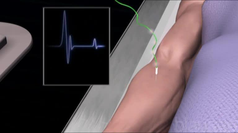
Electromyography
In electromyography (EMG), a small needle is inserted into a muscle to record the electrical activity of the muscle when the muscle is at rest and when it is contracting. Normally, resting muscle produces no electrical activity. A slight contraction produces some electrical activity, which increases as the contraction increases.
The record produced by EMG is called the electromyogram. It is abnormal if muscle weakness results from a problem with a spinal nerve root, peripheral nerve, muscle, or neuromuscular junction. Each type of problem produces a distinctive pattern of abnormalities, which can be identified based on the person's symptoms and results of the examination and electromyography.

Unlike CT or EEG, which can be done routinely by technicians, EMG requires the expertise of a neurologist, who chooses the appropriate nerves and muscles to test and interprets the results.
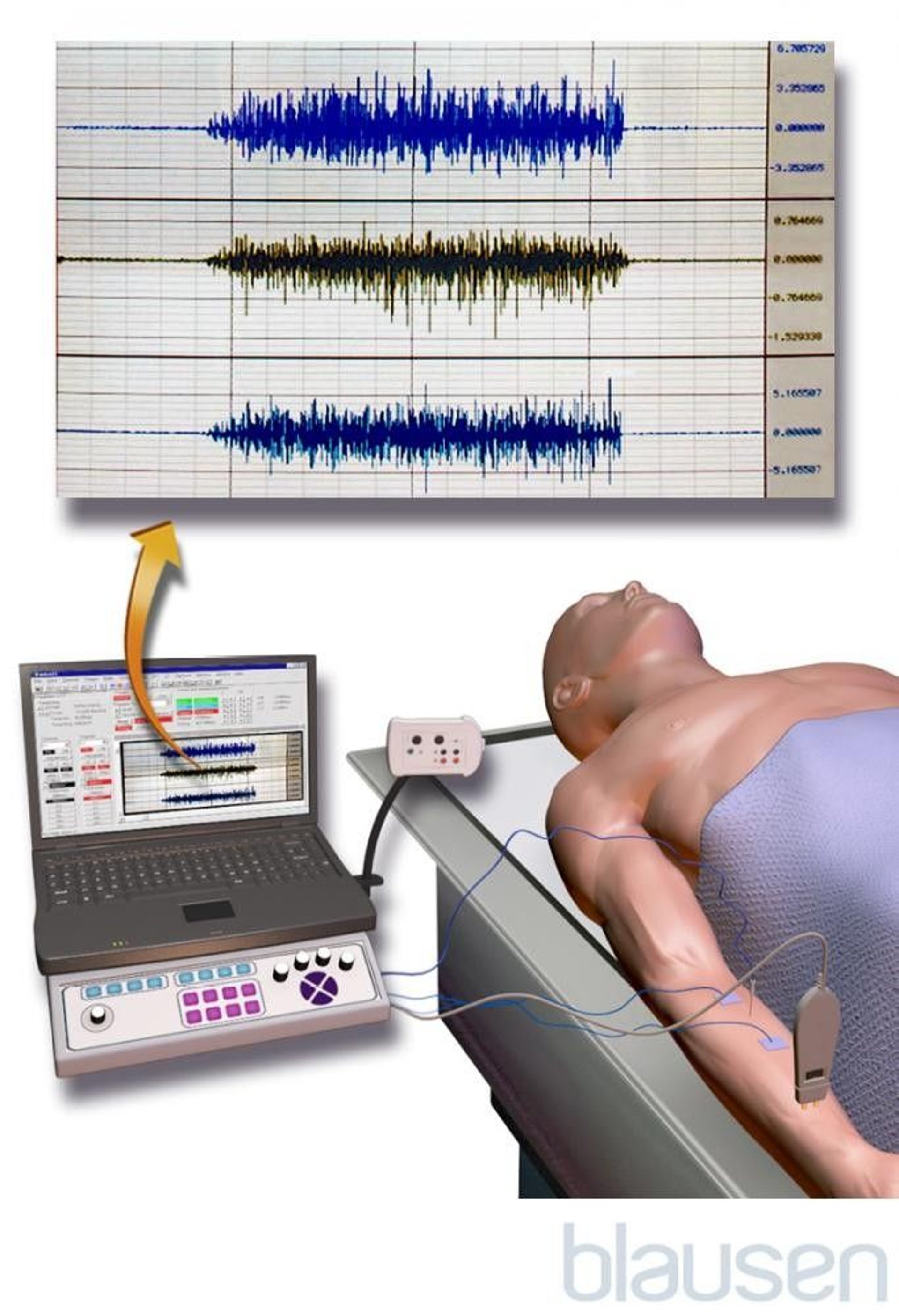
Nerve conduction studies
Nerve conduction studies measure the speed at which motor or sensory nerves conduct impulses. A small electrical current stimulates an impulse along the nerve being tested. The current may be delivered by several electrodes placed on the surface of the skin or by several needles inserted along the pathway of the nerve. The impulse moves along the nerve, eventually reaching the muscle and causing it to contract. By measuring the time the impulse takes to reach the muscle and the distance from the stimulating electrode or needle to the muscle, doctors can calculate the speed of nerve conduction. The nerve may be stimulated once or several times (to determine how well the neuromuscular junction is functioning).
Results are abnormal only if the symptom results from a problem with a nerve or neuromuscular junction. For example,
If nerve conduction is slow, the cause may be a disorder that affects one nerve, such as carpal tunnel syndrome (painful compression of a nerve in the wrist). Or the cause may be a disorder that affects nerves throughout the body (a polyneuropathy), as when diabetes damages nerves throughout the body, starting with those in the feet.
If the muscle’s response is progressively weaker after repeated stimulation, a problem with the neuromuscular junction (as occurs in myasthenia gravis) may be the cause.
However, the speed of nerve conduction may be normal if the affected nerves are small and do not have a myelin sheath (the outer layer of tissues that helps nerves conduct impulses faster). Speed is also normal if the disorder involves only the brain, spinal cord, spinal nerve roots, or the muscle. Such disorders do not affect the speed of nerve conduction.
Evoked Responses
For this test, doctors use stimuli for sight, sound, and touch to activate specific areas of the brain, that is, to evoke responses. EEG is used to detect the response evoked by the stimuli. Based on these responses, doctors can tell how well those areas of the brain are working. For example, a flashing light stimulates the retina of the eye, the optic nerve, and the nerve pathway to the back part of the brain where vision is perceived and interpreted.
Evoked responses are particularly useful in testing how well the senses are functioning in infants and children. For example, doctors can test an infant’s hearing by checking for a response after a clicking sound is made at each ear.
Evoked responses are also useful in identifying the effects of multiple sclerosis and other disorders on areas of the optic nerve, brain stem, and spinal cord. Such effects may or may not be detected by MRI.
Evoked responses can also help predict the prognosis for people who are in a coma. If stimuli do not evoke typical brain activity, the prognosis is likely to be poor.
Myelography
In myelography, x-rays of the spinal cord are taken after a radiopaque contrast agent is injected into the subarachnoid space via a spinal tap. Myelography has been largely replaced by MRI, which usually produces more detailed images and is simpler and safer to do.
Myelography with computed tomography (CT) is used when doctors need more detail of the spinal cord and surrounding bone than MRI can provide. Myelography with CT is also used when MRI is not available or cannot be done safely (for example, when a person has a heart pacemaker).
Other Tests for Brain, Spinal Cord, and Nerve Disorders
Biopsy
Muscle and nerve
Occasionally, doctors cannot determine the cause of the nerve damage or muscle weakness based on results of blood tests, imaging tests, electromyography (EMG), or nerve conduction studies. In such cases, doctors typically refer the person to a specialist, who may remove a small sample of muscle tissue and/or a nerve to examine under a microscope (biopsy). The sample is removed from an area of the body where symptoms occur. The sample is stained to help doctors identify the pattern of muscle or nerve damage and to determine whether white blood cells (which indicate inflammation) are present.
Skin
Often, the sensory nerve examination and EMG do not detect damage of the nerves that sense pain or that automatically regulate body processes (called autonomic nerves). Doctors may suspect such damage if people have less sensitivity to pain, have burning pain in their feet, feel dizzy or light-headed when they stand up, or sweat too much or too little. To check for this damage, doctors may use a small round cutter to remove a sample of skin (punch skin biopsy) and send it to a laboratory to be examined under a microscope.
If the nerve endings in the skin sample have been destroyed, the cause may be a disorder (such as vasculitis) that affects small nerve fibers, including pain-sensing and autonomic nerves fibers.
Echoencephalography
Echoencephalography uses ultrasound waves to produce an image of the brain. This simple, painless, and relatively inexpensive procedure can be used in children younger than 2 years because their skull is thin enough for ultrasound waves to pass through. It can be done quickly at the bedside to detect hydrocephalus (previously called water on the brain) or bleeding.
CT and MRI have largely replaced echoencephalography in older children and adults because they produce much better images in these age groups.
Genetic Testing
Genetic abnormalities cause many neurologic disorders—particularly movement disorders, including those that cause tremor or problems walking. Genetic tests can sometimes help doctors diagnose certain nerve and muscle disorders.
When genetic testing is recommended, people are usually referred to a genetic counselor. If they are not, people may request an appointment with one.


