

Ultrasound scanning uses sound waves to produce pictures of internal organs (see also Ultrasound).
An ultrasound scan can show the size and shape of many organs, such as the liver and pancreas, and can also show abnormalities within them, such as cysts and some tumors. It can also show fluid in the abdominal cavity (ascites). Ultrasound scanning with a probe placed on the outside of the abdomen is not a good method for examining the lining or the wall of the digestive tract. Endoscopic ultrasound, however, shows the wall of the digestive tract or some abdominal organs more clearly because the probe is placed on the tip of an endoscope.

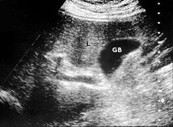
This photo shows a normal ultrasound scan of the liver (L) and gallbladder (GB).
An ultrasound scan is painless and poses no risk of complications.
Endoscopic ultrasound poses a similar risk of complications as endoscopy.


