

Nevi (singular nevus, moles) are flesh- to brown-colored macules, papules, or nodules composed of nests of melanocytes or nevus cells. Their main significance (other than cosmetic) is their resemblance and/or risk of transformation to melanoma. Pigmented lesions are assessed for characteristics of concern (known as the ABCDEs of melanoma and include Asymmetry, irregular Borders, multiple Colors within one lesion or unusual colors such as blue, black or red, Diameter > 6 mm, and Evolution (ie, change in an existing mole or a new mole that could suggest an atypical nevus or melanoma).
Almost everyone has a few nevi, which usually appear in childhood or adolescence. There are different types of nevi (see table Classification of Nevi). During adolescence, more nevi often appear, and existing ones may enlarge or darken. Nevus cells may be eventually replaced with fat or fibrous tissue in older adults, and these nevi typically change consistency, becoming softer and more boggy, or firmer and less pigmented. Nevi may also darken during pregnancy.
An individual nevus is unlikely to become malignant (lifetime risk is approximately 1 in 3000 for men and 1 in 10,000 for women) (1). However, patients who have a large numbers of nevi (> approximately 50), atypical nevi, or a family history of melanoma have an increased risk of developing melanoma. Specifically, there appears to be an approximately 4-fold greater risk of melanoma in patients who have ≥ 25 moles or ≥ 4 atypical moles (2).
Blue nevi are benign nevi that appear as bluish gray macules or thin papules. The depth and density of pigment in the skin account for the apparent blue color.


Copyright © 2025 Merck & Co., Inc., Rahway, NJ, USA and its affiliates. All rights reserved.

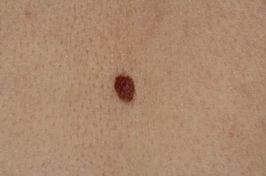
Nevi are flesh- to brown-colored macules, papules, or nodules composed of nests of melanocytes or nevus cells. Nevi develop on nearly everybody, and are significant primarily because they can become dysplastic or malignant and need to be differentiated from melanoma.
Nevi are flesh- to brown-colored macules, papules, or nodules composed of nests of melanocytes or nevus cells. Nevi dev
Image provided by Thomas Habif, MD.

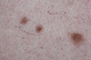
Junctional nevi are well-demarcated evenly pigmented brown macules and patches.
Junctional nevi are well-demarcated evenly pigmented brown macules and patches.
Image courtesy of Marie Schreiner, PA-C.
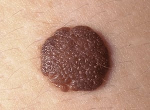
A compound nevus is a well-demarcated brown papule.
A compound nevus is a well-demarcated brown papule.
Image courtesy of Marie Schreiner, PA-C.


Intradermal nevus presenting as a skin-colored raised nodule on the scalp.
Intradermal nevus presenting as a skin-colored raised nodule on the scalp.
© Springer Science+Business Media

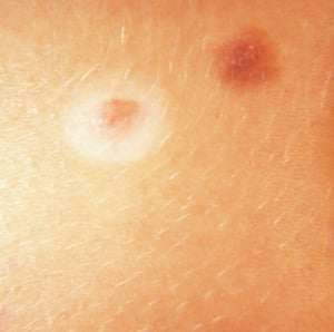
This photo shows a halo nevus surrounded by a ring of depigmented skin next to a nevus without a halo.
This photo shows a halo nevus surrounded by a ring of depigmented skin next to a nevus without a halo.
DermPics/SCIENCE PHOTO LIBRARY

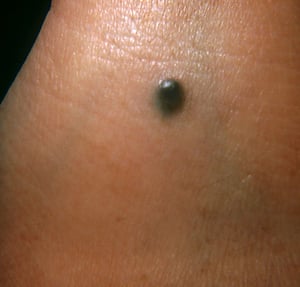
This photo shows a blue nevus composed of dark pigmented cells (melanocytes).
This photo shows a blue nevus composed of dark pigmented cells (melanocytes).
DermPics/SCIENCE PHOTO LIBRARY
Nevi are flesh- to brown-colored macules, papules, or nodules composed of nests of melanocytes or nevus cells. Nevi develop on nearly everybody, and are significant primarily because they can become dysplastic or malignant and need to be differentiated from melanoma.
Nevi are flesh- to brown-colored macules, papules, or nodules composed of nests of melanocytes or nevus cells. Nevi dev
Image provided by Thomas Habif, MD.
Junctional nevi are well-demarcated evenly pigmented brown macules and patches.
Junctional nevi are well-demarcated evenly pigmented brown macules and patches.
Image courtesy of Marie Schreiner, PA-C.
A compound nevus is a well-demarcated brown papule.
A compound nevus is a well-demarcated brown papule.
Image courtesy of Marie Schreiner, PA-C.
Intradermal nevus presenting as a skin-colored raised nodule on the scalp.
Intradermal nevus presenting as a skin-colored raised nodule on the scalp.
© Springer Science+Business Media
This photo shows a halo nevus surrounded by a ring of depigmented skin next to a nevus without a halo.
This photo shows a halo nevus surrounded by a ring of depigmented skin next to a nevus without a halo.
DermPics/SCIENCE PHOTO LIBRARY
This photo shows a blue nevus composed of dark pigmented cells (melanocytes).
This photo shows a blue nevus composed of dark pigmented cells (melanocytes).
DermPics/SCIENCE PHOTO LIBRARY
General references
1. Tsao H, Bevona C, Goggins W, et al: The transformation rate of moles (melanocytic nevi) into cutaneous melanoma: A population-based estimate. Arch Dermatol139(3):282-288, 2003. doi: 10.1001/archderm.139.3.282
2. Titus-Ernstoff L, Perry AE, Spencer SK, et al. Pigmentary characteristics and moles in relation to melanoma risk. Int J Cancer. 2005;116(1):144-149. doi:10.1002/ijc.21001
Diagnosis of Nevi
Primarily history and physical examination
Sometimes biopsy
Nevi are extremely common; therefore, the diagnosis of nevi is primarily clinical. However, biopsy and histologic evaluation should be considered if nevi have certain characteristics of concern (known as the ABCDEs of melanoma) (1):
A: Asymmetry—asymmetric appearance
B: Borders—irregular borders (ie, not round or oval)
C: Color—color variation within the mole, unusual colors, or a color significantly different or darker than the patient's other nevi
D: Diameter—> 6 mm
E: Evolution—a new mole in a patient > 30 years of age or a changing mole
If a mole becomes painful or itchy or bleeds or ulcerates, biopsy can also be considered.
The biopsy specimen must be deep enough for accurate microscopic diagnosis and should encompass the entire lesion if possible, especially if the concern for malignancy is strong. However, wide primary excision should not be the initial procedure, even for highly abnormal-appearing lesions. Many such lesions are not melanomas and, even with melanoma, the proper treatment margin and recommendation for lymph node sampling is determined based on histopathologic features. Excisional biopsy does not increase the likelihood of metastasis if the lesion is malignant, and it avoids extensive surgery for a benign lesion.
Diagnosis reference
1. Rigel DS, Friedman RJ, Kopf AW, et al. ABCDE--an evolving concept in the early detection of melanoma. Arch Dermatol. 2005;141(8):1032-1034. doi:10.1001/archderm.141.8.1032
Treatment of Nevi
Sometimes excision
Because the vast majority of nevi remain benign and the risk of malignant transformation is extremely low, systematic removal of all nevi would provide little benefit and would expose patients to unnecessary morbidity and increased healthcare costs. However, nevi should be excised if they exhibit clinical or dermatoscopic features suspicious for melanoma, or if there are other specific concerns such as difficulty monitoring or other bothersome symptoms (1). Nevi can be removed by shaving or excision. All removed nevi should be examined histologically.
Patients with a large number of nevi should be taught to self-monitor for warning signs (for eg, the ABCDEs of melanoma) and have skin surveillance as part of their primary care to aid in the early detection of melanoma.
Treatment reference
1. Kim CC, Swetter SM, Curiel-Lewandrowski C, et al. Addressing the knowledge gap in clinical recommendations for management and complete excision of clinically atypical nevi/dysplastic nevi: Pigmented Lesion Subcommittee consensus statement. JAMA Dermatol. 2015;151(2):212-218. doi:10.1001/jamadermatol.2014.2694
Key Points
Almost everyone has nevi, but patients with > approximately 50 are at increased risk of melanoma.
Consider biopsy if nevi have ABCDE characteristics: Asymmetry; irregular Borders; high-risk Colors (variations within or between nevi or unusual colors); Diameter > 6 mm; Evolution (new nevi after age 30 or changes in existing nevi).
Consider excision if a nevus is a significant cosmetic problem.


