

Computed tomography (CT) provides rapid, noninvasive imaging of the brain and skull. CT is superior to magnetic resonance imaging (MRI) in visualizing fine bone detail in (but not the contents of) the posterior fossa, base of the skull, and spinal canal.

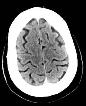
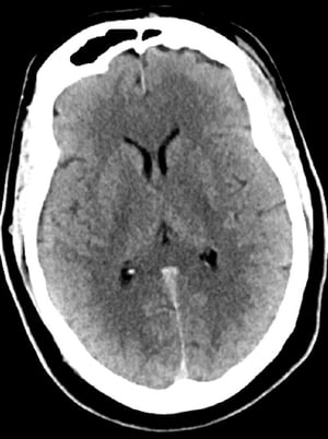
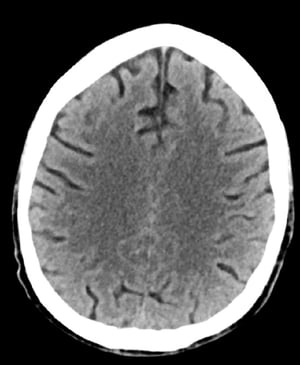
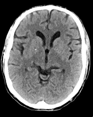
This image is a normal CT scan of the head of a young adult aged 30. There is no intra- or extra-axial fluid or hemorrhage. Grey-white differentiation is preserved. The sulcal pattern is normal.
This image is a normal CT scan of the head of a young adult aged 30. There is no intra- or extra-axial fluid or hemorrh
© 2017 Elliot K. Fishman, MD.

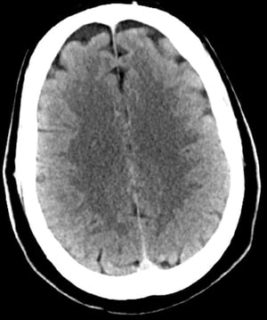
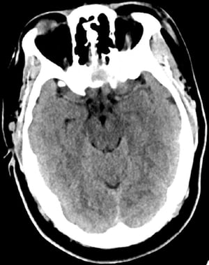
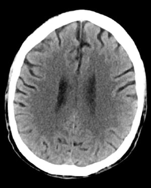
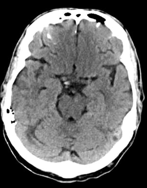
This image is a normal CT scan of the head of a young adult aged 30. There is no intra- or extra-axial fluid or hemorrhage. Grey-white differentiation is preserved. Ventricular size and sulcal pattern are normal.
This image is a normal CT scan of the head of a young adult aged 30. There is no intra- or extra-axial fluid or hemorrh
© 2017 Elliot K. Fishman, MD.
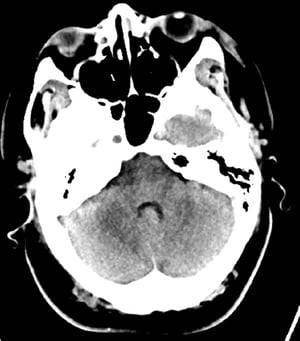
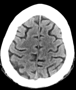
This image is a normal CT scan of the head of a young adult aged 30. There is no intra- or extra-axial fluid or hemorrhage. Grey-white differentiation is preserved. The sulcal pattern is normal.
This image is a normal CT scan of the head of a young adult aged 30. There is no intra- or extra-axial fluid or hemorrh
© 2017 Elliot K. Fishman, MD.
This image is a normal CT scan of the head of a young adult aged 30. There is no intra- or extra-axial fluid or hemorrhage. Grey-white differentiation is preserved. Ventricular size and sulcal pattern are normal.
This image is a normal CT scan of the head of a young adult aged 30. There is no intra- or extra-axial fluid or hemorrh
© 2017 Elliot K. Fishman, MD.

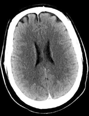
This image is a normal CT scan of the head of an adult aged 74. When compared with the normal CT scan of the head of a 30-year-old, the sulci are larger. These findings are normal in this age group.
This image is a normal CT scan of the head of an adult aged 74. When compared with the normal CT scan of the head of a
© 2017 Elliot K. Fishman, MD.

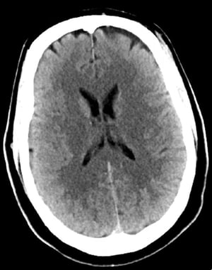
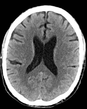
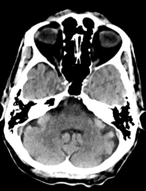
This image is a normal CT scan of the head of an adult aged 74. When compared with the normal CT scan of the head of a 30-year-old, the ventricles and sulci are larger. These findings are normal in this age group.
This image is a normal CT scan of the head of an adult aged 74. When compared with the normal CT scan of the head of a
© 2017 Elliot K. Fishman, MD.
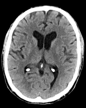
This image is a normal CT scan of the head of an adult aged 74. When compared with the normal CT scan of the head of a 30-year-old, the sulci are larger. These findings are normal in this age group.
This image is a normal CT scan of the head of an adult aged 74. When compared with the normal CT scan of the head of a
© 2017 Elliot K. Fishman, MD.
This image is a normal CT scan of the head of an adult aged 74. When compared with the normal CT scan of the head of a 30-year-old, the ventricles and sulci are larger. These findings are normal in this age group.
This image is a normal CT scan of the head of an adult aged 74. When compared with the normal CT scan of the head of a
© 2017 Elliot K. Fishman, MD.
Noncontrast CT is used to rapidly detect acute hemorrhage and various gross structural changes without concern about contrast allergy or kidney failure.
A radiopaque contrast agent, given IV, helps detect abnormal structural lesions such as brain tumors, brain abscesses, and some vascular anomalies, including arteriovenous malformations. With an intrathecal agent, CT can outline abnormalities encroaching on the brain stem, spinal cord, or spinal nerve roots (eg, meningeal carcinoma, herniated disk) and may detect a syrinx in the spinal cord. However, an intrathecal agent is rarely used because of the availability of MRI.
CT angiography using a contrast agent can show the cerebral blood vessels, obviating the need for MRI or conventional angiography. CT angiography is commonly performed in patients with suspected stroke, to allow early interventional therapy when indicated.
Adverse effects of contrast agents include allergic reactions and contrast nephropathy.


