


Calcaneal (heel bone) fractures often result from great force. Diagnosis is by x-rays and, if needed, CT. Treatment requires orthopedic consultation and includes casting and sometimes surgery.
(See also Overview of Fractures.)
Calcaneal fractures are serious but uncommon injuries; they account for only 1 to 2% of all fractures. However, if not diagnosed and treated promptly, they can result in long-term disability. Up to 10% of these fractures are missed at initial presentation in an emergency department.
Typically, these fractures result from a high-energy axial load to the foot (eg, a fall from a height onto the heels). Because these fractures require great force, they are often accompanied by other serious injuries; 10% of patients with a calcaneal fracture have a thoracolumbar compression fracture.
Stress fractures may also occur in the calcaneus, particularly in athletes, such as long-distance runners.
Calcaneal fractures may be intra-articular.
Symptoms and Signs of Calcaneal Fractures
Usually, the area around the heel and the hindfoot is tender and very swollen. Patients cannot put weight on their foot.
Acute compartment syndrome occurs in up to 10% of patients.
Diagnosis of Calcaneal Fractures
X-rays
Sometimes CT
If a calcaneal fracture is suspected, x-rays that include axial and lateral views should be taken.
CT is done if
X-rays are negative but clinical findings suggest a calcaneal fracture.
The Bohler angle is < 20°.
More detail about the fracture is needed.

Image courtesy of Danielle Campagne, MD.
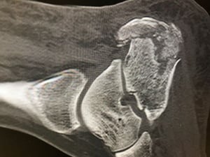
Image courtesy of Danielle Campagne, MD.

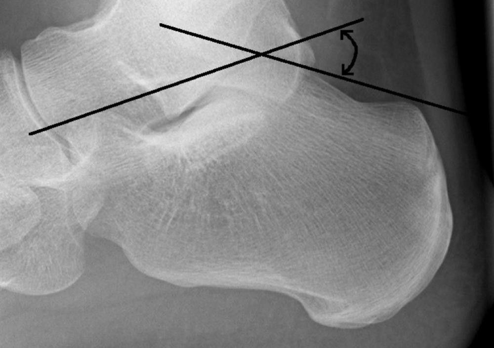
Image courtesy of Danielle Campagne, MD.


Image courtesy of Danielle Campagne, MD.
The Bohler angle is determined on the lateral x-ray. This angle is formed by the intersection of a line drawn from the superior aspect of the posterior calcaneal tuberosity to the superior subtalar articular surface and a line drawn from the superior subtalar articular surface to the superior aspect of the anterior calcaneal process. Normally, the angle is 20 to 40°. An angle of < 20° suggests a fracture.
Pearls & Pitfalls
|
Clinicians should also check for other injuries, such as thoracolumbar compression fractures and compartment syndrome.
Treatment of Calcaneal Fractures
Orthopedic consultation
Casting or possibly surgery, depending on the type of fracture
Orthopedic consultation is necessary.
Whether intra-articular calcaneal fractures should be treated surgically or nonsurgically is much debated.
Extra-articular calcaneal fractures are treated symptomatically with protection, rest (avoiding weight bearing), a compression dressing (which also provides protection), ice, and elevation (PRICE). When the swelling resolves, a cast is applied.
Key Points
If calcaneal fractures are not diagnosed and treated promptly, they can result in long-term disability.
Because these fractures usually result from a high-energy axial load to the foot, other injuries (eg, thoracolumbar compression fracture) are often also present; other complications include compartment syndrome (in up to 10%).
Diagnose based on x-rays and, if needed, CT.
Whether intra-articular calcaneal fractures should be treated surgically or nonsurgically is controversial.
When diagnosing a calcaneal fracture, always check for a thoracolumbar fracture.
Treat extra-articular calcaneal fractures symptomatically with PRICE, followed by casting.


