


Trisomy 13 is caused by an extra chromosome 13 and causes abnormal forebrain, midface, and eye development; severe intellectual disability; heart defects; and small birth size. Diagnosis is with cytogenetic testing. Treatment is supportive.
(See also Overview of Chromosomal Abnormalities.)
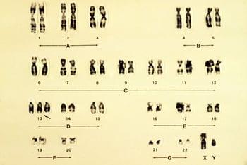
Trisomy 13 occurs in approximately 1.7/10,000 pregnancies (based on data from induced abortion for fetal anomalies, stillbirths, and live births) (1); approximately 80% of cases are complete trisomy 13. Advanced maternal age increases the likelihood, and the extra chromosome is usually maternally derived.
Infants tend to be small for gestational age. Midline anomalies are common and include holoprosencephaly (failure of the forebrain to divide properly), facial anomalies such as cleft lip and cleft palate, microphthalmia, colobomas (fissures) of the iris, and retinal dysplasia. Supraorbital ridges are shallow, and palpebral fissures usually are slanted.
The ears are abnormally shaped and usually low-set. Hearing loss is common. Scalp defects and dermal sinuses are also common. Loose folds of skin often are present over the back of the neck.
A single transverse palmar crease, polydactyly, and hyperconvex narrow fingernails are also common. Approximately 80% of cases have severe congenital cardiovascular anomalies; dextrocardia is common.
Genitals are frequently abnormal in both sexes; cryptorchidism and an abnormal scrotum occur in boys, and a bicornuate uterus occurs in girls.
Apneic spells in early infancy are frequent. Intellectual disability is severe.
General reference
1. Goel N, Morris JK, Tucker D, et al: Trisomy 13 and 18-Prevalence and mortality-A multi-registry population based analysis. Am J Med Genet A 179(12):2382-2392, 2019. doi: 10.1002/ajmg.a.61365
Diagnosis of Trisomy 13
Cytogenetic testing by karyotyping, fluorescent in situ hybridization (FISH) analysis, and/or chromosomal microarray analysis
(See also Next-generation sequencing technologies.)
Diagnosis of trisomy 13 may be suspected postnatally by appearance or prenatally by abnormalities on ultrasonography (eg, intrauterine growth restriction), or by increased risk noted on multiple marker screening or noninvasive prenatal screening (NIPS) using cell-free fetal DNA analysis on a maternal blood sample. Management decisions, including termination of pregnancy, should not be made based on NIPS testing alone (1).
Confirmation is by cytogenetic testing (karyotyping, FISH analysis, and/or chromosomal microarray analysis) of samples obtained by chorionic villus sampling or amniocentesis. Postnatally, confirmation is by cytogenetic testing usually of a blood sample.

Diagnosis reference
1. American College of Obstetricians and Gynecologists’ Committee on Practice Bulletins—Obstetrics; Committee on Genetics; Society for Maternal-Fetal Medicine: Screening for fetal chromosomal abnormalities: ACOG Practice Bulletin, Number 226. Obstet Gynecol 136(4):e48-e69, 2020. doi: 10.1097/AOG.0000000000004084
Treatment of Trisomy 13
Supportive care
The underlying genetic abnormality cannot be cured.
Support for the family is critical.
Prognosis for Trisomy 13
In the past, most infants died during the neonatal period; however, 5-year survival has improved in recent times (1).
Prognosis reference
1. Meyer RE, Liu G, Gilboa SM, et al: Survival of children with trisomy 13 and trisomy 18: A multi-state population-based study. Am J Med Genet A 170A(4):825-837, 2016. doi: 10.1002/ajmg.a.37495
More Information
The following English-language resources may be useful. Please note that THE MANUAL is not responsible for the content of these resources.
American College of Obstetricians and Gynecologists Committee on Practice Bulletins–Obstetrics, Committee of Genetics, and the Society for Maternal–Fetal Medicine: Screening for fetal chromosomal abnormalities: ACOG practice bulletin, number 226 (2020)
SOFT (Support Organization for Trisomy 18, 13, and Related Disorders): An organization providing resources, research information, and community and support services to people caring for others who have trisomy 18, 13, or another related chromosome disorder


