


Glucose-6-phosphate dehydrogenase (G6PD) deficiency is an X-linked enzymatic defect common in people with African ancestry that can result in hemolysis after acute illnesses or intake of oxidant drugs (including salicylates and sulfonamides). Diagnosis is based on assay for G6PD, although test results are often falsely negative during acute hemolysis due to the presence of reticulocytes, which are richer in G6PD than older cells. Treatment is supportive.
(See also Overview of Hemolytic Anemia.)
G6PD deficiency, a defect in the hexose monophosphate shunt pathway, is the most common disorder of red blood cell (RBC) metabolism. The G6PD gene is located on the X chromosome and exhibits a high amount of variation (polymorphism), resulting in a range of G6PD activity from normal to severely deficient. Variants are classified I through V by the amount of activity of the G6PD enzyme. Because the gene is X-linked, males are more likely to present with clinically significant hemolysis. Females who are homozygous, or who are heterozygous with skewed X inactivation that results in a high proportion of affected X chromosomes may also be affected.
In the US, this defect is most common in people with African ancestry, occurring in about 10% of male African Americans and in < 10% of female African Americans. It occurs in lower frequencies among people from the Mediterranean basin (eg, Italian, Greek, Arab, or Sephardic Jewish ancestry) and people with Asian ancestry.
Pathophysiology of G6PD Deficiency
G6PD deficiency renders RBCs susceptible to oxidative stress, which shortens RBC survival. Hemolysis occurs following an oxidative challenge, commonly after fever, acute viral or bacterial infections, and diabetic ketoacidosis. Hemolysis is episodic and self-limited, although rare patients have chronic, ongoing hemolysis in the absence of oxidative challenge.
Symptoms and Signs of G6PD Deficiency
In most cases, hemolysis affects < 25% of RBC mass and causes transient jaundice and dark urine. Some patients have back and/or abdominal pain. However, when the deficiency is more severe, profound hemolysis may lead to hemoglobinuria and acute kidney injury.
Diagnosis of G6PD Deficiency
Peripheral smear
G6PD assay
The diagnosis is considered in patients with evidence of acute hemolysis, particularly males with a direct antiglobulin–negative hemolytic anemia (see Diagnosis of Hemolytic Anemia). Anemia, jaundice, and reticulocytosis develop during hemolysis.

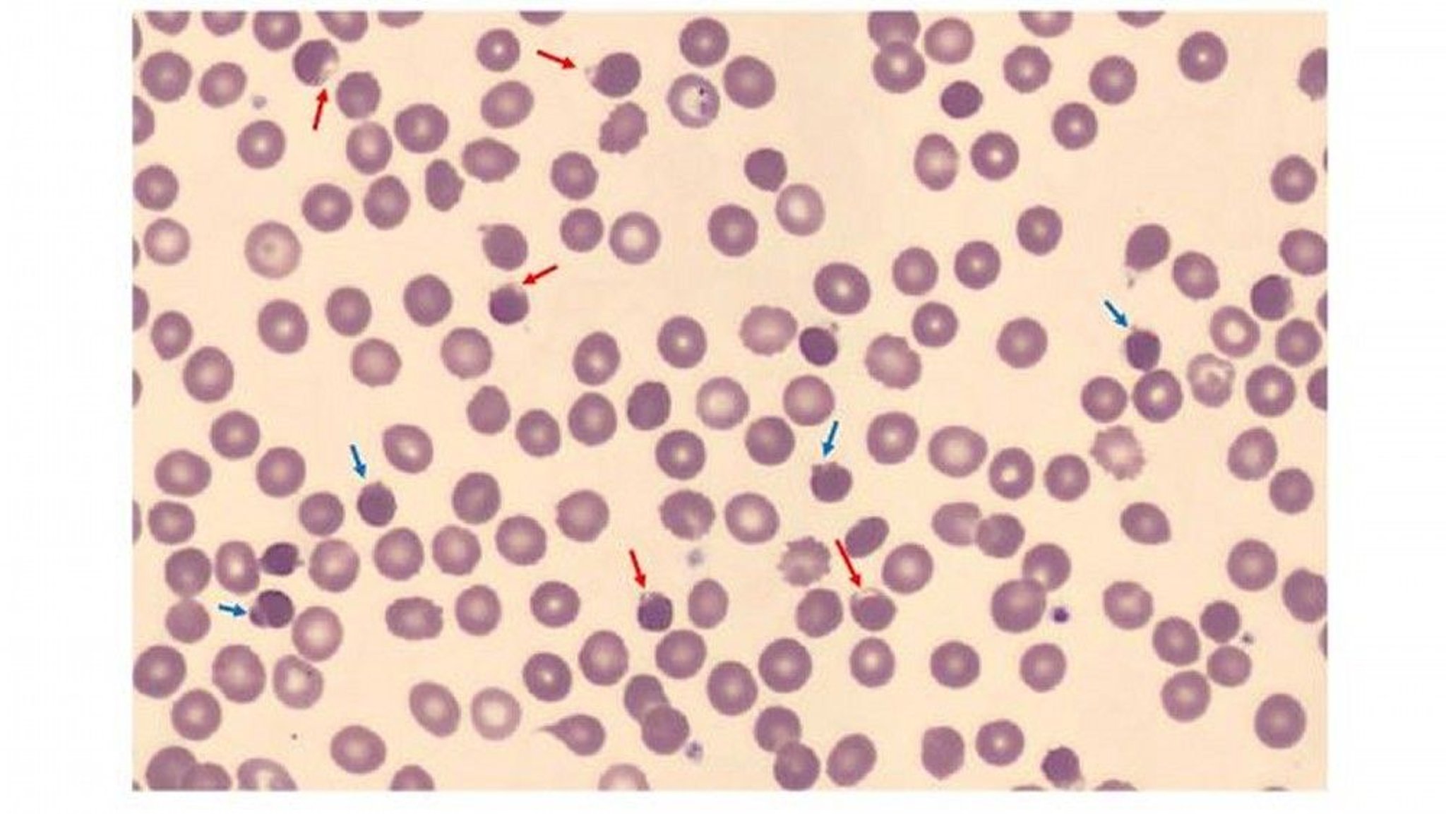
Image courtesy of Jerry L. Spivak, MD.
The peripheral smear may reveal RBCs that appear to have a blister (blister cells) or have one or more "bites" (1-micron wide) taken from the cell periphery (bite cells) and RBCs with inclusions termed Heinz bodies, which are particles of denatured hemoglobin, which can be recognized only by special stains. These cells may be visible early during the hemolytic episode but do not persist in patients with an intact spleen, which removes them.
Testing for G6PD activity is available. However, during and immediately after a hemolytic episode, tests may yield false-negative results because of destruction of the older, more deficient RBCs and the production of reticulocytes, which are rich in G6PD. Thus, testing may need to be repeated several weeks after the acute event. Several screening tests are available, including point-of-care tests; positive results should be confirmed with a quantitative test.
Treatment of G6PD Deficiency
Avoidance of triggers, removal of offending drug or agent, and supportive care
During acute hemolysis, treatment is supportive; transfusions are rarely needed. Patients are advised to avoid drugs or substances that initiate hemolysis.
Key Points
Glucose-6-phosphate dehydrogenase (G6PD) deficiency is the most common inherited disorder of red blood cell metabolism and can cause hemolysis in the presence of triggers.
Incidence is higher in certain ethnic groups (eg, people with African, Mediterranean, or Asian ancestry).
Triggers include acute illnesses (eg, infections), drugs (eg, salicylates) and other substances (eg, fava beans) that cause oxidative stress.
Diagnose using peripheral smear and G6PD assay; false negative G6PD assays are possible during acute hemolysis so repeat testing after several weeks if initial G6PD assay is negative.
Avoid triggers to limit hemolytic episodes.


