


View Patient Education
Laryngoceles are evaginations of the mucous membrane of the laryngeal ventricle.
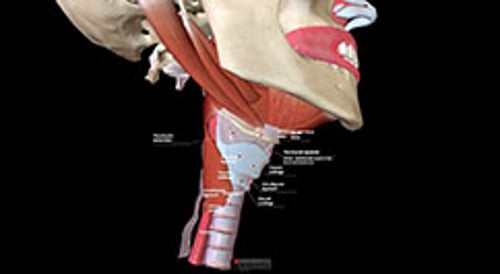
Internal laryngoceles displace and enlarge the false vocal cords, resulting in hoarseness and airway obstruction. External laryngoceles extend through the thyrohyoid membrane, causing a mass in the neck. Laryngoceles tend to occur in musicians who play wind instruments. Laryngoceles are filled with air and can be expanded by the Valsalva maneuver.
Laryngoceles appear on CT as smooth, ovoid, low-density masses. They may become infected (laryngopyocele) when filled with mucoid fluid.
Treatment of laryngoceles is excision.
(See also Overview of Laryngeal Disorders.)
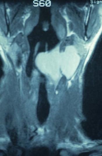
Combined Laryngocele

Hide Details
This coronal CT scan shows a combined laryngocele that begins inside the larynx and extends through the thyrohyoid membrane, causing a mass in the neck.
Image provided by Clarence T. Sasaki, MD.
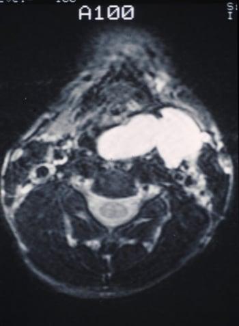
Laryngocele

Hide Details
This CT scan shows a smooth mucus-filled laryngocele bulging into the throat and outward into the neck.
Image provided by Clarence T. Sasaki, MD.


Test your KnowledgeTake a Quiz!


