

Dermatofibromas are firm, red-to-brown, small papules or nodules composed of fibroblastic tissue. They most commonly occur on the thighs or legs but can occur anywhere.

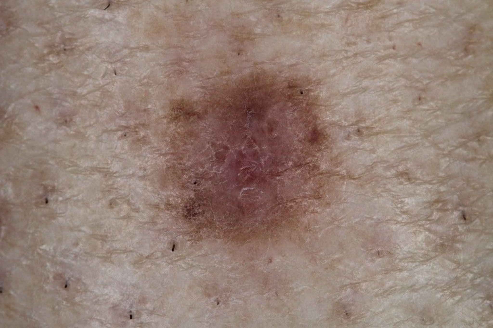
This photo shows a small hyperpigmented lesion consistent with dermatofibroma.
Image courtesy of Marie Schreiner, PA-C.
Dermatofibromas are common among adults, more so in women (1). Their cause is probably genetic (2). Lesions are usually 0.5 to 1 cm in diameter, firm, and may dimple inward with gentle pinching. Most lesions are asymptomatic, but some itch or ulcerate after minor trauma.
Dermatofibrosarcoma protuberans is a rare soft tissue sarcoma that may appear similar to a dermatofibroma; however, it is distinguished from a dermatofibroma by its rapidity of growth. It can eventually grow significantly in size and depth, infiltrating into the subcutaneous and deeper tissues (ie, fascial and muscular planes). Early lesions are typically not painful, but if lesions progress, may ulcerate, leading to pain and tenderness.
References
1. Şenel E, Yuyucu Karabulut Y, Doğruer Şenel S. Clinical, histopathological, dermatoscopic and digital microscopic features of dermatofibroma: a retrospective analysis of 200 lesions. J Eur Acad Dermatol Venereol. 2015;29(10):1958-1966. doi:10.1111/jdv.13092
2. Endzhievskaya S, Hsu CK, Yang HS, et al. Loss of RhoE Function in Dermatofibroma Promotes Disorganized Dermal Fibroblast Extracellular Matrix and Increased Integrin Activation. J Invest Dermatol. 2023;143(8):1487-1497.e9. doi:10.1016/j.jid.2023.01.019
Diagnosis of Dermatofibromas
Primarily physical examination
Occasionally biopsy
The diagnosis of dermatofibromas can often be made clinically. The dimple sign, also known as Fitzpatrick's sign, is a characteristic feature of dermatofibromas, where a skin lesion indents or dimples inward when squeezed from the sides.
There are several described dermatoscopic patterns of dermatofibromas (1). Lesions are sometimes biopsied to exclude melanocytic proliferation (eg, nevus, solar lentigo, melanoma) or other tumors. Dermatofibromas may appear hyperpigmented in dark skin tones.
Diagnosis reference
1. Zaballos P, Puig S, Llambrich A, et al: Dermoscopy of dermatofibromas: A prospective morphological study of 412 cases. Arch Dermatol 144(1):75-83, 2008. doi: 10.1001/archdermatol.2007.8
Treatment of Dermatofibromas
Excision if troublesome
Dermatofibromas that result in bothersome symptoms may necessitate excision. Treatment with cryosurgery may alleviate symptoms.


