


Food and a variety of other swallowed objects can become impacted in the esophagus. Esophageal foreign bodies cause dysphagia and sometimes lead to perforation. Diagnosis is clinical, but imaging studies and endoscopy may be needed. Some objects pass spontaneously, but endoscopic advancement or removal is often required.
(See also Overview of Foreign Bodies in the Gastrointestinal Tract.)
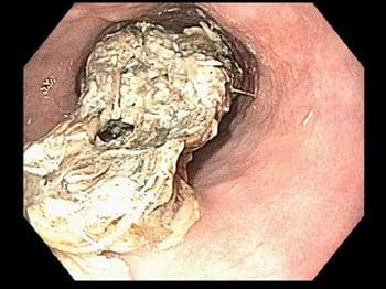
The esophagus is the most common site of foreign body impaction. Food impactions are the most common cause of esophageal foreign bodies. Large, smooth food pieces (eg, steak, hot dogs) are particularly easy to swallow inadvertently before being chewed sufficiently. Bones, particularly fish bones, may be swallowed if the meat in which they are embedded is not chewed sufficiently.
Infants and toddlers do not have fully mature oropharyngeal coordination and often inadvertently swallow small, round foods (eg, grapes, peanuts, candies), which may become impacted. In addition, infants and toddlers often swallow a wide variety of inedible objects (eg, coins, batteries) accidentally or because they are curious, and some of these objects become impacted in the esophagus. Impacted disk batteries are particularly worrisome because they may cause esophageal burns, perforation, or tracheoesophageal fistula.
Foreign bodies in the esophagus usually lodge in areas where physiologic or pathologic luminal narrowing exists. Luminal narrowing may be caused by sphincters (lower esophageal sphincter, upper esophageal sphincter), external vascular structures (eg, vascular rings), webs, strictures, benign tumors, cancerous tumors, achalasia, distal esophageal spasm, previous surgery, and eosinophilic esophagitis.
Complications
The main complications of esophageal foreign bodies are:
Obstruction may be partial (eg, patients can swallow liquids or at least their oral secretions) or complete. Partial obstruction is less emergent unless it involves a sharp object embedded in the wall, which can lead to perforation. Complete obstruction is poorly tolerated clinically, and even a smooth object, if tightly impacted, may cause pressure necrosis and risk of perforation if allowed to remain in the esophagus for more than about 24 hours.
Complications also depend on the nature of the object involved. Despite their small size, impacted disk or button batteries are objects of particular concern because liquefaction necrosis and perforation can occur rapidly.
Symptoms and Signs of Esophageal Foreign Bodies
The main presenting symptom is acute dysphagia. Patients with complete obstruction of the esophagus hypersalivate and are unable to swallow oral secretions. Other symptoms include retrosternal fullness, regurgitation, odynophagia, blood-stained saliva, and gagging and choking. Hyperventilation resulting from anxiety and discomfort often gives the appearance of respiratory distress, but actual dyspnea or auscultatory findings of stridor or wheezing strongly suggest the foreign body is in the airway rather than the esophagus.
Sometimes, foreign bodies scratch the esophagus but do not become lodged. In such cases, patients may report a foreign body sensation even though no foreign body is present.
Diagnosis of Esophageal Foreign Bodies
Clinical evaluation
Sometimes imaging studies
Often endoscopic evaluation
Many patients give a clear history of ingestion; those with significant symptoms suggesting complete obstruction should have immediate therapeutic endoscopy. Patients with minimal symptoms and no high-risk factors (eg, ingestion of sharp objects, disk or button batteries, or packets of illicit drugs) who are able to swallow normally may not have an impacted foreign body and can be observed for resolution of symptoms. Other patients may require imaging studies.
Some patients, such as young children, mentally impaired adults, older adults, and patients with psychiatric illness, may not be able to give an adequate history of ingestion. These patients may present with choking, refusal to eat, vomiting, drooling, wheezing, blood-stained saliva, or respiratory distress. Imaging studies also may be needed in these patients.
Some foreign bodies can be detected with plain radiographs (2 views preferred). These radiographs are best for detecting metallic foreign objects and steak bones as well as for detecting signs of perforation (eg, free air in the mediastinum or peritoneum). Other objects such as fish bones and even some chicken bones, wood, plastic, glass, and thin metal objects can be difficult to identify on plain radiographs. If there is any suspicion of a foreign body or of intentional or dangerous (eg, packets of illicit drugs) foreign body ingestion, imaging studies, such as CT, should be performed, sometimes before endoscopy, to confirm and localize the foreign body. Endoscopic evaluation is typically required in patients with suspected foreign body ingestions and ongoing symptoms despite negative imaging results.

An oral contrast imaging study typically should not be performed because of the risk of aspiration and of contrast leakage in patients with perforation. Also, the presence of residual contrast material can make subsequent endoscopic retrieval more difficult.
Treatment of Esophageal Foreign Bodies
Sometimes trial of observation and/or IV glucagon
Often endoscopic removal
Some foreign bodies pass spontaneously into the stomach, after which they typically pass completely through the gastrointestinal tract and are expelled. Patients without symptoms of high-grade obstruction and without ingestion of sharp objects or disk or button batteries typically can safely be observed for up to 24 hours to await passage, which is indicated by relief of symptoms (1). Administration of glucagon has been used to relax the distal esophagus but does not seem to be particularly effective relative to other methods (2, 3). Other methods, such as use of effervescent agents, meat tenderizer, and bougienage, are not recommended.
Foreign bodies that do not pass within 24 hours (4) should be removed because delay increases the risk of complications, including perforation, and decreases the likelihood of successful removal.
Endoscopic therapy, either with advancement of the bolus into the stomach or removal, is the treatment of choice. Endoscopic advancement is preceded by trying to pass the endoscope around the food bolus and examining the esophagus distal to the bolus (eg, for luminal narrowing or obstructing lesions) and then accomplished by applying gentle pressure to the middle of the food bolus. To minimize the risk of perforation, this procedure should be performed only by an experienced endoscopist. Removal is best achieved using a forceps, multiprong graspers, net, basket, or snare, preferably with an overtube placed in the esophagus or orotracheal intubation to prevent aspiration and protect the airway (5).
Emergent endoscopy is required for sharp-pointed objects, disk or button batteries, and any obstruction causing significant symptoms.
Follow-up care for the evaluation of structural and functional abnormalities is recommended for patients with esophageal food impaction.
Treatment for obstruction due to packets of illicit drugs is performed by surgical intervention, as endoscopic intervention is contraindicated due to risk of rupture of the packaging and drug overdose. In select cases, endoscopic removal can be considered in a multidisciplinary setting with surgical backup available if any package rupture occurs.
Treatment references
1. Ismail A, Beran A, Saadat S, Veraza DI, Guardiola JJ: Glucagon for Esophageal Foreign Body Impaction: A Systematic Review and Meta-analysis of Randomized Controlled Trials. Am J Ther 31(5):e605–e607, 2024. doi:10.1097/MJT.0000000000001747
2. Bodkin RP, Weant KA, Baker Justice S, Spencer MT, Acquisto NM: Effectiveness of glucagon in relieving esophageal foreign body impaction: a multicenter study. Am J Emerg Med 34(6):1049–1052, 2016. doi:10.1016/j.ajem.2016.03.016
3. Peksa GD, DeMott JM, Slocum GW, Burkins J, Gottlieb M: Glucagon for Relief of Acute Esophageal Foreign Bodies and Food Impactions: A Systematic Review and Meta-Analysis. Pharmacotherapy 39(4):463–472, 2019. doi:10.1002/phar.2236
4. ASGE Standards of Practice Committee, Ikenberry SO, Jue TL, Anderson MA, et al: Management of ingested foreign bodies and food impactions. Gastrointest Endosc 73:1085–1091, 2011. doi: 10.1016/j.gie.2010.11.010
5. Fung BM, Sweetser S, Wong Kee Song LM, Tabibian JH: Foreign object ingestion and esophageal food impaction: An update and review on endoscopic management. World J Gastrointest Endosc 11(3):174–192, 2019. doi: 10.4253/wjge.v11.i3.174
Key Points
The esophagus is the most common site of ingested foreign body impaction.
The main presenting symptom is acute dysphagia; patients with complete obstruction of the esophagus hypersalivate and are unable to swallow oral secretions.
Complete obstruction may cause pressure necrosis and increases the risk of perforation if present for more than about 24 hours.
Emergent endoscopy is required for sharp-pointed objects, disk or button batteries, and any obstruction causing symptoms, suggesting complete obstruction.
Drug Information for the Topic


