

Vulvar inclusion cysts contain epithelial tissue; vulvar epidermal cysts develop from sebaceous glands. Both cysts eventually enlarge with cellular debris and sometimes become infected.
Inclusion cysts are common vulvar cysts; they may also occur in the vagina. They may result from trauma (eg, laceration, episiotomy repair) that entraps viable epithelial tissue below the surface, or they may develop spontaneously.
Epidermal cysts (sebaceous cysts) result from obstruction of sebaceous gland ducts.
Uninfected cysts are usually asymptomatic but occasionally cause irritation; they are white or yellow and usually < 1 cm. Infected cysts may be red and tender and cause dyspareunia.

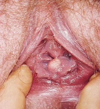
Vaginal inclusion cysts are usually small and asymptomatic.

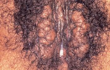
Multiple, bilateral vulvar inclusion cysts, previously referred to as sebaceous cysts, are shown.
Diagnosis
Pelvic examination
Sometimes biopsy
Diagnosis of vulvar cysts is by physical examination (1). A vulvar biopsy may be performed if the patient has symptoms (eg, persistent vulvar pruritus) or findings (whitening or thickening of labia minora) or the mass is concerning for malignancy (a mass that is hyperpigmented or has an irregular border or is solid, fixed, increasing in size, bleeding, or has other concerning features).
Diagnosis reference
1. Maldonado VA. Benign vulvar tumors. Best Pract Res Clin Obstet Gynaecol. 2014;28(7):1088-1097. doi:10.1016/j.bpobgyn.2014.07.014
Treatment
Excision
Treatment of vulvar cysts, indicated only for symptomatic cysts, is excision. A local anesthetic can be used for a single lesion. For multiple lesions, regional or general anesthesia may be preferred.


