Fractures may occur in the long bones in the middle of the foot (metatarsal bones).
There are several different kinds of metatarsal fractures, including stress fractures, Lisfranc fracture-dislocations, and fractures of the 5th metatarsal bone. (See also Overview of Fractures.)
Stress Fractures of the Metatarsals
Stress fractures are incomplete breaks in the bone caused by repeated stress rather than a single injury.
Walking or running for a long time can result in a stress fracture of the metatarsal.
These fractures cause tenderness in the middle of the foot and pain when full weight is put on the foot.
Stress fractures may not be visible on x-rays for 2 to 3 weeks after the injury, so doctors sometimes do computed tomography or magnetic resonance imaging, or they may treat the foot as if it is fractured and repeat x-rays in 2 weeks.
Usually, stopping activities that caused or aggravate the fracture and using crutches are the only treatment needed.
Stress fractures of the metatarsals can occur when people walk or run a long time, as when they suddenly start exercising longer or more intensely.
Symptoms of Stress Fractures
Stress fractures of the metatarsal bones are tender to the touch. Putting full weight on the foot increases the pain.
Diagnosis of Stress Fractures
X-rays
Sometimes computed tomography (CT) or magnetic resonance imaging (MRI)
Stress fractures may not be seen on x-rays if they are small or if the x-rays are taken soon after the fracture occurred (before the bone starts to repair itself). About 2 to 3 weeks after the injury, a stress fracture can be seen on x-rays as new bone is formed to repair the fracture (see How bones heal).
Sometimes CT or MRI is done to check for stress fractures. (See also Diagnosis of Fractures.)
Treatment of Stress Fractures
Avoidance of activities that caused or aggravate the stress fracture
Crutches
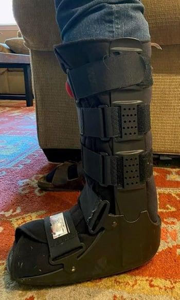
Sometimes a protective shoe or boot or a cast
When a developing stress fracture is recognized early, stopping activities that caused or aggravate the fracture and using crutches may be the only treatment necessary.
People may need to wear a special protective walking shoe or boot. Sometimes a cast is necessary.

Lisfranc Fracture-Dislocation
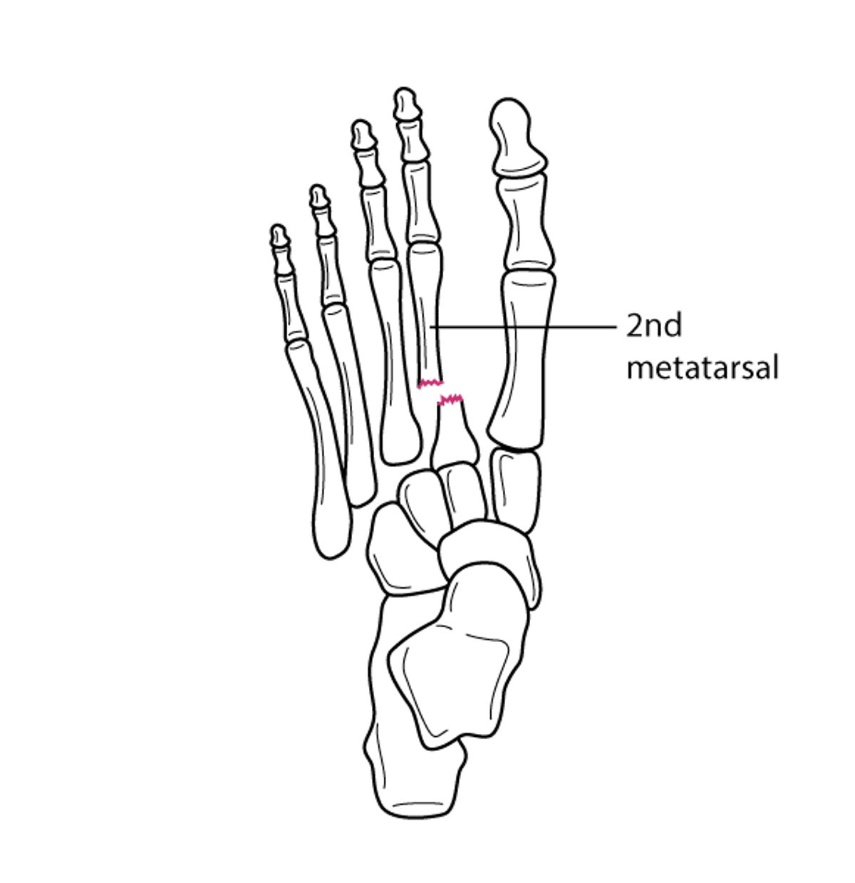
A Lisfranc fracture is a complete break in the 2nd metatarsal bone, which connects the 2nd toe to the bones at the back of the foot. A Lisfranc fracture-dislocation occurs when the broken pieces of bone are separated from each other (dislocated).
Lisfranc fracture-dislocations usually result from a fall on a foot that is flexed or from severe force.
The middle of the foot is painful, swollen, and tender, and the bottom of the foot may be bruised.
Doctors typically take x-rays from several different angles, but often, computed tomography is also needed to identify the injury.
Typically, people with a Lisfranc fracture-dislocation are referred to an orthopedic surgeon for surgery to put the broken pieces of bone back in place and keep them in place or to fuse the broken bones together.
In a Lisfranc fracture-dislocation, the 2nd metatarsal bone is fractured at its base, and the broken pieces may be separated from each other (dislocated). Lisfranc fracture-dislocations usually occur when people fall on a foot that is flexed or when the foot is hit with severe force. This injury is common among football players, motorcyclists, and horseback riders.
Fracture of the 2nd Metatarsal Bone
The 2nd metatarsal is broken near its base, and the broken pieces are separated from each other (dislocated). This injury is called Lisfranc fracture-dislocation. |

Symptoms of Lisfranc Fracture-Dislocation
Symptoms of Lisfranc fracture-dislocation may be mild or severe.
The middle of the foot becomes painful, swollen, and tender. If the injury is severe, the foot may look short, and the bottom of the foot may be bruised. Sometimes the area feels numb.
Lisfranc fracture-dislocation is serious and can result in permanent pain and arthritis. Compartment syndrome can develop. People may have problems participating in strenuous activities for the rest of their life.
Diagnosis of Lisfranc Fracture-Dislocation
X-rays
Often computed tomography (CT)
X-rays are taken from several different angles, but the injury may be difficult to see. Often, CT is also needed to diagnose Lisfranc fracture-dislocation. CT can provide more detailed, three-dimensional images of the injury.
Sometimes, when CT is unavailable, doctors ask the person to stand on the injured foot, and then x-rays are taken. If the bone is fractured, the person's weight causes the bones to separate more so that the space between the broken bones shows on x-rays and the injury can be diagnosed. (See also Diagnosis of Fractures.)
Treatment of Lisfranc Fracture-Dislocation
Referral to an orthopedic surgeon
Surgery to realign the broken bones or fusion of the bone in the foot
People with Lisfranc fracture-dislocation are usually admitted to a hospital and seen by an orthopedic surgeon as soon as possible.
Usually, one of the following is required:
Open reduction and internal fixation (ORIF): ORIF involves putting the broken pieces of bone back in place and keeping them in place with screws and metal plates.
Fusion of the bones in the middle of the foot: Fusion is similar to ORIF. The pieces of broken bone are put back into place and held in place, as in ORIF, but they are placed so that the damaged bones in the midfoot can grow together into one solid piece.
But these procedures do not always restore the foot to its previous condition.
If surgery is not required, a cast is used to immobilize the foot, and people are instructed not to put any weight of the injured foot for at least 6 weeks.
Fractures of the 5th Metatarsal Bone
Fractures of the 5th metatarsal bone occur in the bone that connects the little toe to the bones at the back of foot.
The 5th metatarsal bone can be fractured at its base (near the ankle) or in the middle (shaft).
These fractures can result from inward turning of the foot or a crush injury (for the base) or from repeated stress or a single injury (for the shaft).
If the base of this bone is fractured, the outside edge of the foot is painful, swollen, and tender.
Fractures of the base usually heal relatively quickly.
If the shaft is fractured, the blood supply to the bone may be disrupted, sometimes slowing healing or preventing the broken bones from growing back together.
To diagnose 5th metatarsal fractures, doctors take x-rays from several angles.
Treatment of fractures of the base usually consists of crutches and a protective hard-soled walking shoe or boot.
Treatment of fractures of the shaft may involve a short leg cast and crutches with no weight put on the injured foot or sometimes surgery.
The 5th metatarsal bone is the most commonly fractured bone in the foot. These fractures usually occur
At the base of the metatarsal (near the ankle)
In its shaft (the long middle part) of the metatarsal
Jones and Pseudo-Jones Fractures

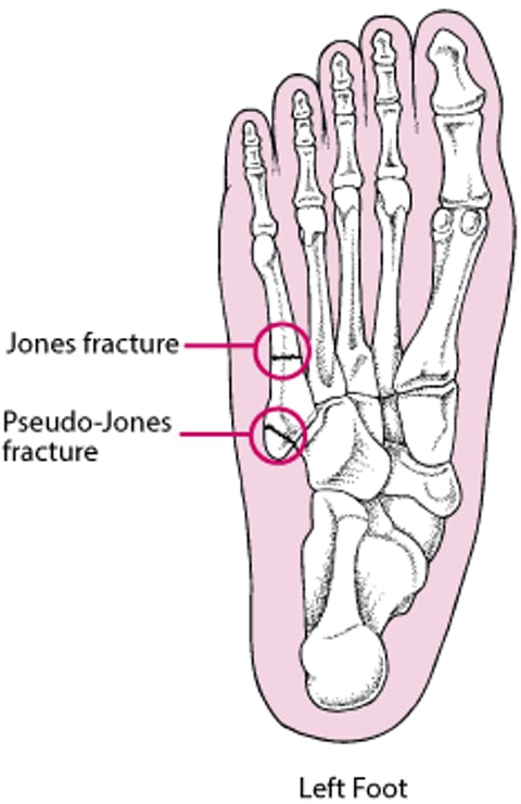
Fractures of the base
The base of the 5th metatarsal can be broken when the foot turns inward or is crushed. These fractures are sometimes called dancer's or pseudo-Jones fractures.
The outside edge of the foot is painful, swollen, and tender. A bruise may develop.
If doctors suspect a fracture of the base, they take x-rays from several different angles.
Crutches and a protective hard-soled walking shoe or boot may be needed for a few days. A cast is not usually necessary. People are encouraged to walk as soon as they can tolerate it.
Usually, these fractures heal relatively quickly.
Fractures of the shaft
Fractures of the shaft can result from repeated stress (stress fractures) or a single injury.
These fractures, called Jones fractures, are less common than fractures of the base.
Because these fractures can disrupt the blood supply to the bone, complications are more likely. For example, the bone may not grow back together (called nonunion), or it may grow back very slowly (called delayed union).
If doctors suspect a fracture of the shaft, they take x-rays from several different angles.
Usually, a short leg cast is applied to immobilize the ankle, and people must use crutches and not put any weight on the foot for 6 weeks.
Sometimes surgery is needed, and people are referred to an orthopedic surgeon. Open reduction with internal fixation (ORIF) may be done to put the pieces of broken bone back into place.