


Hypertensive retinopathy is retinal vascular damage caused by hypertension. Signs usually develop late in the disease. Funduscopic examination shows arteriolar constriction, arteriovenous nicking, vascular wall changes, flame-shaped hemorrhages, cotton-wool spots, yellow hard exudates, and optic disk edema. Treatment is directed at controlling blood pressure and, when vision loss occurs, treating the retina.
Pathophysiology of Hypertensive Retinopathy
Acute blood pressure elevation typically causes reversible vasoconstriction in retinal blood vessels, and hypertensive crisis may cause optic disk edema. More prolonged or severe hypertension leads to exudative vascular changes, a consequence of endothelial damage and necrosis. Other changes (eg, arteriole wall thickening, arteriovenous nicking) typically require years of elevated blood pressure to develop. Smoking compounds the adverse effects of hypertensive retinopathy.
Hypertension is a major risk factor for other retinal disorders (eg, retinal artery or vein occlusion, diabetic retinopathy). Also, hypertension combined with diabetes greatly increases risk of vision loss. Patients with hypertensive retinopathy are at high risk of hypertensive damage to other end organs.
Symptoms and Signs of Hypertensive Retinopathy
Symptoms usually do not develop until late in the disease and include blurred vision or visual field defects.
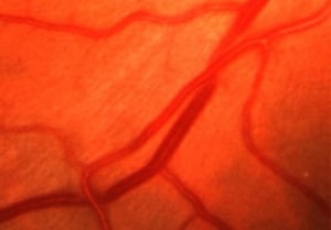
In the early stages, funduscopy identifies arteriolar constriction, with a decrease in the ratio of the width of the retinal arterioles to the retinal venules.
Chronic, poorly controlled hypertension causes the following:
Permanent arterial narrowing
Arteriovenous crossing abnormalities (arteriovenous nicking)
Sometimes total vascular occlusion occurs. Arteriovenous nicking is a major predisposing factor to the development of a branch retinal vein occlusion.
If acute disease is severe, the following can develop:
Superficial flame-shaped hemorrhages
Small, white, superficial foci of retinal ischemia (cotton-wool spots)
Yellow hard exudates
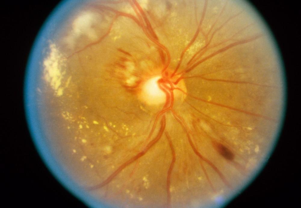
Optic disk edema

RALPH C. EAGLE, JR./SCIENCE PHOTO LIBRARY
© Springer Science+Business Media
© Springer Science+Business Media

RALPH C. EAGLE, JR./SCIENCE PHOTO LIBRARY


© Springer Science+Business Media

© Springer Science+Business Media
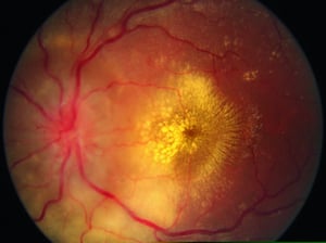
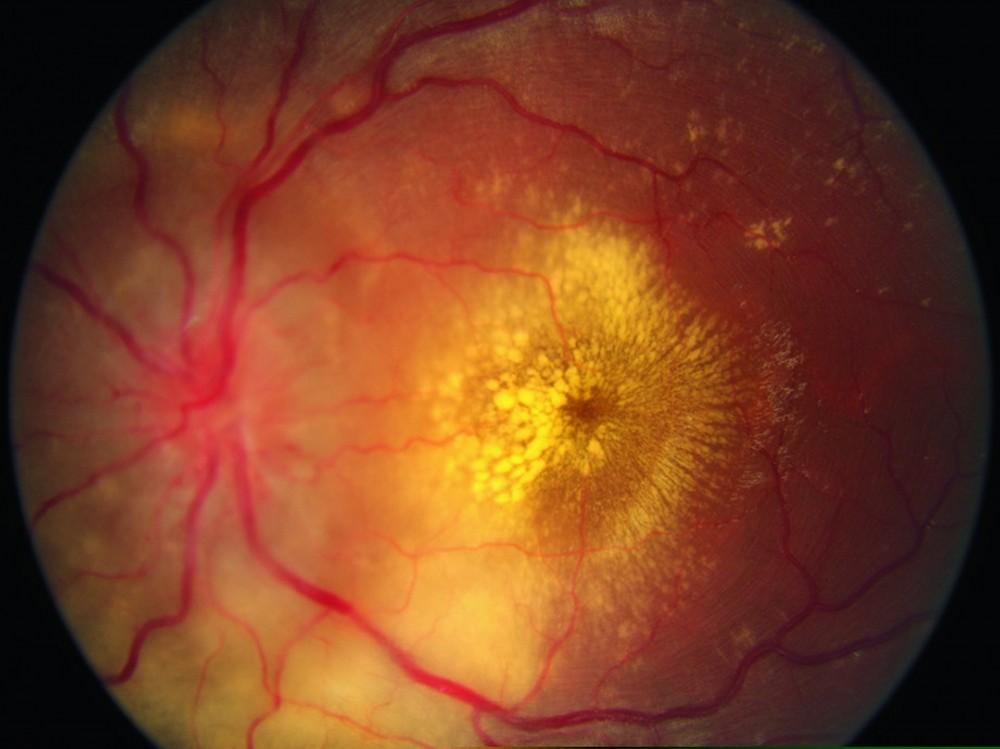
Yellow hard exudates represent intraretinal lipid deposition from leaking retinal vessels. These exudates can develop a star shape within the macula, particularly when hypertension is severe. In severe hypertension, the optic disk becomes congested and edematous (papilledema indicating hypertensive crisis).
Diagnosis of Hypertensive Retinopathy
Diagnosis is by history (duration and severity of hypertension) and funduscopy.
Treatment of Hypertensive Retinopathy
Key Points
Chronic hypertension progressively damages the retina, causing few or no symptoms until changes are advanced.
Hypertensive crisis can cause retinopathy with superficial flame-shaped hemorrhages; small, white, superficial foci of retinal ischemia (cotton-wool spots); yellow hard exudates; and optic disk edema.
Diagnose patients by history and funduscopy.
Treat primarily by controlling blood pressure, and, for retinal edema, sometimes laser or intravitreal injection of corticosteroids or antivascular endothelial growth factor drugs.


