


Craniocervical junction disorders are abnormalities of the bones that join the head and neck. (Cranio- means skull, and cervical means neck.)
These disorders may be present at birth or result from injuries or disorders that occur later.
Most commonly, people have neck pain and headache, but if the spinal cord or lowest part of the brain (brain stem) is affected, people may have difficulty sensing vibration, pain, and temperature and may have weak muscles, dizziness, and impaired vision.
Doctors suspect the diagnosis based on symptoms, and magnetic resonance imaging or computed tomography is done to confirm it.
To relieve pressure on the brain, spinal cord, or nerves, doctors use traction or manipulate the head, then immobilize the neck, but sometimes surgery is needed.
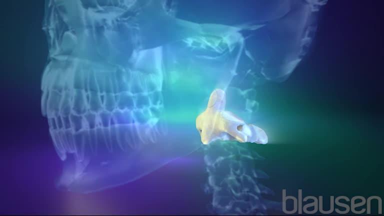
The craniocervical junction consists of the bone that forms the base of the skull (occipital bone) and the first two bones in the spine (which are in the neck): the atlas and axis. Disorders that affect the large opening at the bottom of the occipital bone (called the foramen magnum) are a particular concern because important structures pass through this opening. These structures include the lowest part of the brain (brain stem), which connects to the spine, as well as some nerves and blood vessels.
Craniocervical junction disorders may involve bones that are
Fused together
Abnormally formed or underdeveloped
Out of alignment (misaligned)
Misaligned bones may be completely separated (dislocated) or partially misaligned (subluxed).
Craniocervical junction disorders can put pressure on the lower parts of the brain, the top part of the spinal cord, or nearby nerves. The resulting symptoms can be serious. They include paralysis, weakness, and loss of sensation.
Causes of Craniocervical Junction Disorders
Craniocervical junction disorders may be present at birth (congenital) or acquired later.
Present at birth
Some craniocervical junction disorders—called isolated disorders—affect only the craniocervical junction. Other craniocervical junction disorders result from conditions that also affect many other parts of the body (general or systemic disorders).
Isolated disorders include the following:
Atlantoaxial subluxation or dislocation: The first and second spinal bones are misaligned, sometimes putting pressure on the spinal cord.
Atlas assimilation: The occipital bone and first spinal bone are fused.
Atlas hypoplasia: The first spinal bone develops incompletely.
Basilar invagination: Part of the second spinal bone is pushed up and partly into the skull. As a result, the neck is shortened, and pressure is put on parts of the brain, nerves, and spinal cord.
Klippel-Feil malformation: The first two spinal bones are fused, or the first spinal bone is fused to the skull. As a result, movement of the neck is limited. Usually, this disorder causes no other problems, except sometimes spinal cord damage after a minor injury.
Os odontoideum: Part of the second spinal bone is detached from the rest of the second spinal bone.
Platybasia: The bone at the base of the skull (occipital bone) is flattened. It is often accompanied by Chiari malformation (protrusion of the cerebellum, which controls balance, through the opening in the occipital bone). The protruding cerebellum may put pressure on the brain stem or spinal cord. People with this disorder have a short neck.
General disorders may cause the same abnormalities as isolated disorders, but the abnormalities occur as part of a disorder that affects the body as a whole, as in the following:
Disorders that affect bone formation in children (such as achondroplasia): These disorders affect all newly developing bone, particularly the long bones in the arms and legs. The long bones may be abnormally short and malformed, causing dwarfism. These disorders sometimes also cause the opening in the occipital bone to narrow or the occipital bone to fuse with the first spinal bone (atlas assimilation), putting pressure on the brain stem or spinal cord.
Down syndrome, mucopolysaccharidosis (a rare hereditary disorder that impairs the body's processing of carbohydrates), or osteogenesis imperfecta (a rare hereditary disorder that results in fragile bones): These disorders cause various symptoms, which may include misalignment of the first two spinal bones.
Acquired
Craniocervical junction disorders may occur later in life. They can result from injuries or certain disorders.
Injuries may affect bone, ligaments, or both. They are usually caused by motor vehicle or bicycle accidents, falls, or often diving. Some injuries are immediately fatal.
The most common disorders that affect craniocervical structures are
Tumors can also affect craniocervical structures. If tumors spread to the bones of the neck (metastatic tumors to bone), the first two spinal bones may become misaligned. A rare, slow-growing bone tumor called a chordoma or a common, slow-growing noncancerous brain tumor called a meningioma can develop at the craniocervical junction and press on the brain or spinal cord.
Symptoms of Craniocervical Junction Disorders
Symptoms may start after a slight neck injury or for no apparent reason.
Typically, people with a craniocervical junction disorder have neck pain, often with a headache that starts at the back of the head. Moving the head usually makes neck pain and headache worse, and coughing or bending forward can trigger the pain. Neck pain may spread to the arms.
If there is pressure on the spinal cord, the arms and/or legs may feel weak, and people may have difficulty moving them. People may be unable to sense where their limbs are (called position sense) or feel vibration. When they bend their neck forward, they may feel an electrical shock or a tingling sensation shooting down their back, often into their legs (called Lhermitte sign). Occasionally, people become less sensitive to pain and temperature in their hands and feet.
Depending on the specific disorder, the neck may be short, webbed, or twisted in an abnormal position. Movement of the head may be limited.
Pressure on parts of the brain or cranial nerves (which connect the brain directly to various parts of the head, neck, and trunk) can affect eye movements. People may have double vision or be unable to move their eyes in certain directions, or the eyes may move involuntarily in certain ways (called nystagmus). People may be hoarse and have difficulty swallowing. Speech may be slurred. Coordination may be lost. Some people develop sleep apnea. In this serious disorder, breathing repeatedly stops during sleep, often long enough to temporarily decrease the amount of oxygen and increase the amount of carbon dioxide in the blood.
Changing the position of the head can sometimes put pressure on arteries, cutting off the blood supply to the head. Then, people may faint or feel light-headed, confused, or weak, or they may fall, often without warning. They may have a sensation of spinning (vertigo). Vision and eye movement are sometimes affected.
In many people with Chiari malformation, a cavity (called a syrinx) forms in the spinal cord. These people may lose their ability to feel pain and temperature in the neck, upper arms, and parts of the back. The muscles may feel weak or become paralyzed, particularly in the hands.
Diagnosis of Craniocervical Junction Disorders
Imaging tests
If problems appear suddenly or suddenly worsen, people should see a doctor immediately. Immediate diagnosis and treatment of craniocervical junction disorders are essential and can sometimes reverse symptoms or prevent permanent disability.
Doctors suspect a craniocervical junction disorder if people have
Neck pain or a headache at the back of the head plus problems that are usually caused by pressure on lower parts of the brain or the top of the spinal cord
Certain involuntary movement of the eyes (nystagmus)
The diagnosis can be confirmed by imaging tests, usually magnetic resonance imaging (MRI) or computed tomography (CT). Because problems that appear suddenly or suddenly worsen are an emergency, an imaging test is done immediately. CT shows bone better than MRI and may be done more easily in an emergency. If MRI and CT are unavailable, x-rays are taken.
If MRI and CT are inconclusive, myelography with CT may be done. For this procedure, x-rays are taken after a radiopaque contrast agent—one that can be seen on x-rays—is injected into the space around the spinal cord.
If MRI or CT suggests abnormalities that affect blood vessels, angiography, which provides detailed images of bloods vessels, is done. Doctors may use magnetic resonance angiography (which uses a strong magnetic field and very high frequency radio waves rather than x-rays), CT angiography (CT done after a radiopaque contrast agent is injected), or conventional angiography (which uses x-rays taken after a radiopaque contrast agent is injected).
Treatment of Craniocervical Junction Disorders
Realignment, traction, and immobilization of affected structures
Possibly surgery
Other treatments depending on the cause
If the craniocervical junction structures are putting pressure on the brain, spinal cord, or nerves, doctors try to realign (reduce) the structures by using traction or by manipulating the head into different positions. These techniques may relieve the pressure. After the structures are realigned, a device is used to keep the head and neck from moving (to immobilize them).
Problems that appear suddenly or suddenly worsen require immediate realignment.
Usually, realignment requires traction. Traction typically involves a device that encircles and is fixed to the head (called a halo crown or ring). It may have to remain in place for 5 to 6 days. After the structures are realigned, the halo ring is attached to a brace around the person's torso. The brace, also called a halo vest, immobilizes the neck. It must remain in place for 8 to 12 weeks. After it is applied, x-rays are taken to make sure the structures are being held securely in the correct alignment.

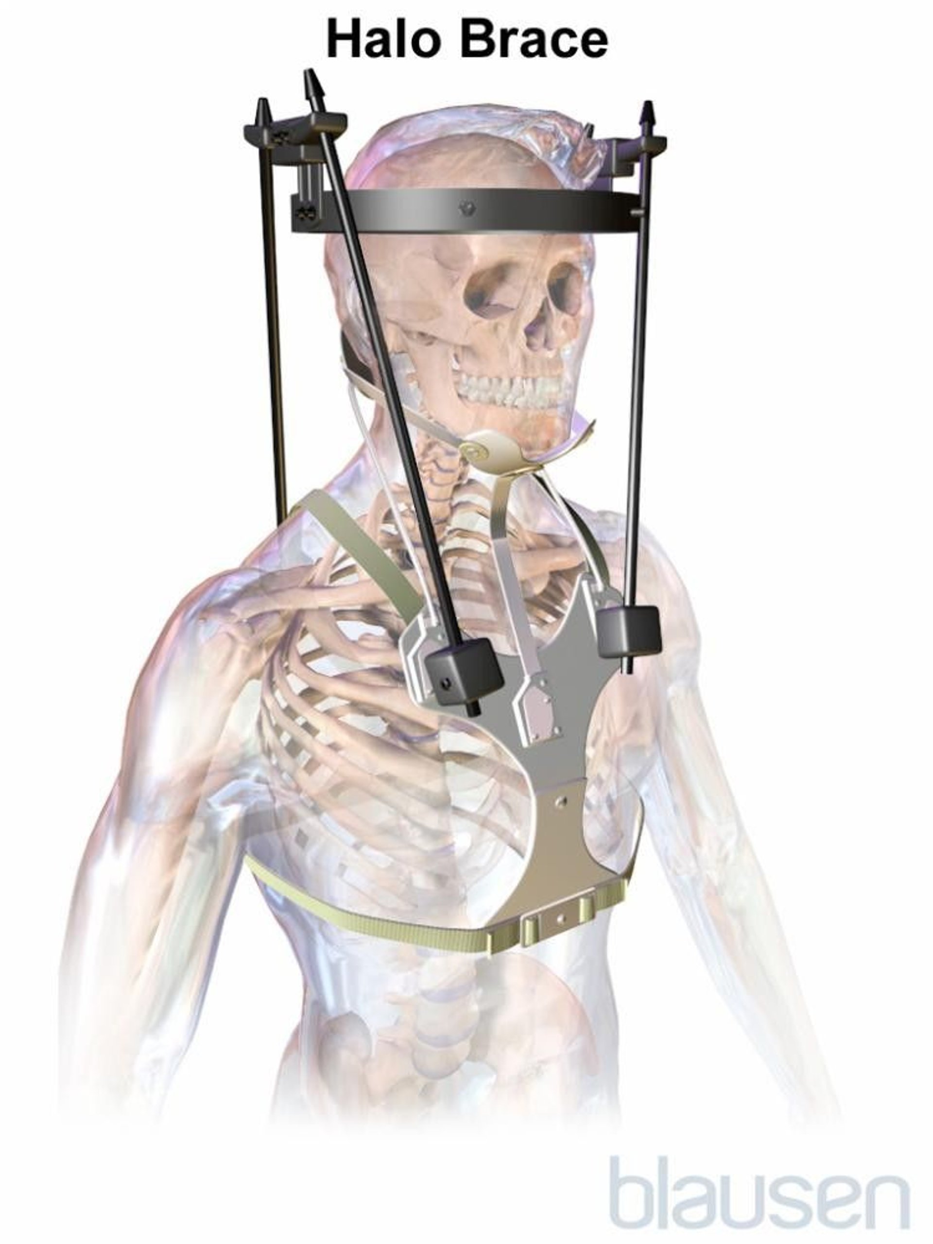

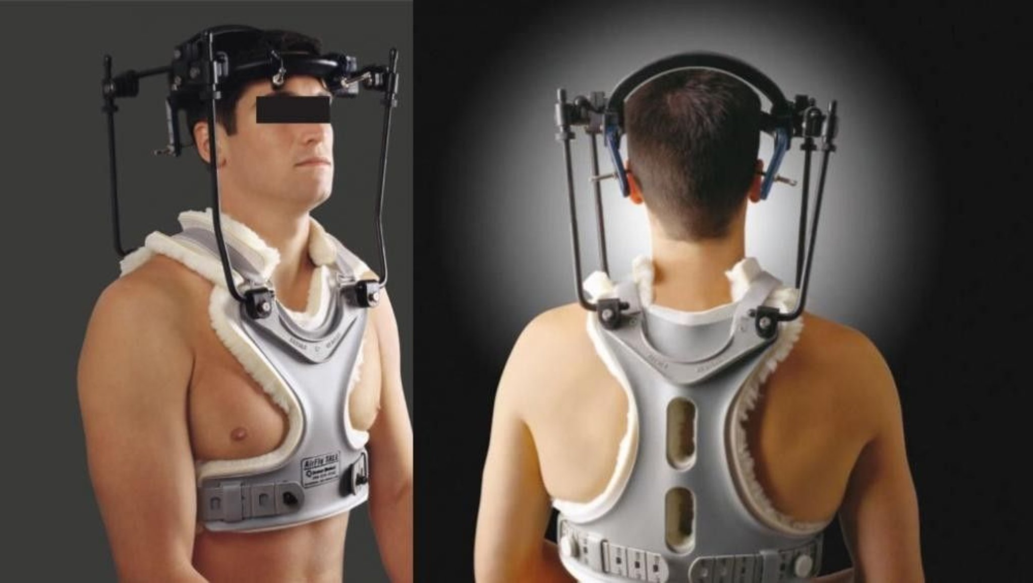
Photo courtesy of Depuy/Synthes.
If traction or manipulation is ineffective, surgery is done to relieve the pressure, stabilize the structures, or both. If the cause is rheumatoid arthritis, surgery is usually needed. Various devices, such as metal plates or rods with screws, are used to securely hold the structures in place until bones fuse and become stable.
If problems are due to a bone tumor that has spread (metastasized), radiation therapy and a rigid collar (neck brace) to keep the neck from moving often help.
If the cause is Paget disease of bone, medications such as bisphosphonates


