


A brain tumor can be a noncancerous (benign) or cancerous (malignant) growth in the brain. It may originate in the brain or have spread (metastasized) to the brain from another part of the body.
Symptoms may include headaches, personality changes (such as becoming depressed, anxious, or uninhibited), weakness, abnormal sensations, loss of balance, trouble concentrating, seizures, and incoordination.
Imaging tests can detect brain tumors, but a biopsy of the tumor is often needed to be certain
Treatment may involve surgery, radiation therapy, chemotherapy, or a combination.
(See also Overview of Tumors of the Nervous System, Some Specific Brain Tumors, and Overview of Brain and Spinal Cord Tumors in Children.)
Brain tumors are slightly more common among men than women. Only meningiomas, which are usually noncancerous, are more common among women. Brain tumors can develop at any age. The most serious type of brain tumor, glioblastoma, is becoming more common among older people as the population ages.
Brain tumors—whether cancerous or not—can cause serious problems because the skull is rigid, providing no room for the tumor to expand. Also, if tumors develop near parts of the brain that control vital functions, they may cause problems, such as weakness, difficulty walking, loss of balance, partial or complete loss of vision, difficulty understanding or using language, and problems with memory.
Brain tumors can cause problems in the following ways:
By directly invading and destroying brain tissue
By directly putting pressure on nearby tissue
By increasing pressure within the skull (intracranial pressure) because the tumor takes up space and the skull cannot expand to accommodate it
By causing fluid to accumulate in the brain
By blocking the normal circulation of cerebrospinal fluid through the spaces within the brain, causing those spaces to enlarge
By causing bleeding
Classification of Brain Tumors
There are two main types of brain tumors:
Primary: These tumors originate in the cells within or next to the brain. They may be cancerous or noncancerous.
Secondary: These tumors are metastases. That is, they originate in another part of the body and spread to the brain. Thus, they are always cancerous.
Primary tumors
The most common primary tumors include
Gliomas (including astrocytomas, oligodendrogliomas, and glioblastomas) and ependymomas
Lymphoma that originates in the central nervous system (primary brain lymphoma)
Gliomas account for 65% of all primary brain tumors.
Did You Know...
|
Noncancerous tumors are named for the specific cells or tissues in which they originate. For example, hemangioblastomas originate in blood vessels (“hema” refers to blood vessels, and hemangioblasts are the cells that develop into blood vessel tissue). Some noncancerous tumors originate in cells of the embryo (embryonic cells), early in the development of the fetus. Such tumors may be present at birth.
Secondary tumors
Brain metastases are much more common than primary tumors. Over 80% of people with brain metastases have more than one metastasis.
Metastases from other parts of the body may spread to a single part of the brain or to several different parts. Many types of cancer can spread to the brain. They include
Lymphomas of the brain are becoming more common among people who have a weakened immune system (such as those who have AIDS), among older people and, for unknown reasons, among people who have a normal immune system. Some lymphomas originate in the brain (called primary central nervous system lymphomas). Other lymphomas originate elsewhere in the body and spread to the brain (metastatic lymphomas).
Symptoms of Brain Tumors
Symptoms can occur whether a brain tumor is noncancerous or cancerous. Noncancerous tumors grow slowly and may become quite large before causing symptoms. Cancerous tumors usually grow rapidly.
A brain tumor can cause many different symptoms, and symptoms may occur suddenly or develop gradually. Which symptoms develop first and how they develop depend on the tumor’s size, growth rate, and location. In some parts of the brain, even a small tumor can have devastating effects. In other parts of the brain, tumors can grow relatively large before any symptoms appear. As the tumor grows, it pushes and stretches but usually does not destroy nerve tissue, which can compensate for these changes very well. Thus, symptoms may not develop at first.
Many symptoms result from increased pressure within the skull:
Headache
Deterioration in mental function
Problems due to pressure on specific structures in or near the brain, such as the nerve to the eye (optic nerve)
Headache is the most common and often the first symptom. However, some people with brain tumors do not get headaches, and most headaches are not caused by brain tumors. A headache due to a brain tumor usually recurs more and more often as time passes. It is often worse when people lie down. The headache may be most intense when people awaken from sleep, then lessen later in the day. If headaches with these characteristics start in people who have not had headaches before, a brain tumor may be the cause.
Often, increased pressure within the skull also causes mental function to deteriorate and mood to worsen. The personality may change. For example, people may become withdrawn, moody, and, often, inefficient at work. They may feel drowsy, confused, and unable to think. Such symptoms are often more apparent to family members and co-workers than to the affected person. Depression and anxiety, especially if either develops suddenly, may be an early symptom of a brain tumor. People may behave bizarrely. They may become uninhibited or behave in ways they never have before. In older people, certain brain tumors cause symptoms that may be mistaken for those of dementia.
Later, as the pressure within the skull increases, nausea, vomiting, lethargy, increased drowsiness, intermittent fever, and even coma may occur. Vision may blur suddenly when people change positions.
Depending on which area of the brain is affected (see page Brain Dysfunction by Location), a tumor can do any of the following:
Cause an arm, a leg, or one side of the body to become weak or paralyzed
Impair the ability to feel heat, cold, pressure, a light touch, or sharp objects
Make people unable to express or understand language
Increase or decrease the pulse and breathing rates if the tumor compresses the brain stem
Reduce alertness
Impair the ability to hear, smell, or see (causing such symptoms as double vision and loss of vision)
For example, a pituitary tumor may press on the nearby optic nerves (2nd cranial nerves), which are involved in vision, and thus impair peripheral vision. Any of the above symptoms suggests a serious disorder and requires immediate medical attention.
Other common symptoms of a brain tumor include vertigo, loss of balance, and incoordination. Some brain tumors, usually primary tumors, cause seizures.
If a tumor blocks the flow of cerebrospinal fluid through the spaces within the brain (ventricles), fluid may accumulate in the ventricles, causing them to enlarge (a condition called hydrocephalus). As a result, pressure within the skull increases. In addition to other symptoms of increased pressure, hydrocephalus makes turning the eyes upward difficult. In infants and very young children, the head enlarges.
If the pressure within the skull is greatly increased, the brain may be pushed downward because the skull cannot expand. Herniation of the brain may result. Two common types are
Transtentorial herniation: The upper part of the brain (cerebrum) is forced through the narrow opening (the tentorial notch) in the relatively rigid tissue that separates the cerebrum from the lower parts of the brain (cerebellum and brain stem). In people with this type of herniation, consciousness is reduced. The side of the body opposite the tumor may be paralyzed.
Tonsillar herniation: A tumor that originates in the lower part of the brain pushes the lowest part of the cerebellum (cerebellar tonsils) through the opening at the base of the skull (foramen magnum). As a result, the brain stem, which controls breathing, heart rate, and blood pressure, is compressed and malfunctions. If not diagnosed and treated immediately, a tonsillar herniation rapidly results in coma and death.
People with metastases to the brain may also have symptoms related to the original cancer. For example, if the cancer originated in the lungs, people may cough up bloody mucus. With metastases, weight loss is common.
Symptoms worsen over time unless the tumor is treated. With treatment, particularly for benign tumors, some people completely recover. For others, life span is shortened, sometimes greatly. The outcome depends on the type and location of the tumor.
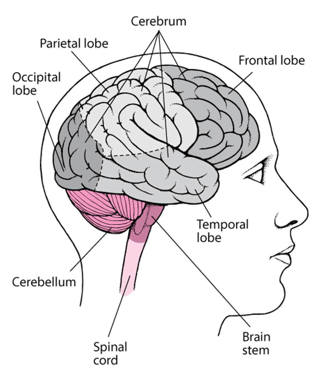
Viewing the Brain
The brain consists of the cerebrum, brain stem, and cerebellum. Each half (hemisphere) of the cerebrum is divided into lobes. |

Diagnosis of Brain Tumors
Magnetic resonance imaging or computed tomography
Sometimes a spinal tap
A biopsy
Doctors consider the possibility of a brain tumor in people who have had a seizure for the first time or who have characteristic symptoms. Although doctors can often detect brain dysfunction during an examination, other procedures are needed to diagnose a brain tumor.
Magnetic resonance imaging (MRI) is the best test for identifying brain tumors. Computed tomography (CT) is a good alternative. It can identify most brain tumors. Before these tests, a substance that makes the tumor easier to see (a contrast agent for MRI or a radiopaque contrast agent for CT) is injected into a vein. These tests can show the tumor’s size and exact position in great detail. When a brain tumor is detected, more diagnostic procedures are done to determine the particular kind.
Sometimes a spinal tap (lumbar puncture) is done to obtain cerebrospinal fluid for examination under a microscope. This procedure is done when doctors suspect that the tumor has invaded the layers of tissues that cover the brain (meninges). Such tumors may block the absorption of cerebrospinal fluid. A spinal tap may also help when the diagnosis or the type of tumor is unclear. Cerebrospinal fluid may contain cancer cells. However, a spinal tap cannot be done in people who have a large tumor that is increasing pressure within the skull. In these people, removing cerebrospinal fluid during a spinal tap may cause the tumor to move, resulting in herniation of the brain.
Specialized tests can sometimes help with the diagnosis. For example, blood and cerebrospinal fluid may also be tested to check for substances secreted by tumors (called tumor markers) and for gene abnormalities that are characteristic of certain tumors. Identifying certain gene abnormalities can help predict which treatments will be most effective.
A biopsy of the tumor (removal of a sample of the tumor for examination under a microscope) is done when results of MRI and other tests cannot definitively identify what the type of tumor is and whether or not it is cancerous. A biopsy may be done during surgery, in which all or part of the tumor is removed. Or if a tumor is difficult to reach, a stereotactic biopsy may be done. For this procedure, a frame is attached to the skull. The frame provides reference points that can be identified on an MRI or a CT scan. These reference points enable doctors to guide the biopsy needle precisely into the tumor.
Treatment of Brain Tumors
Surgery, radiation therapy, medications (such as chemotherapy or immunotherapy) or, most often, a combination
Sometimes medications, usually corticosteroids, to reduce pressure within the skull
Antiseizure medications to treat seizures
Treatment of a brain tumor depends on its location and type.
Specific treatments for brain tumors
Treatments for brain tumors include the following:
Craniotomy (brain surgery)
Radiation therapy
Implants
Shunts
Stereotactic techniques
Craniotomy (surgical removal of the tumor) is done when possible. This procedure involves opening the skull. Some brain tumors can be removed with little or no damage to the brain. However, many grow in areas that make removal by traditional surgery difficult or impossible without destroying essential structures.
For craniotomy, part of the scalp is shaved. Then, an incision is made through the skin. A high-speed drill and a special saw are used to remove a small piece of bone above the tumor. The tumor is located and removed using one of the following:
A scalpel may be used to cut out the tumor.
A laser may be used to vaporize the tumor.
A device that emits ultrasound waves may be used to break the tumor apart, so that the pieces can be suctioned out (aspirated).
Lasers and ultrasound devices are used to remove tumors that would be difficult to cut out. Usually, the bone is then replaced, and the incision stitched closed.
Traditional surgery sometimes causes brain damage that can lead to symptoms such as partial paralysis, changes in sensation, weakness, and impaired mental function. Nevertheless, removing a tumor—whether cancerous or noncancerous—is essential if its growth threatens important brain structures. Even when a cure is impossible, surgery may be useful to reduce the tumor’s size, relieve symptoms, and help doctors determine whether other treatments, such as radiation therapy or chemotherapy, are warranted.
Radiation therapy includes the following:
Whole brain radiation
Stereotactic radiation that targets the tumor (SRS)
Whole brain radiation delivers radiation to the entire brain. It is used most often in people who have cancer that has developed in other organs and spread to the brain. Most people with metastatic brain cancer have multiple metastases. Whole brain radiation is designed to kill cancer cells. However, it can affect normal brain cells and is therefore given in small doses over 2 to 3 weeks.
Radiosurgery uses stereotactic techniques to locate the tumor precisely. Then very focused beams of radiation (gamma rays or photon beams) are used to destroy the tumor. Radiosurgery is not really surgery because no incision is required. Radiosurgery may be done with a gamma knife or a linear accelerator. Both use photon radiation.
When a gamma knife is used, an imaging frame is attached to the person’s skull. The person lies on a sliding bed, and a large helmet with holes in it is placed over the frame. The head of the bed is then slid into a globe that contains radioactive cobalt. Radiation passes through the holes in the helmet and is aimed precisely at the tumor.
When a linear accelerator is used the head is immobilized in a fixed frame or molded mask. CT is used to make a 3-dimensional map of the tumor so that radiation delivered from different angles can be aimed to precisely match the shape of the tumor (called conformal radiation).
Traditionally, radiosurgery is done when a person has four or fewer tumors, and radiation of the whole brain is done when a person has 5 or more tumors. However, some recent evidence suggests that radiosurgery can be done when a person has as many as 10 tumors. Radiosurgery is also useful for brain metastases.
Damage due to radiation sometimes occurs, despite doctor's efforts to prevent it.
Stereotactic techniques can also be used to do the following:
Locate a site for biopsy
Determine where to insert radioactive implants or to direct a laser to destroy tumor cells
Computers are used to produce a three-dimensional image. The three-dimensional image can be obtained by attaching a light-weight metal imaging frame with a series of rods to the person’s skull. A local anesthetic is given to numb the area, and the pins are attached to the skull, piercing the skin. A computed tomography (CT) scan shows the rods as dots, providing reference points, which help locate the tumor. In a similar procedure, a plastic frame is used, and magnetic resonance imaging (MRI) is used to show where the tumor is.
Techniques that do not involve attaching a frame may be used instead. For examples, special markers can be taped to the skull to provide reference points. The location of these markers is entered into a computer that contains images of the brain tumor.
Implants are occasionally inserted into the brain. The implants consist of wafers soaked with a chemotherapy drug, After a tumor is removed and before the skull and incision are closed, these wafers may be placed in the space where the tumor was. As the wafers gradually dissolve, they release the drug to destroy any remaining cancer cells.
Shunts may be surgically placed if a tumor causes pressure within the skull to increase. A shunt is a thin piece of tubing that is inserted into one of the spaces of the brain (ventricles) or sometimes into the space around the spinal cord that contains cerebrospinal fluid (subarachnoid space). The other end of the tubing is threaded under the skin from the head usually into the abdomen. Excess cerebrospinal fluid is drained from the brain into the abdomen, where it is absorbed. The shunt contains a one-way valve that opens when there is too much fluid in the brain. Shunts may be temporary (until the tumor is removed) or permanent.
Noncancerous tumors
Surgical removal is often safe and cures the person. However, very small tumors and tumors in older people may be left in place as long as they are not causing symptoms. Sometimes radiation therapy is given after surgery to destroy any remaining tumor cells.
Radiosurgery using stereotactic techniques is effective for treating noncancerous tumors such as meningiomas and vestibular schwannomas. It is often used instead of traditional surgery for these tumors.
Cancerous brain tumors
Usually, a combination of surgery, radiation therapy, and chemotherapy is used. As much of the tumor as can be removed safely is removed, and then radiation therapy is begun. Radiation therapy is given over a course of several weeks. Radiosurgery is used when traditional surgery cannot be, especially for the treatment of metastases.
For very aggressive tumors, chemotherapy is given with radiation therapy. Radiation therapy plus chemotherapy rarely cures but may shrink a tumor enough to keep it under control for many months or even years.
After radiation therapy, ongoing chemotherapy is used to treat some types of cancerous brain tumors. Chemotherapy appears to be particularly effective in treating anaplastic oligodendrogliomas.
Increased pressure within the skull
This extremely serious condition requires immediate medical attention. If people are in a coma or have difficulty breathing, herniation of the brain may be developing. To help such people breathe, doctors insert a plastic tube through the nose or mouth into the windpipe (trachea) and attach it to a ventilator
If the tumor is blocking the flow of cerebrospinal fluid through the spaces within the brain, a device may be used to drain the cerebrospinal fluid and thus reduce the risk of herniation. The device consists of a small tube (catheter) connected to a gauge that measures the pressure within the skull. The tube is inserted through a tiny opening drilled in the skull. A local anesthetic (usually plus a sedative) or a general anesthetic may be used. The tube is removed or converted to a permanent drain (shunt) after a few days. During this time, doctors surgically remove all or part of the tumor or use radiosurgery or whole-brain radiation therapy to reduce the size of the tumor and thus relieve the blockage.
Metastases
Treatment depends largely on where the cancer originated. Radiation therapy directed at the metastases in the brain is often used. Surgical removal before radiation therapy may benefit people who have only a single metastasis. Sometimes radiosurgery is used. Chemotherapy and immunotherapy may help treat metastases from certain types of cancer.
End-of-life issues
People with cancerous brain tumors have a limited life expectancy and are likely to become unable to make decisions about medical care. Consequently, establishing advance directives is advisable. Advance directives can help a doctor determine what kind of care people want if they become unable to make decisions about medical care. Treatment that focuses on relieving symptoms as fully as possible (palliative care), rather than on curing the person, may be more appropriate.
Many cancer centers, especially those with palliative care and hospice facilities, provide counseling and home health services.


