


Osteogenesis imperfecta is a hereditary disorder that disrupts the proper formation of bones and makes bones abnormally fragile.
This disorder is caused by mutations in certain genes.
Typical symptoms include weak bones that break easily.
The diagnosis is based on x-rays.
The type that occurs in infancy is lethal.
Certain medications can help strengthen bones and injections of growth hormone can help some children.
Osteogenesis imperfecta is an osteodysplasia. Osteodysplasias are disorders that disturb bone growth. Osteogenesis imperfecta is the most well known osteodysplasia.
In osteogenesis imperfecta, synthesis of collagen, one of the normal components of bone, is impaired in most affected people because of mutations in the genes that play an important role in the development of collagen. The bones become weak and break (fracture) easily.
There are 4 main types of osteogenesis imperfecta (I, II, III, and IV) along with other rare types.
Symptoms of Osteogenesis Imperfecta
Osteogenesis imperfecta can range from mild to severe.
Most people with osteogenesis imperfecta have fragile bones, and about 50 to 65% have hearing loss.
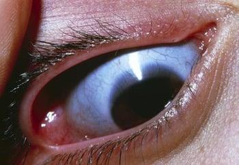
Osteogenesis imperfecta causes the whites of the eyes (sclerae) in some people to turn blue. The blue color appears because the veins beneath the abnormally thin sclerae show through. The sclerae are thinner than normal because collagen has not been formed correctly.
Children may have discolored and poorly developed teeth (called dentinogenesis imperfecta) depending on the type of osteogenesis imperfecta.
Sometimes heart or lung diseases develop in children with osteogenesis imperfecta.

Type I osteogenesis imperfecta is the mildest type. Some children may have only symptoms of blue sclerae and muscle and joint pain caused by loose joints. Children with this type may have increased risk of fractures during childhood.
Type II osteogenesis imperfecta is the most severe type and causes death. Infants are usually born with many broken bones. The skull may be so soft that the brain is not protected from pressure applied to the head during childbirth. These infants have shortened arms and legs and blue sclerae. Infants with this type can die before childbirth or within the first few days or weeks of life.
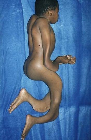
Type III osteogenesis imperfecta is the most severe type that does not cause death. Children with this type are very short and have curving of the spine and frequent fractures. This type causes bones to often break after very minor injuries, usually when children begin to walk. These children also have a large skull and a triangular face shape caused by overdevelopment of the head and underdevelopment of the face bones. Chest deformities are common. The color of the sclerae varies.

Type IV osteogenesis imperfecta ranges widely in severity and can cause deformities. Children with this type have bones that fracture easily during childhood before puberty. The sclerae are typically white. Children are short. Children with this type may benefit from treatment, and the survival rate is high.
Diagnosis of Osteogenesis Imperfecta
Before birth, prenatal ultrasonography
After birth, a doctor's evaluation
Sometimes analysis of cells or genetic testing
Before birth, the most severe and lethal form of osteogenesis imperfecta can be detected in pregnant women by an ultrasound.
After birth, doctors base the diagnosis of osteogenesis imperfecta on the symptoms and on a physical examination.
If the diagnosis is not clear, doctors may remove a sample of skin for examination under a microscope (biopsy) to analyze a type of connective tissue cell (fibroblasts) or they may take a sample of blood to analyze certain genes.
X-rays may show abnormal bone structures that suggest osteogenesis imperfecta.
A test called audiometry is done often throughout childhood to monitor hearing.
Treatment of Osteogenesis Imperfecta
Growth hormone
Bisphosphonates
Denosumab
There is no cure for osteogenesis imperfecta, but treatments are available to manage symptoms and some complications.
Growth hormone injections can help growth and bone strength in children with types I and IV.
Treatment of broken bones is similar to that for children who do not have the disorder. However, broken bones can become deformed or fail to grow. As a result, body growth can become permanently stunted in children with many broken bones, and deformities are common. Bones may require stabilization with internal metal rods.
Physical therapy and occupational therapy help prevent fractures and improve function. Taking measures to avoid even minor injuries can help prevent fractures.
Some children with hearing loss may be helped by a cochlear implant (a device that converts sound waves to electrical signals that it sends to electrodes implanted in the inner ear).
More Information
The following English-language resource may be useful. Please note that THE MANUAL is not responsible for the content of this resource.
Osteogenesis Imperfecta (OI) Foundation: An organization providing support, education, and research information about OI


