


Tinea corporis is a dermatophyte infection of the face, trunk, and extremities. Diagnosis is by clinical appearance and by examination of skin scrapings on potassium hydroxide wet mount. Treatment involves topical or oral antifungals.
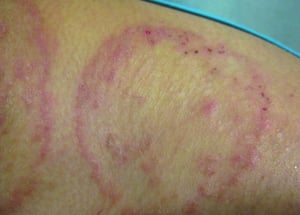
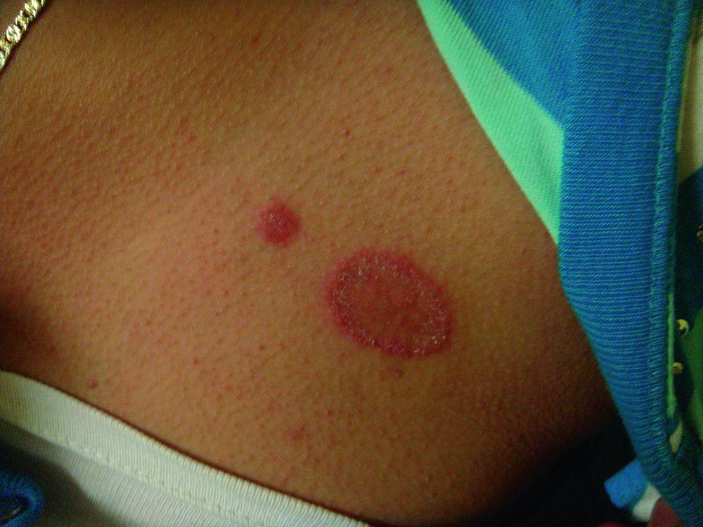
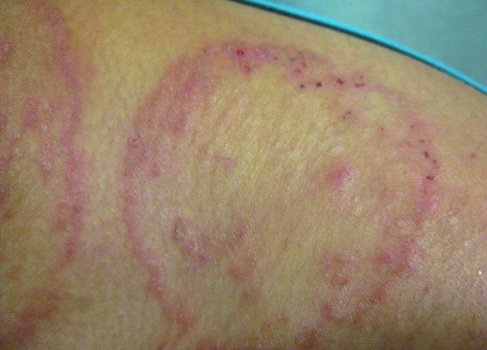
Tinea corporis is a dermatophytosis that causes pink-to-red annular (O-shaped) patches and plaques with raised scaly borders that expand peripherally and tend to clear centrally. Postinflammatory hyperpigmentation can make the centers appear less clear on dark skin.
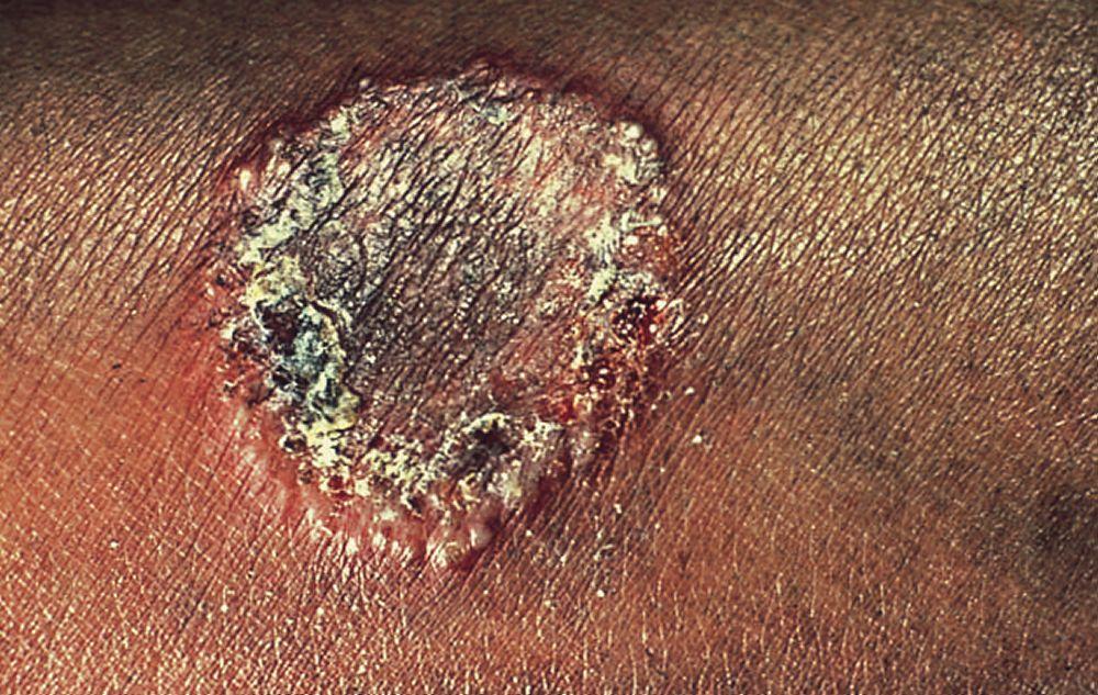
A rare variant form appears as nummular (circle- or round-shaped) scaling patches studded with small papules or pustules that have no central clearing.
Common causes are Trichophyton mentagrophytes, T. rubrum, and Microsporum canis.

© Springer Science+Business Media

© Springer Science+Business Media
© Springer Science+Business Media

Image courtesy of Karen McKoy, MD.

© Springer Science+Business Media

© Springer Science+Business Media

© Springer Science+Business Media

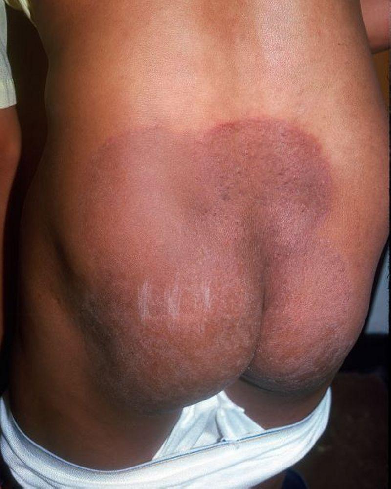
Image courtesy of Karen McKoy, MD.
Diagnosis of Tinea Corporis
Clinical evaluation
Potassium hydroxide wet mount
Tinea corporis is diagnosed by clinical appearance and by potassium hydroxide wet mount of skin scrapings.
Differential diagnosis of tinea corporis includes
Treatment of Tinea Corporis
Topical or oral antifungals
(See table Options for Treatment of Superficial Fungal Infections.)
Extensive and resistant lesions occur in patients infected with T. rubrum
Key Points
Tinea corporis typically causes pink-to-red annular (O-shaped) patches and plaques with raised scaly borders that expand peripherally and tend to clear centrally.
Diagnose based on appearance and potassium hydroxide wet mount.


