

Stones composed of calcium salts often obstruct salivary glands, causing pain, swelling, and sometimes infection. Diagnosis is made clinically or with CT, ultrasound, or sialography. Treatment involves stone expression with saliva stimulants, manual manipulation, a probe, or surgery.
Salivary stones are hardened mineral deposits that form within the salivary glands or ducts and obstruct the flow of saliva.
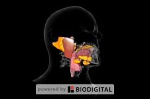
The major salivary glands are the paired parotid, submandibular, and sublingual glands. Stones in the salivary glands are most common among adults. Most (80%) stones originate in the submandibular glands and obstruct the Wharton duct (1). Most of the rest originate in the parotid glands and block the Stensen duct. Only approximately 1% originate in the sublingual glands. Multiple stones occur in approximately 25% of patients.
General reference
1. Pachisia S, Mandal G, Sahu S, Ghosh S. Submandibular sialolithiasis: A series of three case reports with review of literature. Clin Pract. 2019;9(1):1119. Published 2019 Mar 20. doi: 10.4081/cp.2019.1119
Etiology of Salivary Stones
Most salivary stones are composed of calcium phosphate with small amounts of magnesium and carbonate. Patients with gout may have uric acid stones. Stone formation requires a nidus on which salts can precipitate during salivary stasis. Stasis occurs in patients who are debilitated, dehydrated, have reduced food intake, or take anticholinergics. Persisting or recurrent stones predispose to infection of the involved gland (sialadenitis).
Symptoms and Signs of Salivary Stones
Obstructing stones cause glandular swelling and pain, particularly after eating, which stimulates saliva flow. Symptoms may subside after a few hours. Relief may coincide with a gush of saliva. Some stones cause intermittent or no symptoms.
If a stone is lodged distally, it may be visible or palpable at the duct’s outlet.
Diagnosis of Salivary Stones
Physical examination
Imaging (eg, CT, ultrasound, sialography)
The diagnosis of salivary stones is based on physical examination findings and imaging studies. Stones can occasionally be palpated on bimanual examination of the floor of the mouth. If a salivary stone is not apparent on examination, the patient can be given a sialagogue (eg, lemon juice, hard candy, or some other substance that triggers saliva flow). Reproduction of symptoms is almost always diagnostic of a stone.
CT and ultrasound are highly sensitive and are used if clinical diagnosis is equivocal. Contrast sialography is also highly sensitive but is no longer commonly performed. Because 90% of submandibular calculi are radiopaque and 90% of parotid calculi are radiolucent, plain radiographs are not always accurate (1). Ultrasound is increasingly being used and has reported sensitivities for all (radiopaque and radiolucent) stones of approximately 60 to 95% and specificities between 85 and 100% (2). The role of MRI is evolving; reported sensitivities and specificities are > 90%, and in limited studies, MRI has been found to be more sensitive in detecting small stones and distal duct stones than ultrasound or contrast sialography (3).
Diagnosis references
1. Kraaij S, Karagozoglu KH, Forouzanfar T, et al. Salivary stones: Symptoms, aetiology, biochemical composition and treatment. Br Dent J. 2014;217(11):E23, 2014. doi: 10.1038/sj.bdj.2014.1054
2. Kim DH, Kang JM, Kim SW, et al. Utility of ultrasonography for diagnosis of salivary gland sialolithiasis: a meta-analysis. Laryngoscope. 2022;132(9):1785-1791. doi: 10.1002/lary.30020
3. Jäger L, Menauer, F Holzknecht N, et al. Sialolithiasis: MR sialography of the submandibular duct--An alternative to conventional sialography and US? Radiology. 2000;216(3):665–671. doi: 10.1148/radiology.216.3.r00se12665
Treatment of Salivary Stones
Local measures (eg, sialagogues, massage)
Sometimes manual expression or surgical removal
The treatment approach for salivary stones depends primarily on stone size and location. Modalities include conservative (local) measures and surgical intervention, of which sialendoscopy is the increasingly preferred option (1). Sialendoscopy allows direct visualization and stone removal with reduced morbidity compared to gland excision (1). Analgesics, hydration, and massage can relieve symptoms in patients with a salivary stone.
Antistaphylococcal antibiotics can be used to prevent acute sialadenitis if started early.
Stones may pass spontaneously or when salivary flow is stimulated by sialagogues; patients are encouraged to suck a lemon wedge or sour candy every 2 to 3 hours. Stones right at the duct orifice can sometimes be expressed manually by squeezing with the fingertips. Dilation of the duct with a small probe may facilitate expulsion.
Surgical removal of stones succeeds if other methods are ineffective. Stones at or near the orifice of the duct may be removed transorally, whereas those in the hilum of the gland often require complete excision of the salivary gland. Stones up to 5 mm in size may be removed endoscopically (via sialendoscopy) (2, 3).
Treatment references
1. Nahlieli O. Thirty years of experience and current trends in the management of sialolithiasis: a narrative review. Br J Oral Maxillofac Surg. 2025;63(4):270-275. doi:10.1016/j.bjoms.2025.02.011
2. Marchal F, Becker M, Dulguerov P, et al. Interventional sialendoscopy. Laryngoscope. 2000;110:318–320. doi: 10.1097/00005537-200002010-00026
3. Koch M, Zenk J, Iro H. Algorithms for treatment of salivary gland obstructions. Otolaryngol Clin North Am. 2009;42(6):1173–1192. doi: 10.1016/j.otc.2009.08.002
Key Points
Approximately 80% of salivary stones occur in the submandibular glands.
Clinical diagnosis is usually adequate but sometimes CT or ultrasound is needed.
Many stones pass spontaneously or with use of sialagogues and manual expression, but some require endoscopic (via sialendoscopy) or surgical removal.


