


Breast cancers are commonly epithelial histologic types (carcinoma), arising from cells lining the breast ducts or lobules; nonepithelial breast cancers include angiosarcoma, primary stromal sarcoma, and phyllodes tumor. Most patients present with an asymptomatic mass discovered during examination or screening mammography. Diagnosis is made with biopsy. Treatment usually includes surgical excision, often combined with radiation therapy, immunotherapy, targeted therapy, endocrine therapy, and/or chemotherapy.
In the United States, breast cancer is the second most common cancer in women; the most common is skin cancer (1). Breast cancer is the second leading cause of cancer death (the most common is lung cancer) in the overall female population but is the leading cause of cancer death in Black women. Black women are more likely to die from breast cancer than any other race or ethnicity; Asian and Pacific Islander women have the lowest death rate from breast cancer.
In 2026, in women in the United States, it is estimated there will be (1)
321,910 new cases of invasive breast cancer
60,730 new cases of ductal carcinoma in situ (DCIS)
42,140 breast cancer deaths
In 2026, in men in the United States, it is estimated there will be 2670 new cases of invasive breast cancer and 530 deaths from it (2). Manifestations, diagnosis, and management are the same in men and women, although men tend to present at a later stage.
General references
1. American Cancer Society. Key Statistics for Breast Cancer. Revised May 5, 2025. Accessed April 30, 2026.
2. American Cancer Society. Key Statistics for Breast Cancer in Men. Revised October 15, 2025. Accessed April 30, 2026.
Risk Factors for Breast Cancer
For women in the United States, cumulative lifetime risk of developing breast cancer is approximately 13% (1 in 8) (1). The median age at diagnosis is 62 years (see table ). The 5-year survival rate for breast cancer is 90% (2).
The Breast Cancer Risk Assessment Tool (BCRAT), or Gail model, can be used to calculate a women's 5-year and lifetime risk of developing breast cancer. The BCRAT is used for women ages 35 to 85 years. For women under the age of 35, there are other risk models available.

10-Year Risk of Breast Cancer Diagnosis or Mortality
Current Age (years) | 10-year Risk of Breast Cancer Diagnosis (%) | 10-year Risk of Breast Cancer Mortality (%) |
|---|---|---|
20 | 0.1 | < 0.1 |
30 | 0.5 | < 0.1 |
40 | 1.6 | 0.1 |
50 | 2.5 | 0.3 |
60 | 3.6 | 0.5 |
70 | 4.2 | 0.7 |
80 | 3.1 | 1.0 |
Data from American Cancer Society. Breast Cancer Facts & Figures 2024-2025, Table 1. Ten-Year Probability of Breast Cancer Diagnosis (2018-2019, 2021) and Death (2020-2022). Accessed May 11, 2026. | ||
Factors that may affect breast cancer risk include the following:
Age: In the United States, the incidence is highest among women ages 65 to 74 years; median age at diagnosis is 64 years (3). In a United States national database study with data from 1973–2010, age at diagnosis varied by race and ethnicity; the median age (in years) at diagnosis varies by race/ethnicity, as follows: White (59); Black (56); Hispanic (55); Asian (56) (4, 5).
Race and ethnicity: Incidence of breast cancer (per 100,000 persons) in the United States also varies by race and ethnicity: non-Hispanic White (141); non-Hispanic Black (132); non-Hispanic American Indian/Alaska Native (119); non-Hispanic Asian/Pacific Islander (117); Hispanic (108) (3). Rates of triple-negative breast cancer (estrogen, progesterone, and human epidermal growth factor receptor 2 receptors are absent), which has a poorer prognosis than breast cancers with these receptors, which also vary by race and ethnicity (per 100,000 persons): Black (25), White (13), American Indian/Alaska Native (11), Hispanic (11), and Asian/Pacific Islander (6).
Breast cancer gene mutations: Approximately 5 to 10% of women with breast cancer carry a mutation in either the BRCA1 or BRCA2 gene (7). The risk of developing breast cancer by age 80 is approximately 72% with a BRCA1 mutation and approximately 69% with a BRCA2 mutation. Women with BRCA1 mutations also have an approximate 44% lifetime risk of developing ovarian cancer; risk among women with BRCA2 mutations is approximately 17% (7, 8). Men who carry a BRCA mutation have a 1 to 2% lifetime risk of developing breast cancer. The mutations are more common among people with Ashkenazi Jewish ancestry. Women with BRCA1 or BRCA2 mutations require more frequent screening with both mammography and MRI or risk-reducing mastectomy; for patients with a BRCA2 mutation who do not wish to undergo mastectomy, chemoprophylaxis with tamoxifen is an option. Other genetic mutations that increase the risk of developing breast cancer include mutations in CHEK2, PALB2, ATM, RAD51C, RAD51D, BARD1, and TP53.
Family history: Having a first-degree relative (mother, sister, daughter) with breast cancer is associated with a 2-fold increase in the risk, but breast cancer in more distant relatives is associated with only a slight increase in risk (9). When ≥ 2 first-degree relatives have had breast cancer, risk may be 3 to 4 times higher.
Personal history of breast cancer: Having had in situ or invasive breast cancer is associated with increased risk of a new breast cancer. Risk of developing cancer in the contralateral breast after mastectomy is approximately 0.4% per year of follow-up (10).
Lobular carcinoma in situ (LCIS): After a diagnosis of LCIS, the risk of developing invasive breast cancer in either the ipsilateral or contralateral breast is approximately 7 to 11% at 10 years and 20% at 20 years (11, 12).
Benign breast disease: History of a lesion that required a biopsy is associated with a slightly increased risk. Women with multiple breast masses but no histologic confirmation of a high-risk histology should not be considered at high risk. Benign lesions associated with a slightly increased risk of developing invasive breast cancer include complex fibroadenoma, moderate or florid hyperplasia (without atypia), sclerosing adenosis, and papilloma. Risk is approximately 3 to 5 times higher in patients with atypical ductal or lobular hyperplasia (13).
Dense breast tissue: Dense breast tissue seen on screening mammography is associated with an approximately 2-fold increased risk of breast cancer (14).
Radiation therapy: Exposure to radiation therapy of the chest up through 45 years old increases risk, with the highest increase for those exposed between ages 10 to 14 years (15). Mantle-field radiation therapy for Hodgkin lymphoma approximately quadruples risk of breast cancer over the next 20 to 30 years.
Obstetric and gynecologic history: Early menarche or late menopause increases risk. Risk increases with increasing age at first pregnancy (3).
Use of oral contraceptives: Study results vary regarding the use of oral contraceptives and risk of breast cancer. Some studies have found a small increased risk in current or recent users (16).
Menopausal hormone therapy: In the Women's Health Initiative randomized trial, menopausal hormone therapy with estrogen plus a progestin increased risk modestly after only 3 years of use (17). After 5 years of use, the increased risk was approximately 3 additional cases per 1000 women for each year of use (approximately a 24% increase in relative risk). Use of estrogen alone does not appear to increase risk of breast cancer. Selective estrogen-receptor modulators (eg, raloxifene) reduce the risk of developing breast cancer.
Diet: A diet with higher amounts of red or processed meats, foods with a high glycemic index, or eggs appears to be associated with a higher risk of breast cancer, while a diet with higher amounts of vegetables appears to be associated with a lower risk (18).
Obesity: Postmenopausal women with obesity are at increased risk of breast cancer; studies show a 10% increase in risk for each additional 5 body mass index (BMI) units above normal BMI (19).
Smoking and alcohol: Smoking and alcohol use appear to be associated with an increase in breast cancer risk; the increase with alcohol intake is dose-dependent (20, 21).
Risk factor references
1. American Cancer Society. Key Statistics for Breast Cancer. Revised May 5, 2025. Accessed April 30, 2025.
2. American Cancer Society. Survival Rates for Breast Cancer. Revised October 15, 2025. Accessed April 30, 2025.
3. National Cancer Institute. Cancer Stat Facts: Female Breast Cancer. Accessed April 30, 2026.
4. Stapleton SM, Oseni TO, Bababekov YJ, Hung YC, Chang DC. Race/Ethnicity and Age Distribution of Breast Cancer Diagnosis in the United States. JAMA Surg. 2018;153(6):594-595. doi:10.1001/jamasurg.2018.0035
5. Hendrick RE, Monticciolo DL, Biggs KW, Malak SF. Age distributions of breast cancer diagnosis and mortality by race and ethnicity in US women. Cancer. 2021;127(23):4384-4392. doi:10.1002/cncr.33846
6. Sung H, Wiese D, Jatoi I, Jemal A. State Variation in Racial and Ethnic Disparities in Incidence of Triple-Negative Breast Cancer Among US Women. JAMA Oncol. 2023;9(5):700-704. doi:10.1001/jamaoncol.2022.7835
7. Kuchenbaecker KB, Hopper JL, Barnes DR, et al. Risks of breast, ovarian, and contralateral breast cancer for BRCA1 and BRCA2 mutation carriers. JAMA. 317 (23):2402–2416, 2017. doi: 10.1001/jama.2017.7112
8. Breast Cancer Association Consortium; Dorling L, Carvalho S, Allen J, et al. Breast cancer risk genes — Association analysis in more than 113,000 women. N Engl J Med. 4;384 (5):428–439, 2021. doi: 10.1056/NEJMoa1913948
9. Collaborative Group on Hormonal Factors in Breast Cancer. Familial breast cancer: collaborative reanalysis of individual data from 52 epidemiological studies including 58,209 women with breast cancer and 101,986 women without the disease. Lancet. 358(9291):1389-1399, 2001. doi:10.1016/S0140-6736(01)06524-2
10. Giannakeas V, Lim DW, Narod SA. The risk of contralateral breast cancer: a SEER-based analysis. Br J Cancer. 125(4):601-610, 2021. doi:10.1038/s41416-021-01417-7
11. Wong SM, King T, Boileau JF, Barry WT, Golshan M. Population-Based Analysis of Breast Cancer Incidence and Survival Outcomes in Women Diagnosed with Lobular Carcinoma In Situ. Ann Surg Oncol. 2017;24(9):2509-2517. doi:10.1245/s10434-017-5867-6
12. Chuba PJ, Hamre MR, Yap J, et al. Bilateral risk for subsequent breast cancer after lobular carcinoma-in-situ: analysis of surveillance, epidemiology, and end results data. J Clin Oncol. 2005;23(24):5534-5541. doi:10.1200/JCO.2005.04.038
13. Collins LC, Baer HJ, Tamimi RM, et al. Magnitude and laterality of breast cancer risk according to histologic type of atypical hyperplasia: results from the Nurses' Health Study. Cancer. 109(2):180-187, 2007. doi:10.1002/cncr.22408
14. Bodewes FTH, van Asselt AA, Dorrius MD, Greuter MJW, de Bock GH. Mammographic breast density and the risk of breast cancer: A systematic review and meta-analysis. Breast. 2022;66:62-68. doi:10.1016/j.breast.2022.09.007
15. John EM, Kelsey JL. Radiation and other environmental exposures and breast cancer. Epidemiol Rev. 15(1):157-162, 1993. doi:10.1093/oxfordjournals.epirev.a036099
16. American College of Obstetricians and Gynecologists (ACOG) . Hormonal contraception and risk of breast cancer: Practice advisory. Published 2018, reaffirmed 2024. Accessed July 10, 2025.
17. Rossouw JE, Anderson GL, Prentice RL, et al.. Risks and benefits of estrogen plus progestin in healthy postmenopausal women: Principal results from the Women's Health Initiative randomized controlled trial. JAMA. 288 (3):321–333, 2002. doi:10.1001/jama.288.3.321
18. Buja A, Pierbon M, Lago L, Grotto G, Baldo V. Breast Cancer Primary Prevention and Diet: An Umbrella Review. Int J Environ Res Public Health. 2020;17(13):4731. Published 2020 Jul 1. doi:10.3390/ijerph17134731
19. Lauby-Secretan B, Scoccianti C, Loomis D, et al. Body Fatness and Cancer--Viewpoint of the IARC Working Group. N Engl J Med. 375(8):794-798, 2016. doi:10.1056/NEJMsr1606602
20. Gram IT, Park SY, Kolonel LN, et al. Smoking and Risk of Breast Cancer in a Racially/Ethnically Diverse Population of Mainly Women Who Do Not Drink Alcohol: The MEC Study. Am J Epidemiol. 182(11):917-925, 2015. doi:10.1093/aje/kwv092
21. Zhang SM, Lee IM, Manson JE, et al. Alcohol consumption and breast cancer risk in the Women's Health Study. Am J Epidemiol. 165(6):667-676, 2007. doi:10.1093/aje/kwk054
Pathology of Breast Cancer
The most common histologic type of breast cancer is epithelial (carcinoma), which develops from cells lining breast ducts or lobules; nonepithelial cancers of the supporting stroma (eg, angiosarcoma, primary stromal sarcomas, phyllodes tumor) are less common.

Gwen Shockey/SCIENCE PHOTO LIBRARY

Gwen Shockey/SCIENCE PHOTO LIBRARY
Epithelial cancers can be either carcinoma in situ or invasive cancer.
Carcinoma in situ is proliferation of cancer cells within ducts or lobules but without invasion of stromal tissue. There are 2 types:
Ductal carcinoma in situ (DCIS): DCIS refers to neoplastic epithelial lesions that are confined to the breast ducts; lesions may have varying histology and malignant potential. It is usually detected only by mammography. It may involve a small or wide area of the breast; if a wide area is involved, microscopic invasive foci may develop over time. DCIS accounts for approximately 20% of breast cancers (1).
Lobular carcinoma in situ (LCIS): LCIS is often multifocal and is bilateral in approximately 20 to 60% of cases (2). LCIS is classified as classic or nonclassic (pleomorphic or florid). Classic LCIS is not malignant but is a marker of increased risk of developing invasive carcinoma in either breast. This nonpalpable lesion is usually suspected based on calcifications or a mass on mammography or ultrasound and is diagnosed with biopsy. Pleomorphic LCIS is characterized by large cells with marked nuclear pleomorphism. Florid LCIS demonstrates distension of ducts and lobules and may develop into a mass. Upgraded rates (lesions initially diagnosed as LCIS on core biopsy that are found to be invasive carcinoma on surgical excision) for pleomorphic (35%) and florid (33%) LCIS are higher than for classic LCIS (3.5%) (3, 4). Thus, non-classic LCIS is managed with complete excision with negative margins. Patients with any type of LCIS should be followed with close monitoring and offered chemoprevention.
Invasive carcinomas are most frequently adenocarcinoma. Approximately 75% are infiltrating ductal histology and 10% of the remaining cases are infiltrating lobular carcinomas (5).
Epithelial cancers may express hormone receptors (stromal tumors, such as phyllodes tumors, do not express hormone receptors). Approximately 80% of postmenopausal and 20% of premenopausal patients with breast cancer have an estrogen receptor–positive (ER+) tumor (6); approximately 70% of all breast cancers are progesterone receptor-positive (7).
Another cellular receptor found in some breast cancers is human epidermal growth factor receptor 2 (HER2; also called HER2/neu or ErbB2); its presence correlates with a poorer prognosis at any stage of disease. In approximately 15% of patients with breast cancer, HER2 receptors are overexpressed (8).
The majority of breast cancers are hormone receptor-positive and HER2 negative (approximately 70%); 11% are triple negative (hormone receptor-negative and HER2-negative) (8, 9).
Paget disease of the nipple (not to be confused with the metabolic bone disease also called Paget disease) is a form of ductal carcinoma in situ that extends into the skin over the nipple and areola, manifesting with a skin lesion (eg, an eczematous or a psoriaform lesion). Characteristic malignant cells called Paget cells are present in the epidermis. Women with Paget disease of the nipple often have underlying invasive or in situ breast cancer.
Rare epithelial breast tumors that are usually associated with a favorable prognosis include medullary, mucinous, cribriform, and tubular carcinomas (5). Mucinous carcinoma tends to develop in older women and be slow growing. Epithelial malignancies that typically have a poor prognosis include metaplastic and neuroendocrine carcinomas (10, 11).


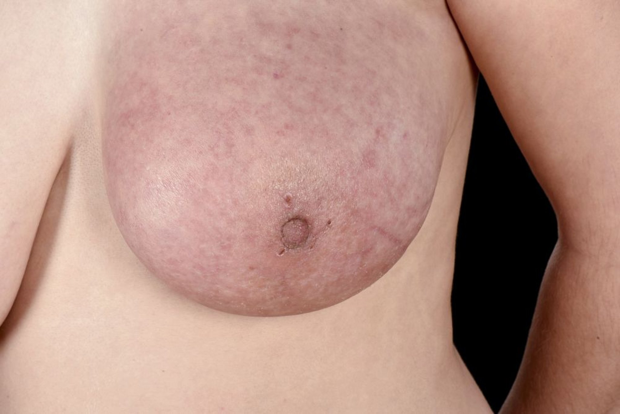
Sometimes in inflammatory breast cancer and very advanced cancers, the breast appears inflamed, and the skin appears thickened, resembling orange peel (peau d’orange).
"Creative Commons Patient with inflammatory breast cancer" by Epidemiology and surgical management of breast cancer in gynecological department of Douala General Hospital (Scientific Figure on ResearchGate) is licensed under Creative Commons 2.0. Available from: https://www.researchgate.net/figure/Patient-with-inflammatory-breast-cancer_fig2_234162338 [accessed 3 Oct, 2019].
Inflammatory breast cancer is a clinical-pathologic syndrome rather than a specific histology, although the majority are invasive ductal carcinomas (12). It is defined by a cancer with rapid-onset erythema, edema, and skin thickening that resembles an orange peel (peau d'orange) involving one-third or more of the breast (13). Usually, inflammatory breast cancer spreads to the lymph nodes in the axilla. The lymph nodes feel like hard lumps. However, often no mass is felt in the breast itself because this cancer is dispersed throughout the breast.

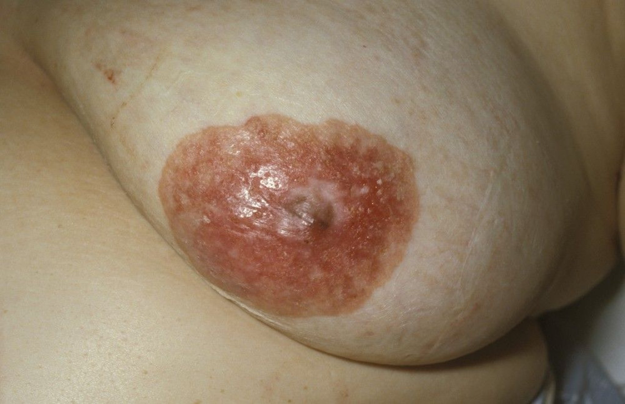
In Paget disease of the nipple, ductal carcinoma in situ extends over the nipple and areola and causes scaling, pruritus, and burning pain in the affected nipple.
DR M.A. ANSARY/SCIENCE PHOTO LIBRARY
Pathology references
1. Giaquinto AN, Sung H, Miller KD, et al. Breast Cancer Statistics, 2022. CA Cancer J Clin. 72(6):524-541, 2022. doi:10.3322/caac.21754
2. Wen HY, Brogi E. Lobular Carcinoma In Situ. Surg Pathol Clin. 11(1):123-145, 2018. doi:10.1016/j.path.2017.09.009
3. Ferrucci M, Passeri D, Milardi F, et al. Pleomorphic and florid lobular carcinoma in situ of the Breast: A systematic review of current evidence and knowledge gaps. Breast. 2026;86:104711. doi:10.1016/j.breast.2026.104711
4. Shehata MN, Rahbar H, Flanagan MR, et al. Risk for Upgrade to Malignancy After Breast Core Needle Biopsy Diagnosis of Lobular Neoplasia: A Systematic Review and Meta-Analysis. J Am Coll Radiol. 2020;17(10):1207-1219. doi:10.1016/j.jacr.2020.07.036
5. American Cancer Society. Breast Cancer Facts & Figures 2024–2025. Accessed January 7, 2026.
6. Anderson WF, Chatterjee N, Ershler WB, et al. Estrogen receptor breast cancer phenotypes in the Surveillance, Epidemiology, and End Results database. Breast Cancer Res Treat. 2002;76(1):27-36. doi:10.1023/a:1020299707510
7. Li Y, Yang D, Yin X, et al. Clinicopathological Characteristics and Breast Cancer-Specific Survival of Patients With Single Hormone Receptor-Positive Breast Cancer. JAMA Netw Open. 3(1):e1918160, 2020. doi:10.1001/jamanetworkopen.2019.18160
8. Howlader N, Altekruse SF, Li CI, et al. US incidence of breast cancer subtypes defined by joint hormone receptor and HER2 status. J Natl Cancer Inst. 106(5):dju055, 2014. Published 2014 Apr 28. doi:10.1093/jnci/dju055
9. National Cancer Institute's Surveillance, Epidemiology, and End Results (SEER) Program . Cancer Stat Facts: Female Breast Cancer Subtypes. Accessed January 7, 2026.
10. Elimimian EB, Samuel TA, Liang H, Elson L, Bilani N, Nahleh ZA. Clinical and Demographic Factors, Treatment Patterns, and Overall Survival Associated With Rare Triple-Negative Breast Carcinomas in the US. JAMA Netw Open. 2021;4(4):e214123. Published 2021 Apr 1. doi:10.1001/jamanetworkopen.2021.4123
11. Pareja F, Bhargava R, Borges VF, et al. Unraveling complexity and leveraging opportunities in uncommon breast cancer subtypes. NPJ Breast Cancer. 2025;11(1):6. Published 2025 Jan 24. doi:10.1038/s41523-025-00719-w
12. Raghav K, French JT, Ueno NT, et al. Inflammatory Breast Cancer: A Distinct Clinicopathological Entity Transcending Histological Distinction. PLoS One. 2016;11(1):e0145534. Published 2016 Jan 11. doi:10.1371/journal.pone.0145534
13. National Comprehensive Cancer Network. NCCN Clinical Practice Guidelines in Oncology (NCCN Guidelines). Breast Cancer, version 3.2026. https://www.nccn.org. Accessed May 17, 2026.
Pathophysiology of Breast Cancer
Breast cancer invades locally and spreads through the regional lymph nodes, bloodstream, or both. Metastatic breast cancer may affect almost any organ in the body—most commonly, lungs, liver, bone, brain, and skin. Most skin metastases occur near the site of breast surgery; scalp metastases are uncommon.

Gwen Shockey/SCIENCE PHOTO LIBRARY
Hormone receptors
Estrogen and progesterone receptors are nuclear hormone receptors that promote DNA replication and cell division when the appropriate hormones bind to them. Thus, medications that block these receptors are useful in treating and preventing tumors with the receptors.
HER2 receptors
HER2 is a transmembrane tyrosine kinase receptor that is a significant driver of cancer cell progression. Tumors in which HER2 receptors are overexpressed are treated with medications that block these receptors.
Breast cancer gene mutations
BRCA1 and BRCA2 gene mutations increase the risk of developing breast cancer to 70% (1). Prophylactic bilateral mastectomy reduces the risk of breast cancer by 90% and should be offered to women with a BRCA mutation.
Other genetic mutations that increase the risk of developing breast cancer include mutations in CHEK2, PALB2, ATM, RAD51C, RAD51D, BARD1, and TP53, which are usually included in panel genetic testing (2).
Pathophysiology references
1. Kuchenbaecker KB, Hopper JL, Barnes DR, et al. Risks of breast, ovarian, and contralateral breast cancer for BRCA1 and BRCA2 mutation carriers. JAMA. 317 (23):2402–2416, 2017. doi: 10.1001/jama.2017.7112
2. Breast Cancer Association Consortium; Dorling L, Carvalho S, Allen J, et al. Breast cancer risk genes — Association analysis in more than 113,000 women. N Engl J Med. 4;384 (5):428–439, 2021. doi: 10.1056/NEJMoa1913948
Symptoms and Signs of Breast Cancer
Most breast cancers are detected by mammography; other modes of detection are self-examination by the patient, clinical breast examination, MRI, and ultrasound (1). Breast pain may be present but is the sole presenting symptom of breast cancer in only approximately 1 to 2% of cases (2). Infrequently, the presenting symptom is overall breast enlargement or a nondescript thickening of the breast.
Patients with metastatic disease may present with associated symptoms and signs (eg, pathologic fracture, abdominal pain, jaundice, dyspnea).
A common finding during breast examination is asymmetry or a dominant mass—a mass distinctly different from the surrounding breast tissue. Diffuse fibrotic changes in a quadrant of the breast, usually the upper outer quadrant, are more characteristic of benign disorders; a slightly firmer thickening in one breast but not the other may be a sign of cancer.
Some types of breast cancer manifest with notable skin changes:
Paget disease of the nipple is associated with an underlying in situ or invasive carcinoma and manifests as skin changes, including erythema, crusting, scaling, and discharge; these changes usually appear so benign that the patient might ignore them, delaying diagnosis. Approximately 50 to 60% of patients with Paget disease of the nipple have a palpable mass at presentation (3).
Inflammatory breast cancer manifests as erythema and enlargement of the breast, often without a mass, and skin may be discolored or appear thickened, resembling orange peel (peau d’orange).
More advanced breast cancers are characterized by 1 or more of the following examination findings:
Fixation of the mass to the chest wall or to overlying skin
Satellite nodules or ulcers in the skin
Matted or fixed axillary lymph nodes suggest tumor spread, as does supraclavicular or infraclavicular lymphadenopathy.


This photo shows an irregular mass on the lateral aspect of the left breast. The mass was later diagnosed as breast cancer.
MID ESSEX HOSPITAL SERVICES NHS TRUST/SCIENCE PHOTO LIBRARY

This photo shows diffuse erythema and edema in the left breast of patient with inflammatory breast cancer.
MID ESSEX HOSPITAL SERVICES NHS TRUST/SCIENCE PHOTO LIBRARY
Signs and symptoms references
1. Basappa SN, Finney Rutten LJ, Hruska CB, Olson JE, Jacobson DJ, Rhodes DJ. Breast Cancer Mode of Detection in a Population-Based Cohort. Mayo Clin Proc. 2023;98(2):278-289. doi:10.1016/j.mayocp.2022.10.010
2. Martín-Díaz M, Maes-Carballo M, Khan KS, Bueno-Cavanillas A. To image or not in noncyclic breast pain? A systematic review. Curr Opin Obstet Gynecol. 2017;29(6):404-412. doi:10.1097/GCO.0000000000000407
3. Günhan-Bilgen I, Oktay A. Paget's disease of the breast: clinical, mammographic, sonographic and pathologic findings in 52 cases. Eur J Radiol. 2006;60(2):256-263. doi:10.1016/j.ejrad.2006.06.010
Diagnosis of Breast Cancer
Initial detection or evaluation of palpable breast mass with mammography and/or other imaging (breast tomosynthesis, ultrasound, MRI)
Biopsy, including analysis for estrogen and progesterone and HER2 receptors
Symptoms, self-breast examination, clinical breast examination, and/or imaging test results may prompt a diagnostic evaluation for breast cancer.
For patients with an abnormal screening mammogram, image-guided biopsy is performed. An abnormal breast MRI is typically evaluated with ultrasound to determine whether biopsy should be performed under guidance with ultrasound or MRI.
Patients who present with breast symptoms (eg, pain, nipple discharge) or a palpable breast mass or other abnormality (eg, skin lesions) on clinical breast examination are typically evaluated initially with breast ultrasound. If ultrasound results are abnormal or indeterminate, mammography is performed. If imaging findings suggest cancer, biopsy is performed.
If a palpable breast mass or other physical findings suggest cancer, a biopsy should be performed even if imaging tests are negative.
If advanced cancer is suspected based on physical examination, biopsy is often performed before imaging; however, a prebiopsy bilateral mammogram may help delineate other areas that should be biopsied and provides a baseline for future reference.
Pearls & Pitfalls
|
Biopsy
Percutaneous core needle biopsy is preferred to surgical biopsy. Core biopsy can be performed guided by imaging or palpation (freehand). Stereotactic biopsy (needle biopsy guided by mammography performed in 2 planes and analyzed by computer to produce a 3-dimensional image) or ultrasound-guided biopsy is commonly used to improve accuracy. Clips are placed at the biopsy site so it can be identified with mammography.
If core biopsy is not possible (eg, the lesion is too posterior), surgical biopsy can be performed; a guidewire or localizing metal seed is inserted, using imaging for guidance, to help identify the biopsy site.
Any skin taken with the biopsy specimen should be examined because it may show cancer cells in dermal lymphatic vessels.
The excised specimen should be radiographed, and the radiograph should be compared with the prebiopsy mammogram to determine whether all of the lesion has been removed. If the original lesion contained microcalcifications, mammography is repeated when the breast is no longer tender, usually 6 to 12 weeks after biopsy, to check for residual microcalcifications. If radiation therapy is planned, mammography should be repeated before radiation therapy begins.
Evaluation after cancer diagnosis
After cancer is diagnosed, a multidisciplinary evaluation is usually performed to plan further testing and treatment. The core multidisciplinary team typically includes a breast surgeon, medical oncologist, and radiation oncologist.
A positive biopsy specimen should be analyzed for estrogen and progesterone and HER2 receptors.
MRI is often used by surgeons for preoperative planning; it can accurately determine tumor size, chest wall involvement, and number of tumors.
Genetic testing
The National Comprehensive Cancer Network (NCCN) recommends that patients with current or a personal history of breast cancer be tested for inherited gene mutations that predispose to breast cancer, if they meet any of the following criteria (1):
Breast cancer diagnosed at ≤ 50 years old
Treatment indications: To aid decisions about use of PARP inhibitors (for metastatic cancer) or olaparib (for high-risk, HER2-negative cancers)
Triple-negative breast cancer (tumor does not have estrogen or progesterone receptors or overexpression of HER2 protein)
Multiple primary breast cancers (synchronous or metachronous)
Lobular breast cancer plus a personal or family history of diffuse gastric cancer
Male breast cancer
Ashkenazi Jewish ancestry
Family history: First-, second-, and third-degree relatives on the same side of the family with breast cancer at ≤ 50 years old, male breast cancer, ovarian cancer, pancreatic cancer, or metastatic or high-risk prostate cancer; ≥ 3 diagnoses of breast and/or prostate cancer on the same side of the family (including the patient); first‑ or second‑degree blood relative meeting any of the criteria listed above
A probability > 5% of a BRCA1/2 pathogenic or likely pathogenic variant based on prior probability models (eg, Tyrer‑Cuzick, BRCAPRO)
Some experts have recommended that genetic testing be offered to all patients with breast cancer (2).
For genetic testing, the best approach is to refer patients to a genetic counselor, who can document a detailed family history, counsel the patient about the risks and benefits of genetic testing, choose the most appropriate tests, and help interpret the results.
Evaluation for metastatic disease
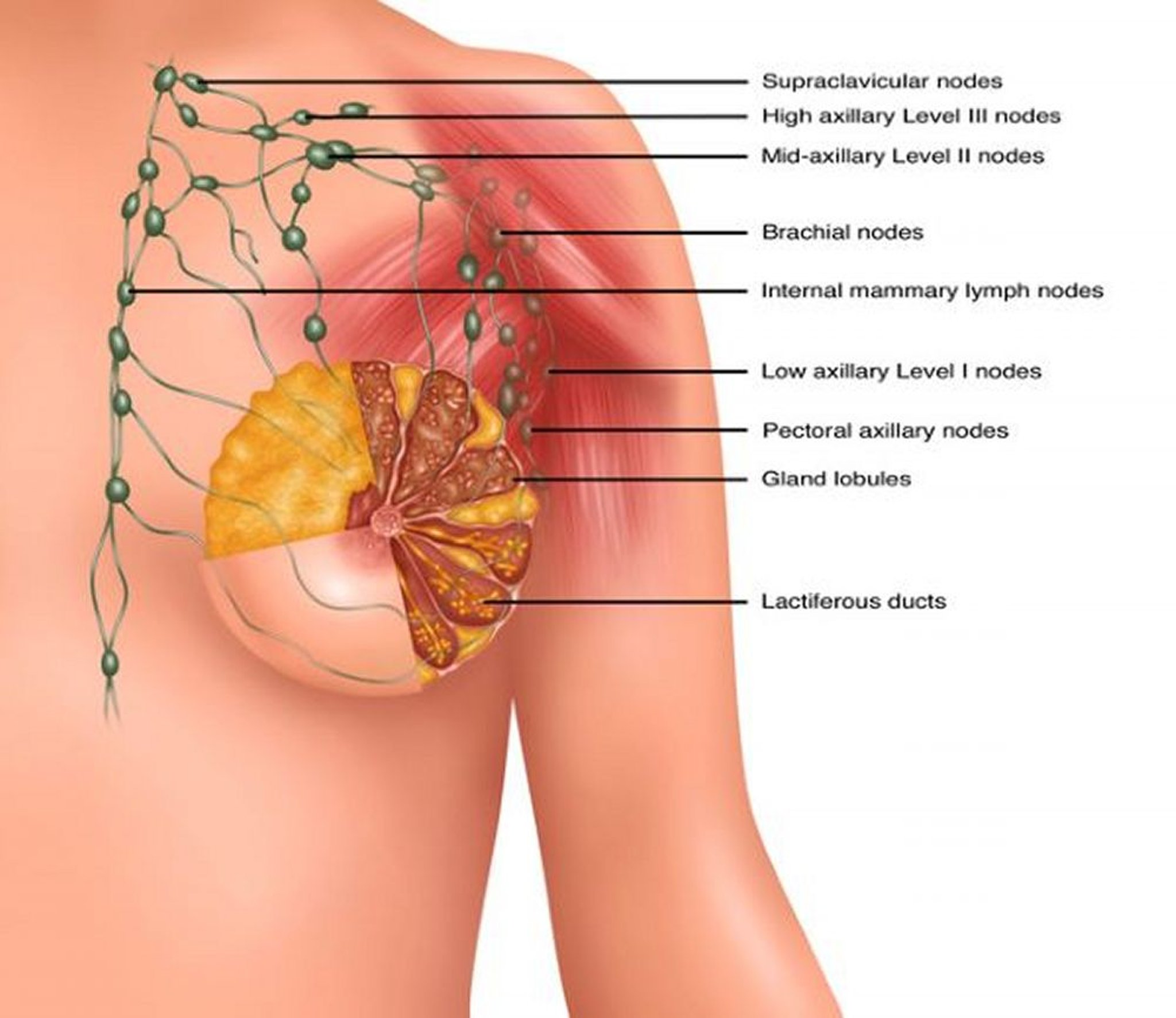
While axillary lymph nodes are the most common site of locoregional spread of breast cancer, the most common sites of distant metastases are bone (51% of metastases), liver/soft tissue (19%), pleura (16%), lung (14%), and brain (4%) (3).
A complete blood count (CBC) and liver function tests can be measured to check for bone marrow and liver metastases, respectively.
An oncologist should determine whether to measure serum carcinoembryonic antigen (CEA), cancer antigen (CA) 15-3, or CA 27-29 and whether bone scanning should be performed.
For bone scanning, common indications include the following:
Bone pain
Elevated serum alkaline phosphatase
Stage III or IV cancer
Abdominal CT to evaluate for liver or other abdominal visceral metastases is performed if patients have any of the following:
Abnormal liver test results
Abnormal abdominal or pelvic examination
Stage III or IV cancer
Chest CT is performed if patients have either of the following:
Pulmonary symptoms such as shortness of breath
Stage III or IV cancer
Diagnosis references
1. Cheng HH, Giri VN, Goggins M, et al. NCCN Guidelines® Insights: Genetic/Familial High-Risk Assessment: Breast, Ovarian, Pancreatic, and Prostate, Version 2.2026. J Natl Compr Canc Netw. 2026;24(2):2-10. doi:10.6004/jnccn.2026.0007
2. Manahan ER, Kuerer HM, Sebastian M, et al. Consensus Guidelines on Genetic Testing for Hereditary Breast Cancer from the American Society of Breast Surgeons. Ann Surg Oncol. 26(10):3025-3031, 2019. doi:10.1245/s10434-019-07549-8
3. Expert Panel on Breast Imaging, McDonald ES, Scheel JR, et al. ACR Appropriateness Criteria® Imaging of Invasive Breast Cancer. J Am Coll Radiol. 2024;21(6S):S168-S202. doi:10.1016/j.jacr.2024.02.021
Staging and Grading
Grading is based on histologic examination of the tissue taken during biopsy. Breast cancers are assigned a Nottingham combined histologic grade based on scoring each of 3 components (tubule formation, nuclear pleomorphism, and mitotic count) to categorize a tumor specimen as low, intermediate, or high grade (1).
Staging follows the TNM (tumor, node, metastasis) classification. Because clinical examination and imaging have poor sensitivity for nodal involvement, staging is refined during surgery, when regional lymph nodes can be evaluated. However, if patients have palpably abnormal axillary nodes, preoperative ultrasound-guided fine needle aspiration or core biopsy may be performed:
If biopsy results are positive, axillary lymph node dissection is typically performed during the definitive surgical procedure. However, use of neoadjuvant chemotherapy may make sentinel lymph node biopsy possible if chemotherapy changes node status from N1 to N0. (Results of intraoperative frozen section analysis determine whether axillary lymph node dissection will be needed.)
If results are negative, a sentinel lymph node biopsy, a less aggressive procedure, may be performed instead.
Staging classification follows these models:
The anatomic staging model, which is based on anatomy of the tumor and which is used in regions of the world where biomarkers cannot be routinely obtained
The clinical prognostic stage, which applies to all patients with breast cancer, is used for clinical classification and staging. It records clinical tumor (T), node (N), and metastases (M) information based on history, physical examination, any imaging performed (although imaging is not required for clinical staging), and biopsies. Genomic profile information is not included under Clinical Prognostic Stage as pathologic information from surgery is necessary to ascertain the prognosis using these tools. (See table .)
The pathological prognostic stage applies to patients treated with surgery as the initial treatment; it does not apply to patients treated with neoadjuvant chemotherapy or radiation. It includes the information used for clinical staging and also findings at surgery and pathological findings from surgical resection.
Breast Cancer Clinical Prognostic Staging
Tumor (T)/Nodes (N) | Metastases (M) | Grade (G) | HER2 | ER/PR | Clinical Prognostic Stage |
|---|---|---|---|---|---|
Tis/N0 | M0 | Any | Any | Any | 0 |
T1*/N0; T0/N1mi; T1*/N1mi | M0 | G1-G2 | Any | ER+/PR+ or ER+/PR- or ER-/PR+ | IA |
" | M0 | G1-G2 | + | ER-/PR- | IA |
" | M0 | G1-G2 | - | ER-/PR- | IB |
" | M0 | G3 | + | Any | IA |
" | M0 | G3 | - | ER+/PR+ | IA |
" | M0 | G3 | - | ER+/PR- or ER-/PR+ or ER-/PR- | IB |
T0/N1†; T1*/N1†; T2/N0 | M0 | G1-G2 | Any | ER+/PR+ | IB |
" | M0 | G1-G2 | Any | ER+/PR- or ER-/PR+ | IIA |
" | M0 | G1-G2 | + | ER-/PR- | IIA |
" | M0 | G1 | - | ER-/PR- | IIA |
" | M0 | G2 | - | ER-/PR- | IIB |
" | M0 | G3 | + | ER+/PR+ | IB |
" | M0 | G3 | + | ER+/PR- or ER-/PR+ or ER-/PR- | IIA |
" | M0 | G3 | - | ER+/PR+ | IIA |
" | M0 | G3 | - | ER+/PR- or ER-/PR+ or ER-/PR- | IIB |
T2/N1‡; T3/N0 | M0 | G1-G2 | + | ER+/PR+ | IB |
" | M0 | G1-G2 | + | ER+/PR- or ER-/PR+ | IIA |
" | M0 | G1-G2 | + | ER-/PR- | IIB |
" | M0 | G1 | - | ER+/PR- or ER-/PR+ or ER-/PR- | IIB |
" | M0 | G2 | - | ER+/PR- or ER-/PR+ | IIB |
" | M0 | G2 | - | ER-/PR- | IIIB |
" | M0 | G1-G2 | - | ER+/PR+ | IIA |
" | M0 | G3 | + | ER+/PR+ | IB |
" | M0 | G3 | + | ER+/PR- or ER-/PR+ or ER-/PR- | IIB |
" | M0 | G3 | - | ER+/PR+ | IIB |
" | M0 | G3 | - | ER+/PR- or ER-/PR+ | IIIA |
" | M0 | G3 | - | ER-/PR- | IIIB |
T0/N2; T1*/N2; T2/N2; T3/N1‡; T3/N2 | M0 | G1 | Any | ER+/PR+ | IIA |
" | M0 | G1 | Any | ER+/PR- or ER-/PR+ | IIIA |
" | M0 | G1 | + | ER-/PR- | IIIA |
" | M0 | G2 | + | ER+/PR+ | IIA |
" | M0 | G2 | + | ER+/PR- or ER-/PR+ or ER-/PR- | IIIA |
" | M0 | G1 | - | ER-/PR- | IIIB |
" | M0 | G2 | - | ER+/PR+ | IIA |
" | M0 | G2 | - | ER+/PR- or ER-/PR+ | IIIA |
" | M0 | G2 | - | ER-/PR- | IIIB |
" | M0 | G3 | + | ER+/PR+ | IIB |
" | M0 | G3 | + | ER+/PR- or ER-/PR+ or ER-/PR- | IIIA |
" | M0 | G3 | - | ER+/PR+ | IIIA |
" | M0 | G3 | - | ER+/PR- or ER-/PR+ | IIIB |
" | M0 | G3 | - | ER-/PR- | IIIC |
T4/N0; T4/N1‡; T4/N2; Any T/N3 | M0 | G1-G2 | Any | ER+/PR+ | IIIA |
" | M0 | G1-G2 | Any | ER+/PR- or ER-/PR+ | IIIB |
" | M0 | G1-G2 | + | ER-/PR- | IIIB |
" | M0 | G1-G2 | - | ER-/PR- | IIIC |
" | M0 | G3 | + | Any | IIIB |
" | M0 | G3 | - | ER+/PR+ | IIIB |
" | M0 | G3 | - | ER+/PR- or ER-/PR+ or ER-/PR- | IIIC |
Any T/Any N | M1 | Any | Any | Any | IV |
Adapted from Hortobagyi GN, Connolly JL, D’Orsi CJ, Edge SB, Mittendorf EA, et al: Breast. In: Amin MB, Edge S, Greene F, et al, eds; American Joint Committee on Cancer. AJCC Cancer Staging Manual. 8th ed. New York, NY: Springer. 2017. | |||||
Tis = ductal carcinoma in situ (DCIS) or Paget disease (Tis [Paget]) of the nipple with no tumor (Paget disease with a tumor is classified by tumor size); T1 = tumor ≤ 2 cm; T1mi ≤ 0.1 cm; T2 ≥ 2 but ≤ 5 cm; T3 ≥ 5 cm; T4 = any size tumor with direct extension to chest wall and/or skin (ulceration or macroscopic nodules) or inflammatory carcinoma. | |||||
HER2 = human epidermal growth factor receptor 2; ER = estrogen receptor; PR = progesterone receptor | |||||
* T1 includes T1mi | |||||
† N1 does not include N1mi. T1 N1mi M0 and T0 N1mi M0 cancers are included for prognostic staging with T1 N0 M0 cancers of the same prognostic factor status. | |||||
‡ N1 includes N1mi. T2, T3, and T4 cancers and N1mi are included for prognostic staging with T2 N1, T3 N1 and T4 N1, respectively. | |||||
Notes: Because N1mi categorization requires evaluation of the entire node, and cannot be assigned on the basis of a fine needle aspiration or core biopsy, N1mi can only be used with Clinical Prognostic Stage when clinical staging is based on a resected lymph node in the absence of resection of the primary cancer, such as when a sentinel node biopsy is performed prior to neoadjuvant chemotherapy or endocrine therapy. For patients with lymph node involvement with no evidence of primary tumor (eg, T0/N1) or with breast ductal carcinoma in situ (eg, Tis/N1), the grade, HER2, ER, and PR information from the tumor in the lymph node should be used for assigning stage group. For cases where HER2 is determined to be equivocal by in situ hybridization testing under the 2013 ASCO/CAP HER2 testing guidelines, the HER2-negative category should be used for clinical prognostic staging. The prognostic value of these prognostic stage groups is based on populations of persons with breast cancer who have been offered and mostly treated with appropriate endocrine and/or systemic chemotherapy (including anti-HER2 therapy). | |||||
Clinical NX (cNX) = Regional lymph nodes cannot be assessed (eg, previously removed). The cNX category is used infrequently and applies only to cases where regional lymph nodes have previously been surgically removed or where there is no documentation of physical examination of the axilla; cN0 = No regional lymph node metastases; cN1 = Metastases to movable ipsilateral Level I, II axillary lymph node(s); cN1mi = Micrometastases (approximately 200 cells > 0.2 mm, but none > 2 mm). cN1mi is rarely used but may be appropriate in cases where sentinel node biopsy is performed before tumor resection, most likely to occur in cases treated with neoadjuvant therapy; cN2 = Metastases in ipsilateral Level I, II axillary lymph nodes that are clinically fixed or matted; or in ipsilateral internal mammary nodes in the absence of axillary lymph node metastases. | |||||
Staging and grading reference
1. National Comprehensive Cancer Network. NCCN Clinical Practice Guidelines in Oncology (NCCN Guidelines). Breast Cancer, version 3.2026. https://www.nccn.org. Accessed May 17, 2026.
Treatment of Breast Cancer
Breast-conserving surgery or mastectomy, usually with lymph node surgery
Radiation therapy
Systemic chemotherapy
Targeted therapy
Immunotherapy
Endocrine therapy
For stages I to III breast cancer, treatment involves surgical removal of the tumor (either with breast-conserving surgery or mastectomy). Radiation therapy is given depending on stage and surgical procedure. Systemic chemotherapy may be given prior to surgery (neoadjuvant) and/or postoperatively (adjuvant); indications for chemotherapy and timing depend upon the feasibility of breast-conserving surgery, patient and tumor characteristics, and presence of lymph node involvement. Stage IV is treated with systemic therapy and with radiation therapy for focal disease if indicated. For all stages, endocrine, targeted, and immune therapies are given based on tumor characteristics responsive to each drug class. (See table .)
Primary Treatment by Type and Stage of Breast Cancer or Carcinoma In-Situ
Stage and/or Histologic Type | Overview of Treatment |
|---|---|
LCIS, nonclassic (pleomorphic or florid) | Surgical excision to negative margins Postoperative follow-up with close monitoring; patients should be offered chemoprevention |
DCIS | BCS vs mastectomy:
Adjuvant endocrine therapy, for ER-positive disease |
Stages I and II (localized) | Neoadjuvant therapy vs primary surgery:
Lymph node evaluation with one or both of the following:
RT is recommended for most patients who undergo BCS (and to selected patients following mastectomy) Adjuvant systemic therapy with chemotherapy, endocrine therapy, targeted therapy, and/or immunotherapy, depending upon prior administration of and response to NACT, patient and tumor characteristics, and lymph node involvement‡ |
Stage III (locally advanced), noninflammatory breast cancer | NACT for most patients†, sometimes combined with other types of neoadjuvant systemic therapies, followed by BCS (or if not feasible, mastectomy) Lymph node evaluation with one or both of the following:
RT for most patients Adjuvant systemic therapy with chemotherapy, endocrine therapy, targeted therapy, and/or immunotherapy, depending upon prior administration of and response to NACT, patient and tumor characteristics, and lymph node involvement‡ |
Stage IV (metastatic) cancer, noninflammatory | Systemic therapy depending upon patient and tumor characteristics‡ RT for brain metastases, local skin recurrences, or isolated symptomatic bone metastases For bone metastases, IV bisphosphonates to reduce bone loss and bone pain |
Inflammatory breast cancer | NACT for most patients Mastectomy (usually modified radical) with ALND RT for most patients Adjuvant systemic therapy with chemotherapy, endocrine therapy, targeted therapy, and/or immunotherapy, depending upon prior administration of and response to NACT, patient and tumor characteristics, and lymph node involvement‡ |
Paget disease of the nipple | BCS vs mastectomy:
Lymph node evaluation with one or both of the following:
Adjuvant systemic therapy with chemotherapy, endocrine therapy, targeted therapy, and/or immunotherapy, depending upon prior administration of and response to NACT, patient and tumor characteristics, and lymph node involvement‡ |
Phyllodes, malignant | Wide local excision to negative margins or mastectomy Lymph node surgery is not indicated Adjuvant RT following BCS and sometimes following mastectomy Adjuvant systemic therapy based on recommendations for soft-tissue sarcomas |
National Comprehensive Cancer Network. NCCN Clinical Practice Guidelines in Oncology (NCCN Guidelines). https://www.nccn.org. Accessed May 17, 202 National Comprehensive Cancer Network. Breast Cancer Screening and Diagnosis. https://www.nccn.org. Accessed May 17, 2026 National Comprehensive Cancer Network. NCCN Clinical Practice Guidelines in Oncology (NCCN Guidelines). Breast Cancer, version 3.2026. https://www.nccn.org. Accessed May 17, 2026 NCCN Clinical Practice Guidelines in Oncology (NCCN Guidelines). Soft Tissue Sarcoma. Version 3.2026. https://www.nccn.org. Accessed May 11, 2026. | |
* Selected low-risk patients (eg, screen-detected, grade 1 to 2, size ≤ 2.5 cm, margins ≥ 3 mm) may be considered for lumpectomy without radiation. | |
† Candidates for NACT include patients with breast cancers that are inoperable breast (inflammatory breast cancer; bulky or matted cN2 axillary nodes; cN3 nodal disease; or cT4 tumors), and selected patients with operable breast cancer (HER2‑positive or TNBC, if ≥ cT2 or ≥ cN1; large primary tumor relative to breast size if breast conservation is desired; cN+ disease likely to become cN0 with NACT; cT1c/cN0 HER2‑positive or TNBC; or patients in whom definitive surgery may be delayed). Patients who are not candidates for NACT includes those with breast cancers with the following characteristics: extensive in situ disease when the extent of invasive carcinoma is not well defined; poorly delineated extent of tumor; or tumors that are not palpable or clinically assessable. | |
‡ Systemic therapy depends on prior administration of and response to NACT, tumor and patient characteristics, and lymph node involvement and may include one or more of the following:
| |
ALND = axillary lymph node dissection; BCS = breast conserving surgery, also referred to as lumpectomy; CDK = cyclin-dependent kinase; DCIS = ductal carcinoma in situ; ER/PR =estrogen receptor/progesterone receptor; HER2 = human epidermal growth factor receptor 2; LCIS = lobular carcinoma in situ; NACT = neoadjuvant (preoperative) chemotherapy; PARP = poly(ADP-ribose) polymerase (PARP); RT = radiation therapy; SNLB = sentinel lymph node biopsy. | |
Surgery
Surgical treatment of breast cancer may involve breast-conserving surgery or mastectomy.
To determine whether breast-conserving surgery (BCS) is feasible, the type and size of the tumor and the required margins (based on the tumor's size relative to the volume of the breast and the distribution [eg, contained within a resectable area versus multifocal]) are considered. For patients with stage I breast cancer, survival and recurrence rates with mastectomy do not differ significantly from those with BCS plus radiation therapy as long as the entire tumor can be removed (1).
The main advantage of BCS plus radiation therapy vs mastectomy is less extensive surgery and opportunity to conserve the breast. However, the need for total removal of the tumor with a tumor-free margin overrides any cosmetic considerations. In some patients, neoadjuvant chemotherapy is given to shrink the tumor with the goal of making BCS feasible.
If BCS is feasible, the surgeon and patient make a decision regarding the type of surgical procedure through shared decision-making. Referral to a plastic or reconstruction surgeon for a preoperative consultation about oncoplastic surgery (which combines cancer removal with reconstruction of the breast) can be helpful.
For BCS, various terms (eg, lumpectomy, wide excision, quadrantectomy) are used to describe how much breast tissue is removed.
Mastectomy is removal of the entire breast and includes the following types:
Skin-sparing mastectomy: Spares the pectoral muscles and enough skin to cover the wound, making breast reconstruction much easier, and spares axillary lymph nodes
Nipple-sparing mastectomy: Same as skin-sparing mastectomy plus spares the nipple and areola
Simple mastectomy: Spares the pectoral muscles and axillary lymph nodes
Modified radical mastectomy: Spares the pectoral muscles and removes some axillary lymph nodes
Radical mastectomy: Removes axillary lymph nodes and the pectoral muscles
Radical mastectomy is rarely performed unless the cancer has invaded the pectoral muscle.
Prophylactic contralateral mastectomy
Contralateral prophylactic mastectomy is considered depending on the risk of developing breast cancer in the contralateral breast. The disadvantage of this surgery is an increase in surgical complication rates. Prophylactic mastectomy is not typically recommended for women at average-risk of a subsequent breast cancer (2).
Patients with heritable risk of breast cancer may consider prophylactic surgery, including those with a gene mutation (eg, BRCA1 or BRCA2 mutation) or a high-risk family history.
In women with lobular carcinoma in situ in one breast, the risk of an invasive cancer is increased and is equally likely to develop in either breast. Thus, some women choose unilateral mastectomy in the ipsilateral breast (rather than BCS) and some also choose bilateral mastectomy.
Contralateral prophylactic mastectomy is not mandatory for patients with the highest risk of developing cancer in the contralateral breast. Close surveillance is a reasonable alternative.
Breast reconstruction
Reconstructive procedures include the following:
Prosthetic reconstruction: Placement of a silicone or saline implant, sometimes after a tissue expander is used
Autologous reconstruction: Muscle flap transfer (using the latissimus dorsi, gluteus maximus, or the lower rectus abdominis) or muscle-free flap transfer
Breast reconstruction can be performed during the initial mastectomy or BCS (eg, if a large area is removed the patient can opt to have oncoplastic reduction with the lumpectomy) or later as a separate procedure. Timing of surgery depends on patient preference as well as the need for adjuvant radiation therapy. If radiation therapy is done prior to reconstructive surgery, this limits the types of reconstructive surgery that can be performed. Thus, consulting a plastic surgeon during treatment planning is recommended.
Breast reconstruction appears to decrease the risk of depression in patients who have undergone mastectomy, although data are variable. Disadvantages of breast reconstruction include surgical complications and possible long-term adverse effects of implants. Patients should be counseled about options, benefits, and risks so they can make an informed decision regarding whether to undergo a breast reconstruction procedure.
Lymph node evaluation
During both mastectomy and BCS, axillary lymph nodes are typically evaluated. Methods include:
Sentinel lymph node biopsy (SLNB)
Axillary lymph node dissection (ALND)
SLNB is the preferred procedure for patients with early stage, clinically node-negative breast cancer (ie, no palpable axillary lymph nodes and/or negative or limited axillary involvement on imaging) (3). The risk of lymphedema is decreased with SLNB compared to ALND. The reported sensitivity of SLNB in breast cancer is approximately 81 to 89% in clinically node-negative patients undergoing primary surgery (4, 5) and 85 to 88% in patients who have received neoadjuvant chemotherapy (6).
ALND is a fairly extensive procedure that involves removal of as many axillary nodes as possible; adverse effects, particularly lymphedema, are common. Risk of lymphedema is increased for patients with a preoperative body mass index ≥ 30 and for those with significant weight gain during and after breast cancer treatment (7).
For SLNB, blue dye and/or radioactive colloid is injected around the breast, and a gamma probe (and when dye is used, direct inspection) is used to locate the nodes the tracer drains into. Because these nodes are the first to receive the tracers, they are considered the most likely to receive any metastatic cells and are thus called sentinel nodes.
If any of the sentinel nodes contain cancer cells, ALND may be necessary, based on numerous factors such as
Tumor stage
Hormone receptor status
Number of involved nodes
Extranodal extension
Patient characteristics (8)
Some surgeons include intraoperative frozen section analysis during mastectomy with SLNB and get prior consent from the patient for ALND in case nodes are positive; others await standard pathology results and do ALND as a second procedure, if needed. Frozen section analysis is not routinely performed during lumpectomy.
Lymphedema
Impaired lymphatic drainage of the ipsilateral arm often occurs after axillary node removal (ALND or SLNB) or radiation therapy, sometimes resulting in substantial swelling due to lymphedema. Magnitude of the effect is roughly proportional to the number of nodes removed; thus, SLNB causes less lymphedema than ALND. The lifetime risk of lymphedema after ALND is approximately 10 to 20%. However, even with SLNB, there is a 5 to 7% lifetime risk of lymphedema (9).
For patients who have undergone axillary SLNB or ALND, to reduce risk of lymphedema, clinicians usually avoid giving IV infusions on the affected side. Avoiding ipsilateral blood pressure measurement and venipuncture is sometimes also recommended, even though supporting evidence is minimal (10).


This photo shows the arms of a patient with lymphedema of the right (top) arm. Lymphedema is an accumulation of lymph fluid in the tissues that occurs when lymphatic vessels are blocked, removed, or damaged. Here, it is a result of surgery for breast cancer.
SCIENCE PHOTO LIBRARY
If lymphedema develops, a specially trained therapist must treat it. Special massage techniques used once or twice a day may help drain fluid from congested areas toward functioning lymph basins; low-stretch bandaging is applied immediately after manual drainage, and patients should exercise daily as prescribed. After the lymphedema lessens, typically in 1 to 4 weeks, patients continue daily exercise and overnight bandaging of the affected limb indefinitely. Wearing compression garments and preventing infection in the affected limbs (eg, by wearing gloves during yard work) can also help manage lymphedema.
Radiation therapy
For women undergoing BCS, adjuvant radiation therapy (RT) significantly reduces incidence of local recurrence in the breast and in regional lymph nodes and appears to result in a modest increase in overall survival (11).
In selected low-risk patients with DCIS (eg, screen-detected, grade 1 to 2, size ≤ 2.5 cm, margins ≥ 3 mm), treatment with BCS without adjuvant radiation may be considered. (These patients should still receive other adjuvant therapies, as appropriate [12]).
In patients with invasive breast cancer, BCS without RT may be considered if adjuvant endocrine therapy is planned and the patient meets 1 of the following sets of criteria: 1) ≥ 70 years, HR+, HER2-negative, cN0, pT1 (≤ 2 cm); 2) ≥ 65 years, HR+, HER2-negative, pN0, pT ≤ 3 cm. In older patients (age threshold was ≥ 65 in one major study and ≥ 70 in another) who have early-stage ER+ (estrogen receptor positive) breast cancer, adjuvant radiation therapy, compared with lumpectomy plus endocrine therapy alone, has not been shown to decrease the rate of mastectomy for local recurrence or occurrence of distant metastases nor increase the survival rate (13, 14).
RT is considered after mastectomy in women with positive lymph nodes and/or for women with high-risk features (tumor > 5 cm, positive margins, or multiple risk factors such as young age, lymphovascular invasion, grade 3, or ER-negative status) (12, 15). When given, post-mastectomy RT targets the chest wall ± comprehensive regional nodal irradiation.
In addition, for primary breast cancer, indications include preoperative RT for locally advanced or inoperable disease that remains unresectable after neoadjuvant systemic therapy, to attempt to achieve operability; inflammatory breast cancer; or locoregional therapy for metastatic disease, if indicated.
Adverse effects of radiation therapy (eg, fatigue, skin changes) are usually transient and mild. Late adverse effects (eg, lymphedema, brachial plexopathy, radiation pneumonitis, rib damage, secondary cancers, cardiac toxicity) are less common.
Adjuvant systemic therapy
Chemotherapy is typically given as neoadjuvant chemotherapy with the goal of making BCS feasible and/or to patients with lymph node involvement, triple negative disease (negative for estrogen receptor [ER] and progesterone receptor [PR] and Human epidermal growth factor 2 [HER2] receptors), or other characteristics associated with high risk of recurrence.
Endocrine, targeted (including HER2-directed), and/or immune therapy are administered based on tumor characteristics, including the presence of particular receptors, gene mutations, or cellular proteins. (See table .)
Adjuvant chemotherapy
Chemotherapy is typically given to patients with breast cancer with high-risk clinical or biological features, including
Triple negative (ER/PR-negative; HER2-negative)
HER2-positive (in combination with HER2-directed therapy)
Positive lymph nodes
Tumor size > 2 cm
High-grade tumors
ER/PR-positive and HER2- with high Oncotype DX score
Inflammatory breast cancer
Metastatic disease
The Oncotype DX test is a multigene assay that analyzes breast tumor tissue (16). The score is both prognostic and predictive, estimating the risk of recurrence and predicting the likely benefit of adjuvant chemotherapy in patients with early-stage, ER-positive, HER2-negative, node-negative (and some patients with node-positive disease). Other genomic tests (eg, Amsterdam 70-gene profile [MammaPrint], 50-gene risk of recurrence score [PAM50 assay]) have been developed, but are less commonly used.
Neoadjuvant chemotherapy is given to selected patients to increase the likelihood of BCS, allow earlier systemic treatment, and provide prognostic information regarding tumor response to chemotherapy (particularly in triple negative and HER2-positive tumors) (17).
Combination chemotherapy regimens are more effective than a single medication. Dose-dense regimens given for 4 to 6 months are preferred; in dose-dense regimens, the time between doses is shorter than that in standard-dose regimens. There are many regimens; a commonly used one is ACT (doxorubicin plus cyclophosphamide followed by paclitaxel). Acute adverse effects depend on the regimen but usually include nausea, vomiting, mucositis, fatigue, alopecia, myelosuppression, cardiotoxicity, and thrombocytopenia. Growth factors that stimulate bone marrow (eg, filgrastim, pegfilgrastim) are commonly used to reduce risk of fever and infection due to chemotherapy. Long-term adverse effects are infrequent with most regimens; death due to infection or bleeding is rare.
Endocrine therapy and other therapies for ER-positive breast cancer
The 2 major endocrine therapy medications used to treat ER-positive breast cancer are aromatase inhibitors and tamoxifen. Aromatase inhibitors are used in postmenopausal women and in some premenopausal women, when combined with ovarian suppression. Tamoxifen is give to premenopausal women or to postmenopausal women who cannot tolerate aromatase inhibitors. With endocrine therapy (eg, tamoxifen, aromatase inhibitors), benefit depends on estrogen and progesterone receptor expression; benefit is greatest with the strongest level of hormone receptor expression (18).
Aromatase inhibitors (anastrozole, exemestane, letrozole) block peripheral conversion of androgens to estrogen in postmenopausal women. These medications have been shown to be more effective than tamoxifen at reducing risk of breast cancer recurrence in postmenopausal women (19). Recommended duration of aromatase inhibitor therapy is 10 years or sequential regimens involving tamoxifen followed by an AI to reach a total of 10 years (20).
Aromatase inhibitors are also considered as therapy for premenopausal women with high-risk disease. These patients also require ovarian suppression, with gonadotropin receptor agonists or antagonists or oophorectomy.
Tamoxifen is a selective estrogen receptor modulator that competitively binds with estrogen receptors. Tamoxifen therapy for 5 years reduces annual odds of breast cancer mortality by approximately 31% in women with ER-positive breast cancer regardless of axillary lymph node involvement (21). Treatment for 10 years appears to be even more effective; it prolongs survival and reduces recurrence risk compared with 5 years of treatment (15-year recurrence reduced to 21% from 25%) (22).
Tamoxifen can induce or exacerbate menopausal symptoms. Tamoxifen increases bone density in postmenopausal women and may reduce risk of fractures. However, it significantly increases risk of developing endometrial cancer in postmenopausal and premenopausal women (23, 24). Thus, if women taking tamoxifen have any vaginal bleeding (including spotting or pink or brown staining), they must be evaluated for endometrial cancer. Nonetheless, the improved survival for women with breast cancer far outweighs increased risk of death due to endometrial cancer. Risk of thromboembolism is also increased.
Fulvestrant is a selective estrogen receptor degrader (SERD) that competitively binds to and downregulates the estrogen receptor. It is used for treatment of ER-positive, metastatic breast cancer alone or in combination with other therapies, such as CDK4/6 inhibitors (25).
Cyclin-dependent kinase 4/6 (CDK4/6) inhibitors (ribociclib, abemaciclib) are targeted therapies used in the treatment of hormone receptor-positive (HR+), HER2-negative breast cancer, particularly in advanced or metastatic disease (26). These agents are typically used in combination with endocrine therapy.
HER2-directed therapies
Women with a HER2-positive breast cancer are treated with anti-HER2 targeted therapy. The monoclonal antibody trastuzumab, in combination with chemotherapy, substantially reduces recurrence and mortality rates. Trastuzumab is continued for a year in the adjuvant setting or until disease progression in the metastatic setting. If lymph nodes are involved, adding pertuzumab to trastuzumab improves disease-free survival. A serious potential adverse effect of both these anti-HER2 medications is a decreased cardiac ejection fraction.
Women with HER2-positive breast cancer are treated with anti-HER2 targeted therapy. The monoclonal antibody trastuzumab, added to chemotherapy, substantially reduces recurrence and mortality. Other drug combinations or drug classes are also given for HER2-positive disease, depending on stage, response to prior therapy, metastases, and recurrence, including trastuzumab combined with pertuzumab (another monoclonal antibody), antibody-drug conjugates (trastuzumab emtansine [T-DM1], trastuzumab deruxtecan [T-DXd]), and tyrosine kinase inhibitors (eg, tucatinib, neratinib).
Treatment of triple-negative disease
Immunotherapy is typically used to treat triple-negative breast cancer. This includes immune checkpoint inhibitors (eg, pembrolizumab), which is used in combination with chemotherapy (27). It is used regardless of programmed death-ligand 1 (PD-L1) status in early stage, triple-negative breast cancer (28). In the metastatic setting, patients with a CPS >10 are eligible to receive pembrolizumab as first-line therapy (29).
Treatment of patients with germline BRCA mutations
Olaparib is a poly (ADP-ribose) polymerase (PARP) inhibitor that prevents cancer cells from repairing DNA damage, particularly in cells with homologous recombination deficiency, particularly those with BRCA mutations. It is used in patients with germline BRCA1 and 2 mutations with HER2-negative breast cancer (30).
Metastatic disease
An evaluation for metastases is performed following the diagnosis of locally advanced (stage III) breast cancer, as well as if suggestive symptoms or signs are noted at any time (12, 31). Treatment of metastases increases median survival by an average of 3 to 6 months (32). These treatments (eg, chemotherapy), although relatively toxic, may palliate symptoms and improve quality of life. Thus, the decision to be treated may be highly personal.
Choice of therapy depends on the following:
Hormone receptor status of the tumor
Length of the disease-free interval (from remission to manifestation of metastases)
Number of metastatic sites and organs affected
Patient’s menopausal status
Systemic endocrine therapy or chemotherapy is usually used to treat symptomatic metastatic disease. Initially, patients with multiple metastatic sites outside the central nervous system (CNS) should be given systemic therapy. If metastases are asymptomatic, there is no proof that treatment substantially increases survival, and it may reduce quality of life.
Endocrine therapy is preferred over chemotherapy for patients with any of the following:
ER+ tumors
A disease-free interval of > 2 years
Disease that is not immediately life threatening
First-line therapy for metastatic, hormone receptor (HR) positive (HR+) breast cancer is CDK4/6 inhibitors in combination with AI or tamoxifen, preferably with ovarian suppression for premenopausal women (33). If the cancer initially responds to endocrine therapy but progresses months or years later, additional forms of endocrine therapy (eg, SERDs such as fulvestrant or elacestrant) may be used sequentially until no further response occurs.
The most effective chemotherapy agents are capecitabine, doxorubicin (including its liposomal formulation), gemcitabine, the taxanes paclitaxel and docetaxel, and vinorelbine. Response rate to a combination of agents is higher than that to a single agent, but survival is not improved and toxicity is increased. Thus, some oncologists use single agents sequentially.
Anti-HER2 monoclonal antibodies (eg, trastuzumab, pertuzumab) are used to treat tumors that are HER2+. These are effective in treating and controlling visceral metastatic sites. Trastuzumab is used alone or with endocrine therapy, chemotherapy, or pertuzumab. Trastuzumab plus chemotherapy plus pertuzumab slows the growth of HER2+ metastatic breast cancer and increases survival more than trastuzumab plus chemotherapy (34). There are 2 antibody drug conjugates (ADC) approved for the treatment of HER2+ metastatic breast cancer, trastuzumab deruxtecan (T-DXd) (35) and ado-trastuzumab emtansine (T-DM1) (36).
Tyrosine kinase inhibitors (eg, lapatinib, neratinib, tucatinib) are being increasingly used in women with HER2+ tumors.
Radiation therapy alone may be used to treat isolated, symptomatic bone lesions or local skin recurrences not amenable to surgical resection. Radiation therapy is the most effective treatment for brain metastases, occasionally providing long-term control.
Palliative mastectomy is sometimes an option for patients with stable metastatic breast cancer.
IV bisphosphonates (eg, pamidronate, zoledronate) decrease bone pain and bone loss and prevent or delay skeletal complications due to bone metastases. Approximately 10% of patients with bone metastases eventually develop hypercalcemia, which can also be treated with IV bisphosphonates.
Fertility preservation
Patients with breast cancer are counseled to avoid becoming pregnant while being treated. All patients who wish to preserve fertility should be referred to a reproductive endocrinologist to discuss fertility preservation before systemic therapy is initiated (37).
Options for fertility preservation include
Assisted reproductive techniques (ART) with ovarian stimulation and oocyte or embryo cryopreservation
Ovarian or testicular tissue cryopreservation
Type of breast cancer, anticipated treatment, and patient preferences affect the type of fertility preservation that can be used. Ovarian suppression (eg, with leuprolide) has been used to minimize the destruction of ova by chemotherapy.
Prognosis of Breast Cancer
Long-term prognosis depends on tumor stage, including nodal status, and grade.
The 5-year survival rate (from the National Cancer Institute's Surveillance, Epidemiology, and End Results [SEER] Program) depends on cancer stage:
Localized (confined to primary site): 100.0%
Regional (confined to regional lymph nodes): 87.5%
Distant (metastasized): 33.8%
Unknown: 70.6%
Patient and additional tumor characteristics associated with poorer prognosis include the following:
Young age: Age ≤ 35 years is associated with increased risk of recurrence and decreased overall survival (1).
Race: In the United States, Black women have the highest death rate from breast cancer compared with other races or ethnicities (2); this may be in part because they have a higher rate of triple-negative breast cancer (negative for estrogen and progesterone receptors and human epidermal growth factor receptor [HER2] oncogene), which has a poorer prognosis than other types. White, Asian, and Pacific Islander women are more likely to be diagnosed with localized breast cancer than Black, Hispanic, American Indian, and Alaska Native women.
Absence of estrogen and progesterone receptors: Patients with ER+ tumors have a somewhat better prognosis and are more likely to benefit from endocrine therapy, although some data suggest that this improved prognosis is limited to women older than 40 (3). Patients with progesterone receptors on a tumor may also have a better prognosis. Patients with both estrogen and progesterone receptors on a tumor may have a better prognosis than those who have only one of these receptors, but this benefit is not clear (4).
HER2 protein overexpression: When the HER2 gene (HER2/neu [erb-b2]) is amplified, HER2 is overexpressed, increasing cell growth and reproduction and often resulting in more aggressive tumor cells. Overexpression of HER2 is an independent risk factor for a poor prognosis; it may also be associated with high histologic grade, ER− tumors, greater proliferation, and larger tumor size, which are all poor prognostic factors (5).
Presence of BRCA gene mutations: For any given stage, patients with the BRCA1 and BRCA2 gene mutations appear to have a worse prognosis than those with sporadic tumors, perhaps because they have a higher proportion of high-grade, hormone receptor–negative cancers (6).
End-of-life issues
For patients with metastatic breast cancer, quality of life may deteriorate, and the chances that further treatment will prolong life may be small. Palliation may eventually become more important than prolongation of life.
Cancer pain can be adequately controlled with appropriate medications, including opioid analgesics. Other symptoms (eg, constipation, difficulty breathing, nausea) should also be treated.
Psychological and spiritual counseling should be offered.
Patients with metastatic breast cancer should be encouraged to prepare advance directives, indicating the type of care they desire in case they are no longer able to make such decisions.
Prognosis references
1. Billena C, Wilgucki M, Flynn J, et al. 10-Year Breast Cancer Outcomes in Women ≤35 Years of Age. Int J Radiat Oncol Biol Phys. 2021;109(4):1007-1018. doi:10.1016/j.ijrobp.2020.10.022
2. American Cancer Society. Key Statistics for Breast Cancer. Accessed July 10, 2025.
3. Sopik V, Sun P, Narod SA. The prognostic effect of estrogen receptor status differs for younger versus older breast cancer patients. Breast Cancer Res Treat. 2017;165(2):391-402. doi:10.1007/s10549-017-4333-2
4. Li Y, Yang D, Yin X, et al. Clinicopathological Characteristics and Breast Cancer-Specific Survival of Patients With Single Hormone Receptor-Positive Breast Cancer. JAMA Netw Open. 2020;3(1):e1918160. Published 2020 Jan 3. doi:10.1001/jamanetworkopen.2019.18160
5. Chia S, Norris B, Speers C, et al. Human epidermal growth factor receptor 2 overexpression as a prognostic factor in a large tissue microarray series of node-negative breast cancers. J Clin Oncol. 2008;26(35):5697-5704. doi:10.1200/JCO.2007.15.8659
6. Liu M, Xie F, Liu M, Zhang Y, Wang S. Association between BRCA mutational status and survival in patients with breast cancer: a systematic review and meta-analysis. Breast Cancer Res Treat. 2021;186(3):591-605. doi:10.1007/s10549-021-06104-y
Guidelines for Breast Cancer
Below is a list of relevant professional medical society or government clinical practice guidelines; this is not a comprehensive list:
National Comprehensive Cancer Network. Genetic/Familial High-Risk Assessment: Breast, Ovarian, Pancreatic, and Prostate Cancer. https://www.nccn.org/
National Comprehensive Cancer Network. Breast Cancer Risk Reduction. https://www.nccn.org/
National Comprehensive Cancer Network. Breast Cancer Screening and Diagnosis. https://www.nccn.org/
National Comprehensive Cancer Network. Breast Cancer. https://www.nccn.org/
Boughey JC, Attai DJ, Chen SL, et al. Contralateral Prophylactic Mastectomy (CPM) Consensus Statement from the American Society of Breast Surgeons: Data on CPM Outcomes and Risks. Ann Surg Oncol. 2016;23(10):3100-3105. doi:10.1245/s10434-016-5443-5
The American Society of Breast Surgeons. Axillary Management for Patients With In-Situ and Invasive Breast Cancer: A Concise Overview. Clinical Consensus Statement. 2022. Accessed January 7, 2026.
Korde LA, Somerfield MR, Carey LA, et al. Neoadjuvant Chemotherapy, Endocrine Therapy, and Targeted Therapy for Breast Cancer: ASCO Guideline. J Clin Oncol. 2021;39(13):1485-1505. doi:10.1200/JCO.20.03399
Burstein HJ, Lacchetti C, Anderson H, et al. Adjuvant Endocrine Therapy for Women With Hormone Receptor-Positive Breast Cancer: ASCO Clinical Practice Guideline Focused Update. J Clin Oncol. 2019;37(5):423-438. doi:10.1200/JCO.18.01160
Moy B, Rumble RB, Come SE, et al. Chemotherapy and Targeted Therapy for Patients With Human Epidermal Growth Factor Receptor 2-Negative Metastatic Breast Cancer That is Either Endocrine-Pretreated or Hormone Receptor-Negative: ASCO Guideline Update. J Clin Oncol. 2021;39(35):3938-3958. doi:10.1200/JCO.21.01374
Korde LA, Somerfield MR, Hershman DL; Neoadjuvant Chemotherapy, Endocrine Therapy, and Targeted Therapy for Breast Cancer Guideline Expert Panel. Use of Immune Checkpoint Inhibitor Pembrolizumab in the Treatment of High-Risk, Early-Stage Triple-Negative Breast Cancer: ASCO Guideline Rapid Recommendation Update. J Clin Oncol. 2022;40(15):1696-1698. doi:10.1200/JCO.22.00503
Key Points
Breast cancer is the second leading cause of cancer death in women; cumulative risk of developing breast cancer by age 95 is 12%.
Factors that greatly increase risk include breast cancer in close relatives (particularly if a BRCA gene mutation is present), atypical ductal or lobular hyperplasia, lobular carcinoma in situ, and significant exposure to chest radiation therapy before age 30.
Factors suggesting a poorer prognosis include younger age, absence of estrogen and progesterone receptors, and presence of HER2 protein or BRCA gene mutations.
For most women, treatment requires surgical removal, lymph node sampling, systemic therapy (endocrine therapy or chemotherapy), and radiation therapy.
Treat with endocrine therapy (eg, tamoxifen, aromatase inhibitor) if tumors have hormone receptors.
Consider treating metastatic disease to relieve symptoms (eg, with chemotherapy, endocrine therapy, or, for bone metastases, radiation therapy or bisphosphonates), even though survival is unlikely to be prolonged.
Drug Information for the Topic


