Anterior and posterior vaginal wall prolapse involve protrusion of an adjacent organ into the vagina. Anterior vaginal wall prolapse is commonly referred to as cystocele (protrusion of the bladder) or urethrocele (urethra). Posterior vaginal wall prolapse is commonly referred to as enterocele (small intestine and parietal peritoneum) and rectocele (rectum). Symptoms include pelvic or vaginal fullness or pressure, urinary incontinence, urinary retention, and/or difficulty passing stool. Diagnosis is clinical. Treatment includes conservative management with pelvic muscle exercises or pessaries, and sometimes surgery.
Many patients have multiple sites of pelvic organ prolapse; a combination of cystocele, enterocele, and rectocele are particularly likely to occur together. Cystocele is often accompanied by urethrocele (cystourethrocele).
Cystocele commonly develops when the pubocervical vesical fascia is weakened. In enterocele, weakness in the pubocervical fascia and rectovaginal fascia allows the apex of the vagina, which contains the parietal peritoneum and small bowel, to descend. Rectocele results from weakening of the levator ani muscles or the connective tissue surrounding these muscles.
Symptoms and Signs of Vaginal Wall Prolapse
Pelvic or vaginal fullness, pressure, and a sensation of organs falling out are common. Organs may bulge into the vaginal canal or through the vaginal opening (introitus), particularly during straining or coughing. Mild prolapse may be asymptomatic.
Anterior vaginal wall prolapse can be accompanied by stress urinary incontinence if there is insufficient support of the urethra. However, stress incontinence symptoms may improve if the degree of prolapse causes a bladder angle that results in urethral obstruction, sometimes even leading to urinary retention.
Posterior vaginal wall prolapse may cause pelvic discomfort, vaginal pressure, and incomplete emptying of the bowels due to obstruction. Patients may have to insert fingers into the vagina and apply pressure to the posterior vaginal wall (called splinting), thus changing the angle of the rectum so that they can defecate.
Pelvic organ prolapse may negatively affect body image, leading to sexual dysfunction. Patients may report dyspareunia, obstructed intercourse, or vaginal laxity, all of which negatively affect sexual activity (1, 2).
Pelvic Organ Prolapse


General references
1. Antosh DD, Kim-Fine S, Meriwether KV, et al. Changes in Sexual Activity and Function After Pelvic Organ Prolapse Surgery: A Systematic Review. Obstet Gynecol. 2020;136(5):922-931. doi:10.1097/AOG.0000000000004125
2. Zielinski R, Miller J, Low LK, Sampselle C, DeLancey JO. The relationship between pelvic organ prolapse, genital body image, and sexual health. Neurourol Urodyn. 2012;31(7):1145-1148. doi:10.1002/nau.22205
Diagnosis of Vaginal Wall Prolapse
Pelvic examination at rest and while a patient strains
Diagnosis of vaginal wall prolapse is made clinically with pelvic examination. Imaging is not needed to diagnose pelvic organ prolapse.
During the examination, to visualize the anterior and posterior vaginal walls separately, the posterior blade of a speculum is inserted into the vagina and used to retract the anterior and then the posterior vaginal wall; the vaginal wall and uterine cervix are noted with the patient at rest and then with the patient straining. Patients can also be examined while standing with 1 knee elevated (eg, on a stool) and straining; sometimes abnormalities are detected only by rectovaginal examination during this maneuver. The Pelvic Organ Prolapse-Quantification (POP-Q) system is usually used to document severity.
Urinary incontinence and/or urinary retention, if present, are also evaluated.

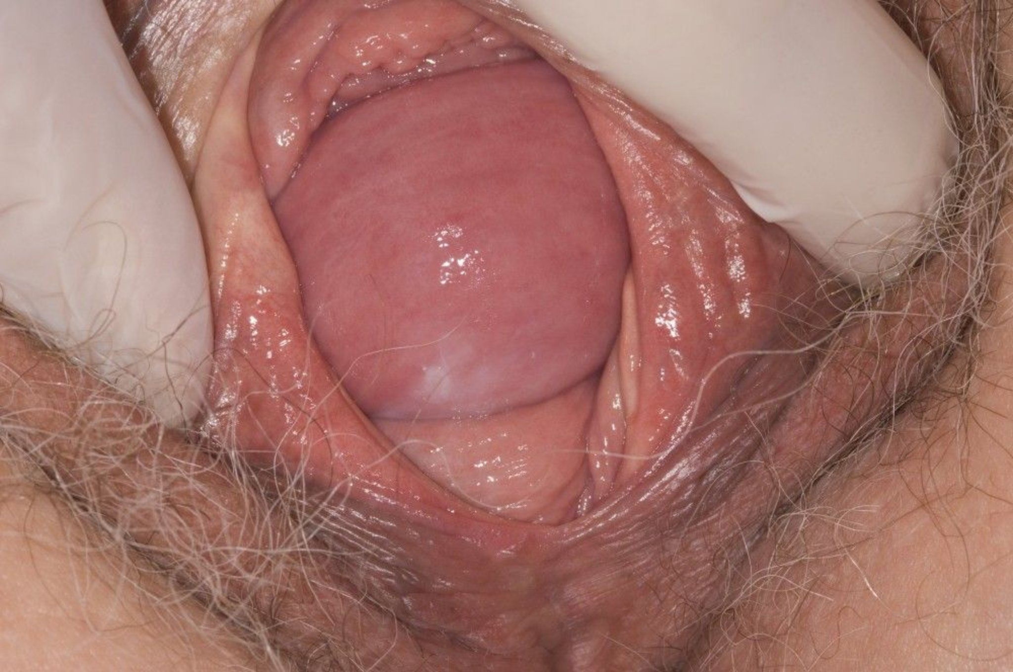
This photo shows prolapse of anterior and posterior vaginal walls.
JIM VARNEY/SCIENCE PHOTO LIBRARY
Treatment of Vaginal Wall Prolapse
Pelvic floor muscle exercises (eg, Kegel exercises)
Pessary
Surgical repair of supporting structures
Treatment of anterior or posterior vaginal wall prolapse is individualized, based on a patient's symptoms, with the goal of improving quality of life (1). Asymptomatic prolapse does not require treatment. Treatment may consist of pelvic floor muscle exercises, a pessary, and, if these measures are unsuccessful or if the patient prefers, surgical repair.
Pelvic floor muscle exercises
Pelvic floor muscle exercises (eg, Kegel exercises) are usually first-line therapy for stage I or II pelvic organ prolapse. Pelvic floor muscle exercises have no or a very low risk of harm to the patient. With consistent use, they can lessen bothersome symptoms of prolapse (and stress incontinence), but they do not appear to reduce the anatomic severity of prolapse (2).
Pelvic floor muscle exercises are isometric contractions of the pubococcygeus muscle. These muscles are contracted tightly for about 1 or 2 seconds, then relaxed for about 10 seconds. Gradually, contractions are held for about 10 seconds each. The exercise is repeated about 10 times in a row. Doing the exercises several times a day is recommended.
Exercises can be facilitated by
Use of weighted vaginal cones (which help patients focus on contracting the correct muscle)
Use of biofeedback devices provide muscle-effort feedback. There are many different types of devices, varying from simple hand-held mirrors to devices that turn patient muscle activity into audio or visual information.
Electrical stimulation, which causes the muscle to contract
Pessaries
Pessaries are devices that are inserted into the vagina to maintain normal anatomy and reduction of the prolapsed structures, resulting in improved objective and subjective benefit (3). Pessaries are typically made of silicone and vary in shape and size; some are inflatable.


To fit a pessary, a clinician should perform a POP-Q examination to guide pessary selection. The pessary is then inserted and the patient is asked to walk around to assess comfort, and empty her bladder to ensure the pessary does not obstruct the urethra. The clinician should provide instructions for removing, cleaning, and reinserting the pessary, and arrange a follow-up visit in 1 month. Proper size, fit, and position are important, because a pessary can cause vaginal ulceration with bleeding if it is does not fit correctly and vaginal discharge if it is not cleaned regularly (at least monthly if not more frequently).
The frequency of follow-up visits for a patient with a pessary is based on the patient's ability to manage the pessary independently. Those patients who are able to remove, clean, and replace the pessary themselves at least weekly may be seen annually, while those who are unable to provide self-care will need more frequent visits (4).
Surgical repair
Surgical repair can help relieve symptoms that are severe or do not resolve with nonsurgical treatment. The surgical approach used depends on the type of prolapse, symptoms, patient age and comorbidities, patient preference, and the surgeon's expertise. Surgery may include 1 (or a combination) of the following procedures
Anterior or posterior colporrhaphy (vaginal repair)
Vaginal apex suspension or repair
Colpocleisis (closure of the vagina after removal of the uterus or with the uterus in place [Le Fort procedure])
Surgical repair of the vagina is usually deferred, if possible, until future pregnancy is no longer desired because subsequent vaginal delivery may disrupt the repair. After surgery, patients should avoid heavy lifting for at least 6 weeks.
Treatment references
1. Pelvic Organ Prolapse. ACOG Practice Bulletin, Number 214. Obstet Gynecol. 2019 (reaffirmed 2024);134(5):e126-e142. doi:10.1097/AOG.0000000000003519
2. Wiegersma M, Panman CM, Kollen BJ, Berger MY, Lisman-Van Leeuwen Y, Dekker JH. Effect of pelvic floor muscle training compared with watchful waiting in older women with symptomatic mild pelvic organ prolapse: randomised controlled trial in primary care. BMJ. 2014;349:g7378. Published 2014 Dec 22. doi:10.1136/bmj.g7378
3. Sansone S, Sze C, Eidelberg A, et al. Role of Pessaries in the Treatment of Pelvic Organ Prolapse: A Systematic Review and Meta-analysis. Obstet Gynecol. 2022;140(4):613-622. doi:10.1097/AOG.0000000000004931
4. Vaginal Pessary Use and Management for Pelvic Organ Prolapse: Developed by the joint writing group of the American Urogynecologic Society and the Society of Urologic Nurses and Associates. Individual writing group members are noted in the Acknowledgments section. Urogynecology (Phila). 2023;29(1):5-20. doi:10.1097/SPV.0000000000001293
Key Points
Anterior and posterior vaginal wall prolapse involve protrusion of an organ into the vagina; many patients have both anterior and posterior prolapse.
Anterior vaginal wall prolapse was commonly referred to as cystocele (protrusion of the bladder) or urethrocele (urethra).
Posterior vaginal wall prolapse was commonly referred to as enterocele (small intestine and parietal peritoneum) and rectocele (rectum).
Symptoms include pelvic or vaginal fullness, pressure, and a sensation of organs falling out. Organs may bulge into the vaginal canal or through the vaginal opening (introitus), particularly during straining or coughing.
Diagnose cystocele on pelvic examination by retracting the posterior vaginal wall and observing the anterior vaginal wall with the patient at rest and then with the patient straining.
Diagnose enterocele or rectocele on pelvic examination by retracting the anterior vaginal wall and observing the posterior vaginal wall with the patient at rest and then with the patient straining, and with a rectovaginal examination.
First-line conservative treatment options include pelvic floor physical therapy (for less severe pelvic organ prolapse) and pessaries, with surgical options available based on patient preference and clinical pelvic organ prolapse severity.