


Hereditary hemochromatosis is a genetic disorder characterized by excessive iron (Fe) accumulation that results in tissue damage. Manifestations can include systemic symptoms, liver disorders, cardiomyopathy, diabetes, erectile dysfunction, and arthropathy. Diagnosis is by elevated serum ferritin, iron, and transferrin saturation levels and confirmed by a gene assay. Treatment is usually with serial phlebotomies.
(See also Overview of Iron Overload.)
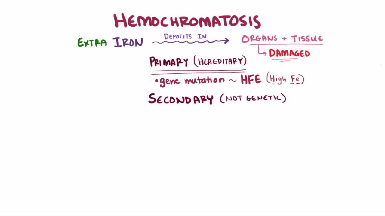
Etiology of Hereditary Hemochromatosis
There are 4 types of hereditary hemochromatosis, types 1 through 4, depending on the gene that is mutated.
Type 1: Mutations of the HFE (human homeostatic iron regulator) gene
Type 2 (juvenile hemochromatosis): Mutations in the HJV (hemojuvelin BMP co-receptor) and HAMP (hepcidin antimicrobial peptide) genes
Type 3: Mutations in the TFR2 (transferrin receptor 2) gene
Type 4 (ferroportin disease): Mutations in the SLC40A1 (solute carrier family 40 member 1) gene
Other much rarer genetic disorders can cause hepatic iron overload, but the clinical picture is usually dominated by symptoms and signs due to failure of other organs (eg, anemia in hypotransferrinemia or atransferrinemia, or neurologic defects in aceruloplasminemia).
Although these types vary markedly in age of onset, clinical consequences of iron overload are the same in all types (1).
Type 1 hereditary hemochromatosis
Type 1 is classic hereditary hemochromatosis, also termed HFE-related hemochromatosis. More than 80% of cases are caused by the homozygous C282Y mutation or the C282Y/H63D compound heterozygote mutation. Homozygous H63D mutations occur rarely and have the same phenotype as homozygous C282y cases. The disorder is autosomal recessive, with a homozygous frequency of 1:200 and a heterozygous frequency of 1:8 in people of northern European ancestry. The C282Y and H63D mutations are uncommon among people with African ancestry and rare among people with Asian ancestry. Of patients with clinical features of hemochromatosis, 83% are homozygous. However, for unknown reasons, phenotypic (clinical) disease is much less common than predicted by the frequency of the gene (ie, many homozygous people do not manifest the disorder).
Type 2 hereditary hemochromatosis
Type 2 hereditary hemochromatosis (juvenile hemochromatosis) is a rare autosomal recessive disorder caused by mutations in the HJV gene that affect the transcription protein hemojuvelin, or mutations in the HAMP gene, which directly codes for hepcidin. It often manifests in adolescents.
Type 3 hereditary hemochromatosis
Mutations in transferrin receptor 2 (TFR2) gene that codes for a protein that appears to control saturation of transferrin, can cause a rare autosomal recessive form of hemochromatosis.
Type 4 hereditary hemochromatosis
Type 4 hereditary hemochromatosis (ferroportin disease) occurs largely in people of southern European ancestry. It results from an autosomal dominant mutation in the SLC40A1 gene and affects the ability of ferroportin to bind hepcidin.
Transferrin and ceruloplasmin deficiency
In transferrin deficiency (hypotransferrinemia or atransferrinemia), absorbed iron that enters the portal system not bound to transferrin is deposited in the liver. Subsequent iron transfer to sites of red blood cell production is reduced because of transferrin deficiency.
In ceruloplasmin deficiency (aceruloplasminemia), lack of ferroxidase causes defective conversion of Fe2+ to Fe3+; such conversion is necessary for binding to transferrin. Defective transferrin binding impairs the movement of iron from intracellular stores to plasma transport, resulting in accumulation of iron in tissues.
Etiology reference
1. Pietrangelo A: Hereditary hemochromatosis: Pathogenesis, diagnosis, and treatment. Gastroenterology 139:393–408, 2010.
Pathophysiology of Hereditary Hemochromatosis
Normal total body iron content is about 2.5 g in women and 3.5 g in men. Because symptoms may be delayed until iron accumulation is excessive (eg, > 10 to 20 g), hemochromatosis may not be recognized until later in life, even though it is an inherited abnormality. In women, clinical manifestations are uncommon before menopause because iron loss due to menses (and sometimes pregnancy and childbirth) tends to offset iron accumulation.
The mechanism for iron overload in both HFE and non-HFE hemochromatosis is increased iron absorption from the gastrointestinal tract, leading to chronic deposition of iron in the tissues. Hepcidin, a liver-derived peptide, is the critical control mechanism for iron absorption. Hepcidin is normally up-regulated when iron stores are elevated and, through its inhibitory effect on ferroportin (which participates in iron absorption), it prevents excessive iron absorption and storage in normal people. Hemochromatosis types 1 through 4 share the same pathogenic basis (eg, lack of hepcidin synthesis or activity) and key clinical features.
In general, tissue injury appears to result from reactive free hydroxyl radicals generated when iron deposition in tissues catalyzes their formation. Other mechanisms may affect particular organs (eg, skin hyperpigmentation can result from increased melanin as well as iron accumulation). In the liver, iron-associated lipid peroxidation induces hepatocyte apoptosis, which stimulates Kupffer cell activation and release of pro-inflammatory cytokines. These cytokines activate hepatic stellate cells to produce collagen, resulting in pathologic accumulation of liver fibrosis and risk of hepatocellular carcinoma.
Symptoms and Signs of Hereditary Hemochromatosis
The clinical consequences of iron overload tend to be similar regardless of the etiology and pathophysiology of the overload.
Historically, experts believed that symptoms did not develop until significant organ damage had occurred. However, organ damage is slow and subtle, and fatigue and nonspecific systemic symptoms and signs often occur early. For example, liver dysfunction can manifest insidiously with fatigue, right upper quadrant abdominal pain, and hepatomegaly. Laboratory abnormalities of iron overload and hepatitis usually precede symptoms.
In type 1 hereditary (HFE) hemochromatosis, symptoms relate to the organs with the largest iron deposits (see table Common Manifestations of Hereditary Hemochromatosis). In men, the initial symptoms may be hypogonadism and erectile dysfunction caused by gonadal iron deposition. Glucose intolerance or diabetes mellitus is another common initial presentation. Some patients present with hypothyroidism.
Liver disease is the most common complication and may progress to cirrhosis; 20 to 30% of patients with cirrhosis develop hepatocellular carcinoma. Liver disease is the most common cause of death. Elevated liver enzymes are one of the most common laboratory abnormalities in patients with type 1 hemochromatosis.
Cardiomyopathy with heart failure is the 2nd most common fatal complication. Hyperpigmentation (bronze diabetes) and porphyria cutanea tarda are common, as is symptomatic arthropathy in the hands.
In type 2 disease, symptoms and signs include progressive hepatomegaly and hypogonadotropic hypogonadism.
In type 3 disease, symptoms and signs are similar to type 1 hereditary (HFE) hemochromatosis.

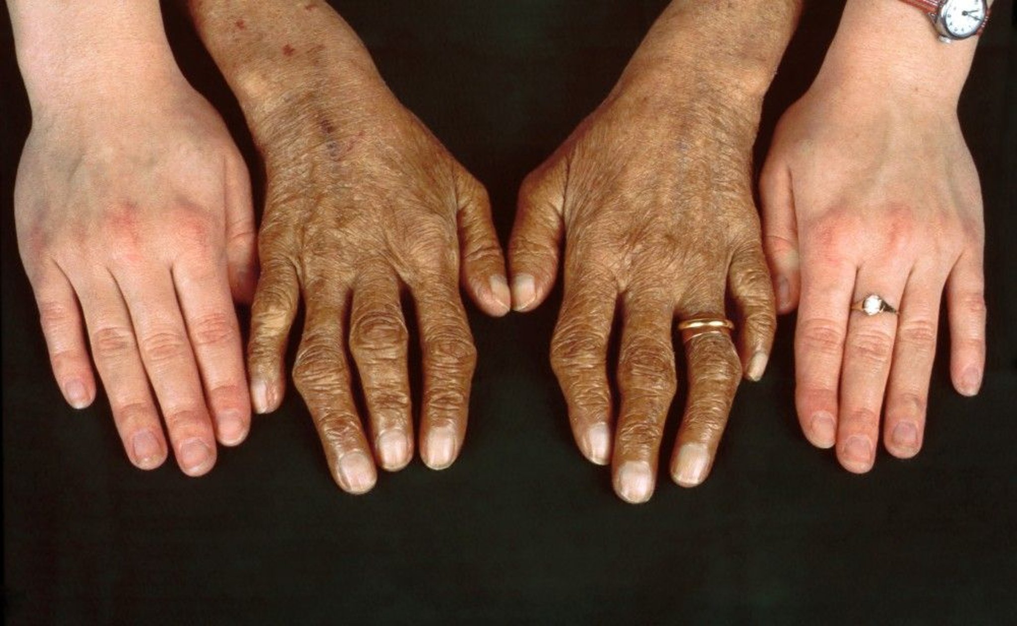
Clinical Photography/SCIENCE PHOTO LIBRARY
Type 4 disease manifests in the first decade of life as increased serum ferritin levels with low or normal transferrin saturation; progressive saturation of transferrin occurs when patients are in their 20s and 30s. Clinical manifestations are milder than in type 1 disease, with modest liver disease and mild anemia.
Diagnosis of Hereditary Hemochromatosis
Serum ferritin, fasting serum iron, and transferrin saturation
Genetic testing
Sometimes liver biopsy
Symptoms and signs may be nonspecific, subtle, and of gradual onset, so that index of suspicion should be high. Primary hemochromatosis should be suspected when typical manifestations, particularly combinations of such manifestations, remain unexplained after routine evaluation. Family history of hemochromatosis, cirrhosis, or hepatocellular carcinoma is a more specific clue. All patients with chronic liver disease should be evaluated for iron overload, regardless of race or ethnicity
Serum ferritin measurement is the simplest and most direct initial test. Elevated levels (> 200 ng/mL [> 200 mcg/L] in women or > 250 ng/mL [> 250 mcg/L] in men) are usually present in hereditary hemochromatosis but can result from other abnormalities, such as inflammatory liver disorders (eg, chronic viral hepatitis, nonalcoholic fatty liver disease, alcoholic liver disease), cancer, certain systemic inflammatory disorders (eg, rheumatoid arthritis, hemophagocytic lymphohistiocytosis), or obesity. Further testing is done if ferritin level is abnormal; testing includes fasting serum iron (usually > 300 mg/dL [> 53.7 mcmol/L]) and iron binding capacity (transferrin saturation; levels usually > 50%). A transferrin saturation of < 45% has a negative predictive value of 97% for iron overload.
In type 2 disease, ferritin levels are >1000 ng/mL (> 1000 mcg/L), and transferrin saturation is >90%.
In transferrin or ceruloplasmin deficiency, serum transferrin (ie, iron-binding capacity) and ceruloplasmin levels are profoundly low or undetectable.
Gene assay is diagnostic of hereditary hemochromatosis caused by HFE gene mutations. About 70% of patients with C282Y homozygous mutations of the HFE gene have an elevated ferritin level, but only about 10% of these patients have evidence of organ dysfunction. Clinically significant iron overload is even less common in patients with heterozygous mutations of the HFE gene (ie, C282Y/H63D). Hemochromatosis types 2 to 4 are suspected in the less common instances in which ferritin and iron blood tests indicate iron overload and genetic testing is negative for the HFE gene mutation, particularly in younger patients. Confirmation of these diagnoses by genetic testing is not routinely available.
When the diagnosis is confirmed, the liver must be tested for fibrosis and cirrhosis. Up to 80% of patients with cirrhosis and a homozygous C282Y mutation will have a ferritin of > 1000 ng/mL, elevated AST (aspartate transaminase) and ALT (alanine transaminase), and platelet count < 200 × 103 /mcL (< 200 × 109/L). Because the presence of cirrhosis affects prognosis, when the ferritin is > 1000 ng/mL, liver biopsy is commonly done and tissue iron content is measured (when available). Liver biopsy is also recommended in patients with serologic evidence of iron overload but negative genetic evaluation. MRI with noncontrast MR elastography (MRE), a noninvasive alternative for estimating hepatic iron content and hepatic fibrosis, is becoming increasingly accurate.
Screening is required for first-degree relatives of people with hereditary hemochromatosis by measuring serum ferritin levels and testing for the C282Y and H63D mutations in the HFE gene.
Treatment of Hereditary Hemochromatosis
Phlebotomy
Treatment is indicated for patients with clinical manifestations, elevated serum ferritin levels (particularly levels > 1000 ng/mL [> 1000 mcg/L]), or elevated transferrin saturation. Asymptomatic patients need only periodic (eg, yearly) clinical evaluation and measurement of serum iron, ferritin, transferrin saturation, and liver enzymes.
Phlebotomy is the simplest and most effective method to remove excess iron. It delays progression of fibrosis to cirrhosis, sometimes even reversing cirrhotic changes, and prolongs survival, but it does not prevent hepatocellular carcinoma. About 500 mL of blood (about 250 mg of iron) is removed weekly or biweekly (every other week) until serum ferritin levels reach 50 to 100 ng/mL. Weekly or biweekly phlebotomy may be needed for many months (eg, if 250 mg of iron are removed per week, 40 weeks will be required to remove 10 g of iron). When iron levels are normal, phlebotomies can be intermittent to maintain ferritin between 50 and 100 ng/mL.
Diabetes mellitus, cardiomyopathy, erectile dysfunction, and other secondary manifestations are treated as indicated. Patients with advanced fibrosis or cirrhosis due to iron overload should be screened for hepatocellular carcinoma every 6 months with a liver ultrasound.
Patients should follow a balanced diet; it is not necessary to restrict consumption of iron-containing foods (eg, red meat, liver). Alcohol should be consumed only in moderation because it can increase iron absorption and, in high amounts, increases the risk of cirrhosis. Vitamin C supplements should be avoided because they increase the absorption of iron in the duodenum.
In patients with type 4 disease, tolerance to vigorous phlebotomy is poor; serial monitoring of hemoglobin level and transferrin saturation is required.
Treatment of transferrin deficiency and ceruloplasmin deficiency is experimental; eg, iron chelators may be better tolerated than phlebotomy because patients typically have anemia.
Key Points
There are 4 types of hereditary hemochromatosis, which all involve mutations that impair the ability of the body to inhibit iron absorption when iron stores are excessive.
The effects of iron overload are similar in all types and include liver disease (leading to cirrhosis), skin pigmentation, diabetes, arthropathy, hypogonadism, and sometimes heart failure.
Diagnose by measuring serum ferritin level; if elevated, confirm by demonstrating elevated serum iron, transferrin saturation, and genetic testing.
Once diagnosis is made, the degree of liver fibrosis should be assessed by liver biopsy or non-invasive imaging such as MRI with MR elastography to i determine prognosis; consider genetic testing and screening of first-degree relatives.
Treat with phlebotomy and moderation of alcohol consumption.


