


Ulnar and radial shaft fractures frequently result from direct trauma to the radius or ulna. Concomitant dislocations are possible.
(See also Overview of Fractures.)
Fractures of the radius and ulna are frequently caused by direct blows to the forearm (eg, during contact sports, falls, or defensive actions during an assault). Concomitant dislocations can result from forces transmitted via the interosseous membrane between the radius and ulna.
Pearls & Pitfalls
|
Isolated midshaft radius or midshaft ulna fractures are common.
Monteggia fractures are proximal ulnar fractures with a radial head dislocation.
Galeazzi fractures are distal radial shaft fractures with a dislocation of the distal radioulnar joint.
Symptoms and Signs of Ulnar and Radial Shaft Fractures
Radial and ulnar shaft fractures can cause pain, deformity, ecchymosis, and swelling at the site of injury.
Diagnosis of Ulnar and Radial Shaft Fractures
Anteroposterior and lateral radiographs
Radial and ulnar shaft fractures are generally diagnosed with anteroposterior and lateral radiographs. If a fracture is suspected, the elbow and wrist should also be examined and, when appropriate, radiographs performed.
The radial head dislocation is easy to overlook in Monteggia fractures because the fracture is so obvious and should be specifically considered if a proximal ulnar fracture is identified. The radial head is typically visible outside of its normal articulation with the capitellum (capitulum). In Monteggia fractures, the radiocapitellar line also is not aligned correctly, suggesting dislocation (see figure ). In Galeazzi fractures (similar to Monteggia fractures), the distal radioulnar dislocation is easy to overlook with distal radial shaft fractures and should be specifically considered.

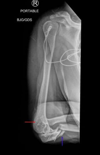
This radiograph shows proximal ulnar fracture (blue arrow) with associated radial head dislocation. The radial head (red arrow) is above the capitellum, its normal articulation.

This radiograph shows proximal ulnar fracture with concomitant radial head dislocation (red arrow).

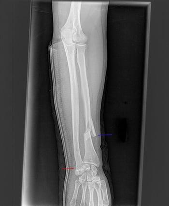
This radiograph shows a fracture of the distal radius (blue arrow) and disruption of the distal radioulnar joint (red arrow). The articular surfaces of the distal ulna and distal radius are not aligned with each other.
Treatment of Ulnar and Radial Shaft Fractures
For isolated radial and ulnar shaft fractures, closed reduction and splinting with outpatient orthopedic followup
For Monteggia and Galeazzi injuries, urgent orthopedic consultation and usually open reduction with internal fixation (ORIF)
Most isolated radial and ulnar shaft fractures can be treated with closed reduction and splinting with subsequent orthopedic referral.
For Monteggia and Galeazzi fractures, urgent orthopedic consultation is required, and ORIF is usually necessary to maintain alignment.
Key Points
Radial and ulnar fractures commonly result from direct forces to the forearm.
Take anteroposterior and lateral radiographs.
For isolated midshaft radial or ulnar fractures, always consider concomitant dislocations involving the wrist and elbow.
Treat with closed reduction and consult orthopedics.


