


Increased intracranial pressure sometimes causes protrusion (herniation) of brain tissue through one of the rigid intracranial barriers (tentorial notch, falx cerebri, foramen magnum).
Brain herniation is classified based on the structure through which tissue is herniated:
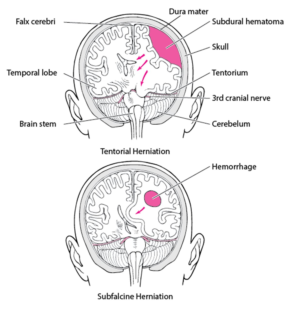
Transtentorial (uncal) herniation: The medial temporal lobe is squeezed by a unilateral mass across and under the tentlike tentorium that supports the temporal lobe.
Subfalcine herniation: The cingulate gyrus is pushed under the falx cerebri by an expanding mass high in a cerebral hemisphere.
Central herniation: Both temporal lobes herniate through the tentorial notch because of bilateral mass effects or diffuse brain edema.
Upward transtentorial herniation: This type can occur when an infratentorial mass (eg, tumor in the posterior fossa, cerebellar hemorrhage) compresses the brain stem, kinking it and causing patchy brain stem ischemia.
Tonsillar herniation: Usually, the cause is an expanding infratentorial mass (eg, cerebellar hemorrhage), forcing the cerebellar tonsils, through the foramen magnum.