


Plantar fasciitis is pain at the site of the attachment of the plantar fascia and the calcaneus (calcaneal enthesopathy), with or without accompanying pain along the medial band of the plantar fascia. Diagnosis is mainly clinical. Treatment involves Achilles tendon and plantar soft-tissue foot-stretching exercises, night splints, orthotics, and shoes with appropriate heel elevation.
(See also Overview of Foot and Ankle Disorders.)
Syndromes of pain in the plantar fascia have been called plantar fasciitis; however, because there is usually no inflammation, plantar fasciosis is more correct. Other terms used include calcaneal enthesopathy pain or calcaneal spur syndrome; however, there may be no bone spurs on the calcaneus. Plantar fasciitis may involve acute or chronic stretching, tearing, and degeneration of the fascia at its attachment site.
Etiology of Plantar Fasciitis
Recognized causes of plantar fasciitis include shortening or contracture of the Achilles tendon and plantar fascia. Risk factors for such shortening include a sedentary lifestyle, occupations requiring sitting, very high or low arches in the feet, and chronic wearing of high-heel shoes. The disorder is also common among runners and dancers and may occur in people whose occupations involve standing or walking on hard surfaces for prolonged periods.
Disorders that may be associated with plantar fasciitis are obesity, rheumatoid arthritis, reactive arthritis, psoriatic arthritis, and other spondyloarthropathies. Multiple injections of glucocorticoids may contribute by causing degenerative changes of the fascia and possible loss of the cushioning subcalcaneal fat pad.
Symptoms and Signs of Plantar Fasciitis
Plantar fasciitis is characterized by pain at the bottom of the heel with weight bearing, particularly when first arising in the morning; pain usually abates within 5 to 10 minutes, only to return later in the day. It is often worse when pushing off the heel (the propulsive phase of gait) and after periods of rest. Acute, severe heel pain, especially with mild local puffiness, may indicate an acute fascial tear. Some patients describe burning or sticking pain along the plantar medial border of the foot when walking.
Diagnosis of Plantar Fasciitis
Primarily history and physical examination
Plantar fasciitis is confirmed if pain is elicited when firm thumb pressure is applied to the calcaneus when the foot is dorsiflexed. Fascial pain along the plantar medial border of the fascia may also be present. If findings are equivocal, demonstration of a heel spur on radiograph may support the diagnosis; however, absence does not exclude the diagnosis, and visible spurs are not generally the cause of plantar fasciitis symptoms. Also, infrequently, calcaneal spurs appear ill defined on radiographs, exhibiting fluffy new bone formation, suggesting spondyloarthropathy (eg, ankylosing spondylitis, reactive arthritis). If an acute fascial tear is suspected, MRI is indicated.

Other disorders causing heel pain can mimic plantar fasciitis:
Throbbing heel pain, particularly when the shoes are removed or when mild warmth and puffiness are present, is more suggestive of calcaneal bursitis.
Acute, severe retrocalcaneal pain, with erythema and warmth, may indicate gout.
Pain that radiates from the low back to the heel may be an S1 radiculopathy due to an L5 disk herniation.


The lateral radiograph shows a combined plantar and posterior calcaneal spur at the Achilles tendon insertion.
Image courtesy of James C. Connors, DPM.

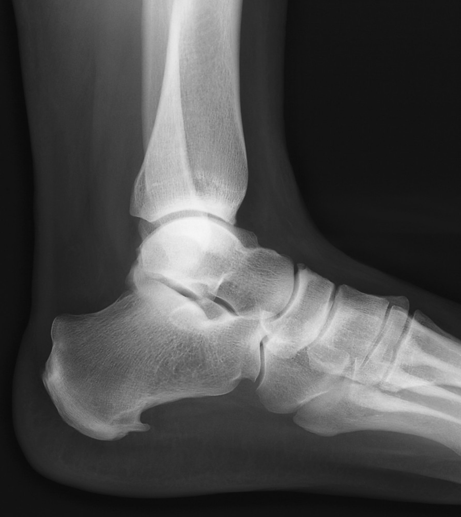
The heel spur is the bony exostosis extending forward at the bottom of the calcaneus.
ZEPHYR/SCIENCE PHOTO LIBRARY
Treatment of Plantar Fasciitis
Activity modification, stretching, splinting, and cushioning or orthotics
Physical therapy
To alleviate the stress and pain on the fascia, the person can take shorter steps and avoid walking barefoot. Activities that involve foot impact, such as jogging, should be avoided. The most effective plantar fasciitis treatments include the use of in-shoe heel cushioning and arch supports with Achilles tendon-stretching exercises and night splints that stretch the Achilles tendon and plantar fascia while the patient sleeps. Prefabricated or custom-made foot orthotics may also alleviate fascial tension and symptoms while the patient is ambulatory. Physical therapy should be used early in the treatment process to develop a dedicated daily Achilles tendon stretching routine to help restore tendon flexibility.
Other treatments may include activity modifications, nonsteroidal anti-inflammatory drugs (NSAIDs), weight loss in patients with obesity, cold and ice massage therapy, and occasional glucocorticoid injections. However, because glucocorticoid injections can predispose to plantar fascial rupture, many clinicians limit these injections (see Considerations for Using Glucocorticoid Injections).
For recalcitrant cases, cast immobilization should be used before surgical intervention is considered. For recalcitrant types of plantar fasciitis, some studies recommend extracorporeal pulse activation therapy (EPAT), in which low-frequency pulse waves are delivered locally using a handheld applicator, may be tried. The pulsed pressure wave is a safe, noninvasive technique that is thought to stimulate metabolism and enhance blood circulation, which in turn may help regenerate damaged tissue and accelerate healing (1).
Treatment reference
1. Auersperg V, Trieb K: Extracorporeal shock wave therapy: an update. EFORT Open Rev 5(10):584-592, 2020. doi: 10.1302/2058-5241.5.190067
Key Points
Plantar fasciitis involves various syndromes that cause pain in the plantar fascia.
Various lifestyle factors and disorders increase risk by leading to shortened calf muscles and plantar fascia.
Pain at the bottom of the heel worsens with weight bearing, particularly when pushing off the heel and over the course of the day.
Confirm the diagnosis by reproducing pain with calcaneal pressure exerted by the thumb during dorsiflexion.
Treat initially with in-shoe heel cushioning, arch supports, Achilles tendon–stretching exercises, physical therapy and splinting devices worn at night.


