


A seizure is an abnormal, unregulated electrical discharge that occurs within the brain’s cortical gray matter and transiently interrupts normal brain function. A seizure typically causes altered awareness, abnormal sensations, focal involuntary movements, or convulsions (widespread violent involuntary contraction of voluntary muscles). Diagnosis may be clinical and involves results of neuroimaging, laboratory testing, and electroencephalography (EEG) for new-onset seizures or levels of antiseizure medications (anticonvulsants) for previously diagnosed seizure disorders. Treatment includes elimination of the cause if possible, antiseizure medications, and surgery (if the medications are ineffective).
(See also Febrile Seizures and Neonatal Seizure Disorders.)
About 2% of adults have a seizure at some time during their life. Two thirds of these people never have another one.
Definitions
Terminology related to seizures can be confusing.
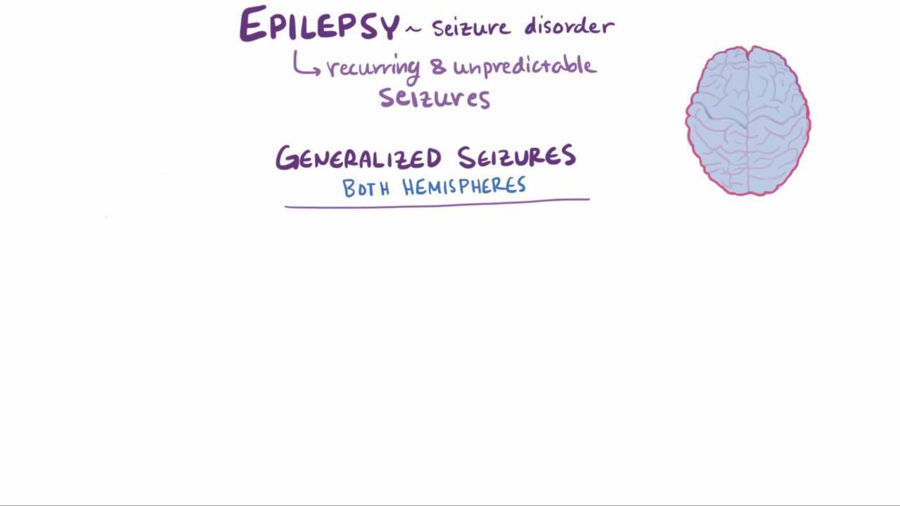
Epilepsy (also called epileptic seizure disorder) is a chronic brain disorder characterized by recurrent (≥ 2) seizures that are unprovoked (ie, not related to reversible stressors) and that occur > 24 hours apart. A single seizure is not considered an epileptic seizure. Epilepsy is often idiopathic, but various brain disorders, such as malformations, strokes, and tumors, can cause symptomatic epilepsy.
Symptomatic epilepsy is epilepsy due to a known cause (eg, brain tumor, stroke). The seizures it causes are called symptomatic epileptic seizures. Such seizures are most common among neonates and older people.
Cryptogenic epilepsy is epilepsy assumed to be due to a specific cause, but whose specific cause is currently unknown.
Nonepileptic seizures are provoked by a temporary disorder or stressor (eg, metabolic disorders, central nervous system (CNS) infections, cardiovascular disorders, medication toxicity or withdrawal, psychogenic disorders). In children, fever can provoke a seizure (febrile seizures).
Psychogenic nonepileptic seizures (pseudoseizures) are symptoms that simulate seizures in patients with psychiatric disorders but that do not involve an abnormal electrical discharge in the brain.
Etiology of Seizure Disorders
Common causes of seizures (see table Causes of Seizures) vary by age of onset:
Before age 2: Fever, hereditary or congenital neurologic disorders, birth injuries, and inherited or acquired metabolic disorders
Ages 2 to 14: Idiopathic seizure disorders
Adults: Cerebral trauma, alcohol withdrawal, tumors, strokes, and an unknown cause (in 50%)
Older people: Tumors and strokes
In reflex epilepsy, a rare disorder, seizures are triggered predictably by an external stimulus, such as repetitive sounds, flashing lights, video games, music, or even touching certain parts of the body.
In cryptogenic epilepsy and often in refractory epilepsy, a rare but increasingly identified cause is anti-NMDA (N-methyl-d-aspartate) receptor encephalitis, especially in young women. This disorder also causes psychiatric symptoms, a movement disorder, and cerebrospinal fluid (CSF) pleocytosis. Ovarian teratoma occurs in almost 60% of women who are > 18 years and have anti-NMDA receptor encephalitis (1). Removal of the teratoma (if present) and immunotherapy control the seizures much better than antiseizure medications.
Etiology reference
1. Florance NR,. Davis RL, Lam C, et al: Anti–N-methyl-D-aspartate receptor (NMDAR) encephalitis in children and adolescents. Ann Neurol 66 (1):11–18, 2009. doi: 10.1002/ana.21756
Classification of Seizure Disorders
In 2017, the International League Against Epilepsy (ILAE) developed a new classification system for seizures (1).
Initial classification is by type of onset:
Generalized onset
Focal onset
Unknown onset
Focal-onset seizures are then classified by level of awareness (knowledge of self and environment). Level of awareness is not used to classify generalized-onset seizures because most of these seizures (but not all) impair awareness.
All seizures are then classified, if possible, as
Motor onset
Nonmotor onset
Responsiveness is not used to classify seizures, but it can be useful as a descriptor. Responsiveness can be intact or impaired whether awareness is impaired or not.
Generalized-onset seizures
In generalized-onset seizures, seizures originate in networks in both hemispheres. Awareness is usually impaired, and consciousness is usually lost.
Generalized-onset seizures are classified as motor and nonmotor (absence) seizures. (However, nonmotor seizures may involve motor activity.) In generalized-onset motor seizures, motor activity is usually bilateral from the onset. When bilateral onset of motor activity is asymmetric, determining whether onset is focal or generalized may be difficult.
Generalized-onset motor seizures may be further classified by type of seizure:
Tonic-clonic seizures (formerly, grand mal seizures)
Clonic seizures (sustained rhythmic jerking)
Tonic seizures (generalized stiffening involving all limbs and without rhythmic jerking)
Atonic seizures (loss of muscle tone)
Myoclonic seizures (rhythmic jerking not preceded by stiffening)
Myoclonic-tonic-clonic seizures (myoclonic jerking followed by tonic and clonic movements)
Myoclonic-atonic seizures (myoclonic jerking followed by atonia)
Epileptic spasms (formerly, infantile spasms)
Generalized-onset nonmotor seizures may be further classified by type of seizure (defined by the earliest prominent feature):
Typical absence seizures
Atypical absence seizures (eg, with less abrupt onset or termination or with abnormal changes in tone)
Myoclonic seizures (myoclonic jerking preceded by brief impairment of consciousness [absence])
Eyelid myoclonia
All absence seizures are generalized-onset seizures. The following can help distinguish absence seizures from focal impaired-awareness seizures, although the distinctions are not absolute:
Absence seizures tend to occur in younger people.
They tend to start and end more suddenly.
Usually, automatisms are less complex in absence seizures than in focal impaired-awareness seizures.
Generalized-onset seizures result most often from metabolic disorders and sometimes from genetic disorders.
Focal-onset seizures
Focal-onset seizures originate in networks in one hemisphere and may originate in subcortical structures. They may be discretely localized or more widely distributed.
Focal-onset seizures may be classified by level of awareness:
Focal aware seizures (formerly, simple partial seizures)
Focal impaired-awareness seizures (formerly, complex partial seizures)
If awareness is impaired during any part of the seizure, the seizure is classified as a focal impaired-awareness seizure.
Focal-onset motor seizures may be further classified by type of seizure:
Automatisms (coordinated, purposeless, repetitive motor activity)
Atonic (focal loss of muscle tone)
Clonic (focal rhythmic jerking)
Epileptic spasms (focal flexion or extension of arms and flexion of trunk)
Hyperkinetic (causing pedaling or thrashing)
Myoclonic (irregular, brief focal jerking)
Tonic (sustained focal stiffening of one limb or one side of the body)
Awareness level is usually not specified for atonic seizures or epileptic spasms.
In focal-onset tonic seizures, stiffening involves only one limb or one side of the body, usually with no loss of consciousness. electroencephalography (EEG) may show contralateral focal epileptiform abnormalities. In contrast, in generalized-onset tonic seizures, stiffening involves all limbs, with or without loss of consciousness, and EEG may show bilateral epileptiform abnormalities.
Focal-onset nonmotor seizures may be further classified based on the earliest prominent feature:
Autonomic dysfunction (autonomic effects such as gastrointestinal (GI) sensations, a sense of heat or cold, flushing, sexual arousal, piloerection, and palpitations)
Behavior arrest (cessation of movement and unresponsiveness as the main feature of the entire seizure)
Cognitive dysfunction (impairment of language or other cognitive domains or positive features such as déjà vu, hallucinations, illusions, or perceptual distortions)
Emotional dysfunction (manifesting with emotional changes, such as anxiety, fear, joy, other emotions, or affective signs without subjective emotions)
Sensory dysfunction (causing somatosensory, olfactory, visual, auditory, gustatory, or vestibular sensations or a sense of heat or cold)
Focal-onset seizures may evolve into a generalized-onset tonic-clonic seizure (called a focal-to-bilateral tonic-clonic seizure; formerly, secondary generalization), which causes loss of consciousness. Focal-to-bilateral tonic-clonic seizures occur when a focal-onset seizure spreads and activates the entire cerebrum bilaterally. Activation may occur so rapidly that the initial focal-onset seizure is not clinically apparent or is very brief.
Unknown-onset seizures
Seizures are usually classified as unknown-onset seizures when information about onset is lacking. If clinicians acquire more information about the seizures, these seizures may be reclassified as focal-onset or generalized-onset.
Seizures of unknown onset can be motor or nonmotor.
Unknown-onset motor seizures may be further classified as
Tonic-clonic
Epileptic spasms
Unknown-onset nonmotor seizures may be further classified as
Behavior arrest
Tonic-clonic seizures with an obscure onset are often classified as unknown-onset seizures. Seizures that are later identified as epileptic spasms or behavior-arrest seizures may initially be classified as unknown-onset seizures.
Detailed video EEG monitoring can help clarify whether onset is focal or generalized; doing so is important because if onset is focal, the cause may be treatable.
Classification reference
1. Fisher RS, Cross JH, D'Souza C, et al: Instruction manual for the ILAE [International League Against Epilepsy] 2017 operational classification of seizure types. Epilepsia 58 (4):531–542, 2017. doi: 10.1111/epi.13671
Symptoms and Signs of Seizure Disorders
An aura may precede seizures. Aura describes how patients feel as a seizure starts. Auras may consist of motor activity or sensory, autonomic, or psychic sensations (eg, paresthesias, a rising epigastric sensation, abnormal smells, a sensation of fear, a déjà vu or jamais vu sensation). In jamais vu, a familiar place or experience feels very unfamiliar—the opposite of déjà vu. In most cases, the aura that patients describe is part of a focal aware seizure.
Most seizures end spontaneously in 1 to 2 minutes.
A postictal state often follows generalized-onset seizures; it is characterized by deep sleep, headache, confusion, and muscle soreness; this state lasts from minutes to hours. Sometimes the postictal state includes Todd paralysis (a transient neurologic deficit, usually weakness, of the limb contralateral to the seizure focus).
Most patients appear neurologically normal between seizures, although high doses of the medications used to treat seizure disorders, particularly sedating antiseizure medications, can reduce alertness. Any progressive mental deterioration is usually related to the neurologic disorder that caused the seizures rather than to the seizures themselves.
Occasionally, seizures are unremitting, as in status epilepticus.
Focal-onset seizures
Focal-onset seizures may be
Focal aware seizures (formerly, simple partial seizures)
Focal impaired-awareness seizures (formerly, complex partial seizures)
During a focal aware seizure, awareness is intact. If awareness is impaired during any part of the seizure, the seizure is classified as a focal seizure with impaired awareness; awareness may be impaired but not completely lost.
Focal aware seizures cause motor, sensory, or psychomotor symptoms. Specific symptoms reflect the affected area of the brain (see table Manifestations of Focal-Onset Seizures by Site). In jacksonian seizures, focal motor symptoms begin in one hand, then march up the arm (jacksonian march). Other focal-onset seizures affect the face first, then spread to an arm and sometimes a leg. Some focal-onset motor seizures begin with an arm raising and the head turning toward the raised arm (called fencing posture).
Epilepsia partialis continua, a rare disorder, is a continuous focal aware motor seizure. It usually involves the arm, hand, or one side of the face; seizures recur every few seconds or minutes for days to years at a time. The cause is usually as follows:
In adults: A structural lesion (eg, stroke)
In children: A focal cerebral cortical inflammatory process (eg, Rasmussen encephalitis), possibly caused by a chronic viral infection or by autoimmune processes
Pearls & Pitfalls
|
Focal impaired-awareness seizures are often preceded by auras. During the seizure, patients may stare. Awareness is impaired, but patients have some awareness of the environment (eg, they purposefully withdraw from noxious stimuli). The following may also occur:
Oral automatisms (involuntary chewing or lip smacking)
Limb automatisms (eg, automatic purposeless movements of the hands)
Utterance of unintelligible sounds without understanding what they say
Resistance to assistance
Tonic or dystonic posturing of the extremity contralateral to the seizure focus
Head and eye deviation, usually in a direction contralateral to the seizure focus
Bicycling or pedaling movements of the legs if the seizure emanates from the medial frontal or orbitofrontal head regions
Motor symptoms subside after 1 to 2 minutes, but confusion and disorientation may continue for another 1 or 2 minutes. Postictal amnesia is common. Patients may lash out if restrained during the seizure or while recovering consciousness if the seizure generalizes. However, unprovoked aggressive behavior is unusual.
Left temporal lobe seizures may cause verbal memory abnormalities; right temporal lobe seizures may cause visual spatial memory abnormalities.
Generalized-onset seizures
Awareness is usually impaired or lost, and motor function is abnormal from the onset. Generalized-onset seizures are classified as motor or nonmotor (absence) seizures.
Typical absence seizures (formerly called petit mal seizures) consist of 10- to 30-second loss of consciousness with eyelid fluttering; axial muscle tone may or may not be lost. Patients do not fall or convulse; they abruptly stop activity, then just as abruptly resume it, with no postictal symptoms or knowledge that a seizure has occurred. Absence seizures are genetic and occur predominantly in children. Usually, they begin between ages 5 and 15 years and do not continue into adulthood. Without treatment, such seizures are likely to occur many times a day. Seizures often occur when patients are sitting quietly, can be precipitated by hyperventilation, and rarely occur during exercise. Neurologic and cognitive examination results are usually normal.
Atypical absence seizures usually occur as part of the Lennox-Gastaut syndrome, a severe form of epilepsy. They differ from typical absence seizures as follows:
They last longer.
Jerking or automatic movements are more pronounced.
Loss of awareness is less complete.
Many patients have a history of damage to the nervous system, developmental delay, abnormal neurologic examination results, and other types of seizures. Atypical absence seizures usually continue into adulthood.
Lennox-Gastaut syndrome is a severe form of epilepsy that causes several types of seizures; the disorder usually begins before the age of 4 years and can continue into adulthood. Periods of frequent seizures may alternate with relatively seizure-free periods. Intellectual function and/or information processing is impaired in most patients with Lennox-Gastaut syndrome; development may be delayed, and behavioral problems may occur. Causes of Lennox-Gastaut syndrome include brain malformations, tuberous sclerosis, perinatal asphyxia, severe head injury, central nervous system infection, and hereditary genetic and degenerative or metabolic disorders. Sometimes no cause is identified.
In myoclonic absence seizures, the arms and shoulders jerk rhythmically (3 times/second), causing progressive lifting of the arms. Typically, these seizures last 10 to 60 seconds. Impairment of awareness may not be obvious. Myoclonic absence seizures are caused by various genetic disorders; sometimes the cause is unknown.
Eyelid myoclonia consists of myoclonic jerks of the eyelids and upward deviation of the eyes, often precipitated by closing the eyes or by light. Eyelid myoclonia can occur in motor as well as nonmotor seizures.
Atonic seizures occur most often in children, usually as part of the Lennox-Gastaut syndrome. Atonic seizures are characterized by brief, complete loss of muscle tone and consciousness. Children fall or pitch to the ground, risking trauma, particularly head injury.
Tonic seizures occur most often during sleep, usually in children. The cause is usually the Lennox-Gastaut syndrome. Tonic (sustained) contraction of axial muscles may begin abruptly or gradually, then spread to the proximal muscles of the limbs. The neck is often stiff. Tonic seizures usually last 10 to 15 seconds. In longer tonic seizures, a few, rapid clonic jerks may occur as the tonic phase ends.
In clonic seizures, sustained rhythmic jerking occurs in the limbs on both sides of the body and often in head, neck, face, and trunk. Clonic seizures usually occur in infants and should be distinguished from jitteriness or shuddering attacks. Clonic seizures are much less common than tonic-clonic seizures.
Tonic-clonic seizures may be
Generalized-onset (formerly primarily generalized)
Focal-to-bilateral tonic-clonic (formerly, secondarily generalized)
Generalized-onset tonic-clonic seizures typically begin with an outcry; they continue with loss of consciousness and falling, followed by tonic contraction, then clonic (rapidly alternating contraction and relaxation) motion of muscles of the extremities, trunk, and head. Urinary and fecal incontinence, tongue biting, and frothing at the mouth sometimes occur. Seizures usually last 1 to 2 minutes. There is no aura.
Focal-to-bilateral tonic-clonic seizures begin with a focal aware or focal impaired-awareness seizure, then progress to resemble other generalized-onset tonic-clonic seizures.
Myoclonic seizures are brief, lightning-like jerks of a limb, several limbs, or the trunk. They may be repetitive, leading to a tonic-clonic seizure. The jerks may be bilateral or unilateral. Unlike other seizures with bilateral motor movements, consciousness is not lost unless the myoclonic seizure progresses into a generalized tonic-clonic seizure.
In myoclonic-atonic seizures, the extremities or trunk jerks briefly, then goes limp (drop attack). Seizures usually begin at age 6 months to 6 years. In two thirds of children, febrile seizures and generalized-onset convulsive seizures precede the myoclonic-atonic seizure. These seizures are more common among males (2:1). Development and cognition are typically normal but may become impaired when or after seizures begin.
The term epileptic spasms has replaced the term infantile spasms, although infantile spasms may be used for epileptic spasms that occur during infancy. Onset of epileptic spasms may be focal, generalized or unknown. They are characterized by sudden flexion and adduction of the arms and forward flexion of the trunk. Seizures last a few seconds and recur many times a day. They occur only in the first 5 years of life, then are replaced by other types of seizures. Developmental defects are usually present.
Juvenile myoclonic epilepsy is a type of generalized myoclonic-tonic-clonic seizure; it is characterized by myoclonic, tonic-clonic, and absence seizures. It typically appears during adolescence. Seizures begin with a few bilateral, synchronous myoclonic jerks, followed in 90% of cases by generalized tonic-clonic seizures. They often occur when patients awaken in the morning, especially after sleep deprivation or alcohol use. Absence seizures occur in about one third of patients.
Febrile seizures occur, by definition, with fever and in the absence of intracranial infection; they are considered a type of provoked seizure. They affect about 4% of children aged 3 months to 5 years (1). Benign febrile seizures are brief, solitary, and generalized tonic-clonic in appearance. Complicated febrile seizures are focal, last > 15 minutes, or recur ≥ 2 times in < 24 hours. Overall, 2% of patients with febrile seizures develop a subsequent seizure disorder. However, incidence of seizure disorders and risk of recurrent febrile seizures are much greater among children with any of the following:
Complicated febrile seizures
Preexisting neurologic abnormalities
Onset before age 1 year
A family history of seizure disorders
Dravet syndrome (severe myoclonic epilepsy of infancy) develops during early childhood; it has focal and generalized components (and thus is not clearly a type of generalized-onset or focal-onset seizure). Fever-induced focal seizures predominate during the first year of life; at about age 2 years, seizures evolve into generalized myoclonic seizures. Generalized myoclonic seizures are characterized by frequent axial-predominant bilateral myoclonic jerks that are accompanied by bursts of bisynchronous spike and wave activity on EEG. Other seizure types that can occur in Dravet syndrome include atypical absence, clonic, atonic, and tonic-clonic seizures. Psychomotor development stagnates or regresses during the second year of life. Mutations in the sodium channel alpha-1 subunit gene (SCN1A) occur in > 80% of patients with Dravet syndrome (2).
Status epilepticus
Status epilepticus is continuous seizure activity; onset can be generalized or focal. Status epilepticus has 2 forms:
Convulsive (with prominent motor symptoms)
Nonconvulsive (without prominent motor symptoms)
Generalized convulsive status epilepticus involves at least one of the following:
Tonic-clonic seizure activity lasting > 5 minutes (3)
≥ 2 seizures between which patients do not fully regain consciousness
The previous definition of > 30-minute duration was revised to encourage more prompt identification and treatment. Untreated generalized seizures lasting > 60 minutes may result in permanent brain damage; longer-lasting seizures may be fatal. Heart rate and temperature increase. Generalized convulsive status epilepticus has many causes, including head trauma and rapid withdrawal of antiseizure medications.
Nonconvulsive status epilepticus includes focal-onset status epilepticus and absence status epilepticus. These seizures often manifest as prolonged episodes of mental status changes. EEG may be required for diagnosis.
Sudden unexpected death in epilepsy
Sudden unexplained death in epilepsy (SUDEP) is a rare complication of seizures; cause is unknown.
SUDEP usually occurs at night or during sleep.
Risk of SUDEP is highest for patients who have frequent seizures, especially generalized tonic-clonic seizures. No measures have been shown to decrease risk of SUDEP, but the best possible control of seizures is recommended.
Symptoms and signs references
1. Nelson KB, Ellenberg JH: Prognosis in children with febrile seizures. Pediatrics 61 (5):720–727, 1928. https://doi.org/10.1542/peds.61.5.720
2. He Z, Li Y, Zhao X, Li B: Dravet syndrome: Advances in etiology, clinical presentation, and treatment. Epilepsy Res188:10704, 20221. doi: 10.1016/j.eplepsyres.2022.107041. Epub 2022 Oct 29.
3. Trinka E, Cock H, Hesdorffer D, et al: A definition and classification of status epilepticus: Report of the ILAE (International League Against Epilepsy) Task Force on Classification of Status Epilepticus. Epilepsia 56 (10):1515–1523, 2015. doi: 10.1111/epi.13121 Epub 2015 Sep 4.
Diagnosis of Seizure Disorders
Clinical evaluation
For new-onset seizures, neuroimaging, laboratory testing, and usually EEG
For known seizure disorders, usually antiseizure medication levels
For new-onset or known seizure disorders, other testing as clinically indicated
Evaluation must determine whether the event was a seizure versus another cause of obtundation (eg, a pseudoseizure, syncope), then identify possible causes or precipitants. Patients with new-onset seizures are evaluated in an emergency department; they can sometimes be discharged after thorough evaluation. Those with a known seizure disorder may be evaluated in a physician’s office.
History
Patients who have had a seizure should be asked about unusual sensations, suggesting an aura and thus a seizure, and about typical seizure manifestations. Patients typically do not remember generalized-onset seizures, so a description of the seizure itself must be obtained from witnesses.
Manifestations of other conditions, such as sudden global brain ischemia (eg, due to ventricular arrhythmia), can resemble those of a seizure, including loss of consciousness and some myoclonic jerks.
Pearls & Pitfalls
|
History should include information about the first and any subsequent seizures (eg, duration, frequency, sequential evolution, longest and shortest interval between seizures, aura, postictal state, precipitating factors). All patients should be asked about risk factors for seizures:
Prior head trauma or CNS infection
Known neurologic disorders
Substance use or withdrawal, particularly of illicit drugs
Alcohol withdrawal
Nonadherence to antiseizure medications
Family history of seizures or neurologic disorders
Patients should also be asked about rare triggers (eg, repetitive sounds, flashing lights, video games, touching certain parts of the body) and about sleep deprivation, which can lower the seizure threshold.
Physical examination
In patients who have lost consciousness, a bitten tongue, incontinence (eg, urine or feces in clothing), or prolonged confusion after loss of consciousness suggest seizure.
In pseudoseizures, generalized muscular activity and lack of response to verbal stimuli may at first glance suggest generalized tonic-clonic seizures. However, pseudoseizures can usually be distinguished from true seizures by clinical characteristics:
Pseudoseizures often last longer (several minutes or more).
Postictal confusion tends to be absent.
Typical tonic phase activity, followed by clonic phase, usually does not occur.
The progression of muscular activity does not correspond to true seizure patterns (eg, pseudoseizure movements may include jerks moving from one side to the other and back and exaggerated pelvic thrusting).
Intensity may wax and wane.
Vital signs, including temperature, usually remain normal.
Patients often actively resist passive eye opening.
Physical examination rarely indicates the cause when seizures are idiopathic but may provide clues when seizures are symptomatic (see table Clinical Clues to the Causes of Symptomatic Seizures).
Testing
Testing is done routinely, but normal results do not necessarily exclude a seizure disorder. Thus, the diagnosis may ultimately be clinical. Testing depends on results of the history and neurologic examination.
If patients have a known seizure disorder but have symptoms or signs of a treatable disorder (eg. trauma, infection, metabolic disorder), additional testing is indicated. However, if examination results are normal or unchanged, little testing is required except for blood levels of antiseizure medications.
If seizures are new-onset or if examination results are abnormal for the first time, neuroimaging is required. Patients with new-onset seizures or atypical manifestations also require laboratory testing, including blood tests (serum electrolytes, blood urea nitrogen (BUN), creatinine, glucose, calcium, magnesium, and phosphate levels), and liver function tests.
Other tests may be done based on disorders that are suspected clinically:
Meningitis or CNS infection with normal neuroimaging results: Lumbar puncture is required.
Unreported use of illicit drugs that can cause or contribute to seizures: Drug screens may be done, although this practice is controversial because positive results do not indicate causality and test results can be inaccurate.
Cryptogenic epilepsy: Testing for the anti-NMDA receptor antibody should be considered, especially in young women (as many as 26% may test positive); a positive result suggests anti-NMDA receptor encephalitis.
Syncope mimicking seizure (eg, with myoclonic jerks): ECG may detect unsuspected cardiac arrhythmias.
Neuroimaging (typically head CT, but sometimes MRI) is usually done immediately to exclude a mass or hemorrhage. CT can be deferred and possibly avoided in children with typical febrile seizures whose neurologic status rapidly returns to normal.
Follow-up MRI is recommended when CT is negative. It provides better resolution of brain tumors and abscesses and can detect cortical dysplasias, cerebral venous thrombosis, and herpes encephalitis. An epilepsy-protocol MRI of the head uses high-resolution coronal T1 and T2 sequences, which can detect hippocampal atrophy or sclerosis. MRI can detect some common causes of seizures, such as malformations of cortical development in young children and mesial temporal sclerosis, traumatic gliosis, and small tumors in adults.
Electroencephalography (EEG) is critical in the diagnosis of epileptic seizures, particularly of focal impaired-awareness seizures or absence status epilepticus, when EEG may be the most definitive indication of a seizure. EEG may detect epileptiform abnormalities (spikes, sharp waves, spike and slow-wave complexes, polyspike and slow-wave complexes). Epileptiform abnormalities may be bilateral, symmetric, and synchronous in patients with generalized-onset seizures and may be localized in patients with focal-onset seizures.
EEG findings may include the following:
Epileptiform abnormalities in temporal lobe foci between seizures (interictal) in focal impaired-awareness seizures originating in the temporal lobe
Interictal bilateral symmetric bursts of 4- to 7-Hz epileptiform activity in primarily generalized tonic-clonic seizures
Focal epileptiform discharges in focal-to-bilateral tonic-clonic seizures
Spikes and slow-wave discharges occurring bilaterally at a rate of 3/second and usually normal background EEG activity in typical absence seizures
Slow spike and wave discharges usually at a rate of < 2.5/second, typically with interictal disorganization of background activity and diffuse slow waves, in atypical absence seizures
Bilateral polyspike and wave abnormality at a rate of 4- to 6-Hz in juvenile myoclonic epilepsy
However, normal EEG cannot exclude the diagnosis of epileptic seizures, which must be made clinically. EEG is less likely to detect abnormalities if seizures are infrequent. The initial EEG may detect an epileptiform abnormality in only 30 to 55% of patients with a known epileptic seizure disorder (1). Serial EEG may detect epileptiform abnormalities in up to 80 to 90% of such patients (2). In general, serial EEG with extended recording times and with tests done after sleep deprivation greatly increases the chance of detecting epileptiform abnormalities in patients with epileptic seizures.
Inpatient combined video-EEG monitoring, usually for 2 to 7 days, records EEG activity and clinical behavior simultaneously. It is the most sensitive EEG testing available and is thus useful in differentiating epileptic from nonepileptic seizures.
Ambulatory EEG can be done while patients are at home. It may be useful if seizures recur in patients who cannot be admitted to the hospital for a long time.
If surgical resection of areas of epileptic foci is being considered, advanced imaging tests to identify such areas are available in epilepsy centers:
Functional MRI can identify functioning cortex and guide surgical resection.
If EEG and MRI do not clearly identify the epileptic focus, magnetoencephalography with EEG (called magnetic source imaging) may localize the lesion, avoiding the need for invasive intraoperative mapping procedures.
Single-photon emission CT (SPECT) during the ictal period may detect increased perfusion in the seizure focus and help localize the area to be surgically removed. Because injection of contrast is required at the time of seizure, patients must be admitted for continuous video-EEG monitoring when SPECT is done during the ictal period.
Neuropsychologic testing may help identify functional deficits before and after surgery and help predict social and psychologic prognosis and capacity for rehabilitation.
Diagnosis references
1. Salinsky M, Kanter R, Dasheiff RM: Effectiveness of multiple EEGs in supporting the diagnosis of epilepsy: An operational curve Epilepsia, 28 (4):331–334, 1987. https://doi.org/10.1111/j.1528-1157.1987.tb03652.x
2. Marsan CA, Zivin LS: Factors related to the occurrence of typical paroxysmal abnormalities in the EEG records of epileptic patients. Epilepsia 11 (4):361–381, 1970. doi: 10.1111/j.1528-1157.1970.tb03903.x
Treatment of Seizure Disorders
Elimination of the cause if possible
Avoidance of or precautions during situations when loss of consciousness could be life threatening
Medications to control seizures
urgery if ≥ 2 medications in therapeutic doses do not control seizures
For information about specific antiseizure medications, see Medications Used to Treat Seizures.
Optimal treatment of seizures is to eliminate the causes whenever possible.
If the cause cannot be corrected or identified, antiseizure medications are often required, particularly after a second seizure; usefulness of antiseizure medications after a single seizure is controversial, and risks and benefits should be discussed with the patient. Because the risk of a subsequent seizure is low, medications may be withheld until a second seizure occurs, particularly in children. In children, certain antiseizure medications cause important behavior and learning problems.
General measures
During a generalized tonic-clonic seizure, injury should be prevented by loosening clothing around the neck and placing a pillow under the head. Attempting to protect the tongue is futile and likely to damage the patient’s teeth or the rescuer’s fingers. Patients should be rolled onto their side to prevent aspiration. These measures should be taught to the patient’s family members and coworkers.
Because focal-onset seizures can become generalized, patients are at risk of losing consciousness and thus should be advised to take certain precautions. Until seizures are controlled, patients should refrain from activities in which loss of consciousness could be life threatening (eg, driving, swimming, climbing, operating power tools, bathing in a bathtub). After seizures are completely controlled (typically for > 6 months), many such activities can be resumed if appropriate safeguards (eg, lifeguards) are used, and patients should be encouraged to lead a normal life, including exercise and social activities.
In a few states, physicians must report patients with seizures to the Department of Motor Vehicles. However, most states allow automobile driving after patients have been seizure-free for 6 months to 1 year.
Patients should be advised to avoid cocaine
Family members must be taught a commonsense approach toward the patient. Overprotection should be replaced with sympathetic support that lessens negative feelings (eg, of inferiority or self-consciousness); invalidism should be prevented.
Institutional care is rarely advisable and should be reserved for severely cognitively impaired patients and for patients with seizures so frequent and violent, despite treatment with antiseizure medications, that they cannot be cared for elsewhere.
Dravet syndrome
Generalized convulsive status epilepticus
Most generalized-onset and focal-to-bilateral tonic-clonic seizures remit spontaneously in several minutes or less and do not require emergency treatment with medications. However, status epilepticus and most seizures lasting > 5 minutes require medications to terminate the seizures, with monitoring of respiratory status. Endotracheal intubation is necessary if there is any indication of airway compromise.
The sooner antiseizure medications are started, the better and the more easily seizures are controlled.
There is no consensus or evidence-based guideline indicating which longer-acting medication is preferred (1). Many experts choose one of the following:
phenytoin equivalents) mg/kg IV, given at a rate of 100 to 150 PE/min
2)
phenytoin
After initial treatment, the cause of status epilepticus must be identified and treated.
Posttraumatic seizures
Medications are given to prevent seizures if head injury causes significant structural injury (eg, large contusions or hematomas, brain laceration, depressed skull fracture) or if a Glasgow Coma Scale (GCS) score is < 10. These medications reduce risk of seizures during the first week after injury but do not prevent permanent posttraumatic epilepsy months or years later. They should be stopped after 1 week unless seizures occur.
If seizures begin > 1 week after head injury, long-term treatment with medications is required.
Long-term antiseizure therapy
Antiseizure medications may be required indefinitely in patients with juvenile myoclonic epilepsy and in patients who require multiple antiseizure medications for seizure control. However, many types of seizures (eg, most febrile seizures, seizures due to alcohol withdrawal, seizures that do not recur) do not require treatment with antiseizure medications.
No single medication controls all types of seizures, and different patients require different medications. Some patients require multiple medications. The medications preferred vary according to type of seizure (see table Choice of Medications for Seizures). For more detailed treatment information, see Antiseizure Medication Choice for Long-Term Treatment.
Surgery
As many as 40% of patients have new-onset seizures that will become refractory to medical treatment; these patients may be candidates for conventional epilepsy surgery (3). If seizures originate from a focal, resectable area in the brain, resection of the epileptic focus usually improves seizure control markedly. If the focus is in the anteromesial temporal lobe, resection eliminates seizures in approximately 60% of patients (4). After surgical resection, some patients remain seizure-free without taking antiseizure medications, but many still require the medications, but in reduced doses and possibly as monotherapy.
Because surgery requires extensive testing and monitoring, these patients are best treated in specialized epilepsy centers.
Vagus nerve stimulation
Intermittent electrical stimulation of the left vagus nerve with an implanted pacemaker-like device (vagus nerve stimulator) is used as an adjunct to medications in patients who have intractable seizures and are not candidates for conventional epilepsy surgery. This procedure reduces the number of focal-onset seizures by ≥ 50% in about 40% of patients (5). After the device is programmed, patients can activate it with a magnet to abort an imminent seizure.
Adverse effects of vagus nerve stimulation include deepening of the voice during stimulation, cough, and hoarseness. Complications are minimal.
Duration of effectiveness is unclear.
Brain responsive neurostimulation
The responsive neurostimulation system is a programmable neurostimulator device that is implanted intracranially and connected to cortical strip leads that are surgically placed in up to 2 seizure foci within the brain (4). When the system detects epileptiform activity, it directly stimulates the seizure focus, with the aim of disrupting epileptiform activity before a seizure can develop. It is an adjunctive surgical therapy to reduce the frequency of seizures in adults with medically intractable focal-onset seizures when they are not candidates for conventional epilepsy surgery.
Brain responsive neurostimulation has been effective in treating different types of drug-resistant seizures (6, 7, 8). In one study, responsive neurostimulation reduced seizures by 75% (median) in patients with mesial temporal lobe or neocortical epilepsy during a mean follow-up period of 7.5 years, and in 35%, seizure frequency was reduced by ≥ 90% [7].
Treatment references
1. Kapur J, Jordan Elm J, Chamberlain JM, et al: Randomized trial of three anticonvulsant medications for status epilepticus. N Engl J Med. 381 (22):2103–2113, 2019. doi: 10.1056/NEJMoa1905795
2. Chamberlain JM, Kapur J, Shinnar S, et alLancet 395 (10231):1217–1224, 2020. doi: 10.1016/S0140-6736(20)30611-5 Epub 2020 Mar 20.
3. Engel J Jr: The current place of epilepsy surgery, Curr Opin Neurol 31 (2):192–197, 2018. doi: 10.1097/WCO.0000000000000528
4. Dalio MTRP, Velasco TR, Feitosa IDF, et al: Long-term outcome of temporal lobe epilepsy surgery in 621 patients with hippocampal sclerosis: Clinical and surgical prognostic factors. Front Neurol 13:833293, 2022. doi: 10.3389/fneur.2022.833293 eCollection 2022
5. Toffa DH, Touma L, El Meskine T, et al: Learnings from 30 years of reported efficacy and safety of vagus nerve stimulation (VNS) for epilepsy treatment: A critical review. Seizure 83:104–123, 2020. doi: 10.1016/j.seizure.2020.09.027. Epub 2020 Oct 10
6. Bergey GK, Morrell MJ, Mizrahi EM et al: Long-term treatment with responsive brain stimulation in adults with refractory partial seizures. Neurology 84:810–817, 2015. doi: 10.1212/WNL.0000000000001280
7. Nair, DR, Laxer KD, Weber PB, et al: Nine-year prospective efficacy and safety of brain-responsive neurostimulation for focal epilepsy. Neurology 95 (9):e1244-e1256, 2020. doi: 10.1212/WNL.0000000000010154 Epub 2020 Jul 20.
8. Geller EB, Skarpaas TL, Gross RE, et al: Brain-responsive neurostimulation in patients with medically intractable mesial temporal lobe epilepsy. Epilepsia 58 (6):994–1004, 2017. doi: 10.1111/epi.13740
Prognosis for Seizure Disorders
With treatment, seizures are eliminated in one third of patients with epileptic seizures, and frequency of seizures is reduced by > 50% in another third (1). About 60% of patients whose seizures are well-controlled by medications can eventually stop the medications and remain seizure-free.
Epileptic seizures are considered resolved when patients have been seizure-free for 10 years and have not taken antiseizure medications for the last 5 years of that time period.
Prognosis reference
1. Brodie MJ, Richens A, Yuen AWLancet 345 (8948):476–479, 1995. doi: 10.1016/s0140-6736(95)90581-2
Key Points
Common causes of seizures include birth injuries, hereditary or congenital neurologic disorders, and metabolic disorders in children < 2 years; idiopathic seizure disorders in children 2 to 14 years; head trauma, alcohol withdrawal, tumors, strokes, and unknown causes (in 50%) in adults; and tumors and strokes in the older people.
Loss of consciousness is likely to be caused by seizures if patients have bitten their tongue, are incontinent (eg, urine or feces in clothing), or are confused for a long time after loss of consciousness.
Evaluate patients with seizures for signs of possible causes (eg, fever, stiff neck, focal neurologic deficits, neuromuscular irritability and hyperreflexia, papilledema), and test accordingly.
Evaluate all patients who have new or unexplained seizures with neuroimaging, EEG, and blood tests.
Talk to patients about how to avoid or minimize seizure triggers and how to reduce risk of seizure complications (eg, by not driving and not swimming alone).
Antiseizure medications may be required indefinitely, but many types of seizures (eg, most febrile seizures, seizures due to alcohol withdrawal, seizures that do not recur) do not require treatment with antiseizure medications.
Consider surgery if therapeutic doses of ≥ 2 antiseizure medications do not control seizures.
Consider vagal nerve stimulation or the responsive neurostimulation system if patients have medically intractable seizures and may not be candidates for neurosurgical procedures.


