


A dislocation is complete separation of the bones that form a joint. In subluxation, the bones in a joint are partly out of position. Often, a dislocated joint remains dislocated until it is put back in place (reduced) by a doctor, but sometimes it moves back in place on its own.
Most dislocations result from acute injuries or overuse.
The dislocated part hurts (especially when it is used) and may be bruised or look distorted, bent, or out of place.
Other injuries, such as fractures, blood vessel and nerve damage, compartment syndrome, infections, and long-lasting joint problems, may also be present or develop.
Doctors can sometimes diagnose dislocations based on symptoms, the circumstances causing the injury, and results of a physical examination, but sometimes x-rays or other imaging tests are needed.
Treatment involves putting the bones back in place (reduction), usually by manipulation, and immobilizing them, but sometimes surgery is needed.
Many dislocations cause no long-lasting problems, but some weaken or tear the ligaments and tendons that stabilize the joint.
Joints may become stiff, and muscles may shorten or waste away when a joint is immobilized.
Joints are part of the musculoskeletal system, which consists of bones, muscles, and the tissues that connect them (ligaments, tendons, and other connective tissue, called soft tissues). The musculoskeletal system gives the body its form, makes it stable, and enables it to move.
In dislocations, the bones in a joint are completely separated. In subluxation, the bones are only partly out of position, not completely separated.
Dislocations may be open (the skin is torn) or closed (the skin is not torn).
Dislocations usually involve a limb but can occur in other parts of the body, such as the jaw, neck, or spine.
The prognosis and treatment of dislocations vary greatly depending on the location and severity of the dislocation.
Dislocations can be accompanied by injuries to other tissues of the musculoskeletal system, such as the following:
Fractures: Bones can be cracked or broken. Usually, the tissues around the fractured bones are also injured.
Sprains: Ligaments (which attach bone to bone) can be torn.
Strains: Muscles can be torn.
Tendon rupture: Tendons (which attach muscle to bone) can be torn.
Dislocations, fractures, sprains, and strains (collectively called musculoskeletal injuries) vary greatly in severity and in the treatment needed.
Causes of Dislocations
Physical trauma is the most common cause of dislocations and other musculoskeletal tissues. Trauma includes:
Direct force, as occurs in falls or motor vehicle crashes
Repeated wear and tear, as occurs during daily activities or results from vibration or jerking movements
Overuse, as may occur when athletes overtrain
How severe a dislocation is depends partly on the type and force of the trauma that caused it.
Some dislocations occur while playing certain sports (see Sports Injuries).
Some disorders make dislocations more likely. An example is Ehlers-Danlos syndrome, a rare hereditary connective tissue disorder that makes joints unusually flexible. People with this disorder are prone to dislocations and sprains.
Symptoms of Dislocations
When a dislocation occurs, the bones may be obviously out of place. The joint may look distorted or bent. A bone may protrude abnormally, causing the skin around it to stretch and bulge.
Dislocations cause the following symptoms:
Pain
Swelling
Inability to use the injured part normally
Bruising or discoloration
Possibly loss of feeling (numbness or abnormal sensations)
The area around the dislocation hurts, especially when people try to put weight on the injured part or use it. It is tender to the touch.
The injured part (such as an arm, a leg, a hand, a finger, or a toe) often cannot be moved normally.
Bruises may develop around the dislocated joint. They appear when bleeding occurs under the skin. At first, the bruise is purplish black, then slowly, over several days, turns green and yellow as the blood is broken down and reabsorbed back into the body.
Because moving the injured part is so painful, some people are unwilling or unable to move it. If people (such as young children) cannot speak, refusal to move a body part may be the only sign of a dislocation.
Complications of Dislocations
Dislocations can be accompanied by or lead to complications. However, serious complications are unusual. The risk of serious complications is increased if the skin is torn or if blood vessels or nerves are damaged. Dislocated joints, unless they are realigned quickly, are more likely to damage blood vessels and nerves than are fractures.
Some complications (such as blood vessel and nerve damage) occur during the first hours or days after the injury. Others (such as problems with joints and healing) develop over time.
Fractures
The injury that causes the dislocation may also have caused a fracture. Rarely, fractures cause nearby injured muscles to swell so much that they reduce or block blood flow to the injured limb. If blood flow is not restored, the limb eventually feels cool and turns blue, and tissues in the limb become damaged or die. This disorder is called compartment syndrome.
Blood vessel damage
A dislocated hip or knee can disrupt blood flow to the leg. Thus, the tissues in the leg may not get enough blood (called ischemia) and may die (called necrosis). A dislocated hip is prone to necrosis, particularly if it is not realigned quickly. When the hip is dislocated,the blood vessels to the upper end of the thighbone (the part of the hip joint called the femoral head) are stretched. As a result, this part of the thighbone does not get enough blood. When the knee is dislocated, the lower leg may not get enough blood. If the lack of blood causes a large amount of tissue to die, part of the leg may have to be amputated. Certain elbow injuries can disrupt blood flow to the forearm, causing similar problems. A disrupted blood supply may not cause any symptoms until several hours after the injury.
Bleeding
Severe or traumatic dislocations can injure tissues around them and cause internal bleeding. A dislocated bone may break the skin and cause external bleeding.
Nerve damage
Sometimes nerves are stretched, bruised, or crushed when a joint is dislocated. A direct blow can bruise or crush a nerve. Crushing causes more damage than bruising. These injuries usually heal on their own over weeks to months to years, depending on the severity of the injury.
Rarely, nerves are completely torn. Torn nerves do not heal on their own and may have to be repaired surgically.
Some nerve injuries never heal completely.
Infections
If the skin is torn when a joint is dislocated, the wound may become infected, and the infection may spread to the bone (called osteomyelitis). Osteomyelitis is very difficult to cure.
Joint problems
Sometimes a dislocation damages the cartilage at the ends of bones in the joint (called joint surfaces). Normally, this smooth, tough, protective tissue enables joints to move smoothly. Damaged cartilage tends to scar, causing osteoarthritis, which makes joints stiff and limits their range of motion. The knee, elbow, and shoulder are particularly likely to become stiff after they are dislocated, especially in older adults. Also, the injury that causes the dislocation may weaken or tear the tissues that stabilize the joint, such as ligaments and tendons.
Physical therapy is usually needed to prevent stiffness and help the joint move as normally as possible. Surgery is often needed to repair the damaged cartilage. After such surgery, the cartilage is less likely to scar, and if scarring occurs, it tends to be less severe. Surgery is sometimes needed to repair torn ligaments or tendons.
Diagnosis of Dislocations
A doctor's evaluation
X-rays to identify fractures
Sometimes magnetic resonance imaging (MRI) or computed tomography (CT)
If a dislocation occurs suddenly, people must decide whether to go to an emergency department, call their doctor, or wait and see whether the problem (pain, swelling, or other symptoms) goes away or lessens on its own.
People should be taken to an emergency department, often by ambulance, if any of the following apply:
The problem is obviously serious (for example, if it resulted from a car crash or if people cannot use the affected body part).
They suspect that they have a severe dislocation or another severe soft-tissue injury.
They suspect that they have a fracture.
They have several injuries.
They have symptoms of a complication—for example, if they lose sensation in the affected body part, they cannot move the affected part normally, the skin feels cool or turns blue or dusky, or the affected part is weak.
They cannot put any weight on or use the affected body part.
An injured joint feels unstable.
People should call a doctor when:
The injury causes pain or swelling, but they do not think the injured part is fractured or severely injured and they can move the joint normally.
If none of the above apply and the injury seems minor, people can call the doctor or wait and see whether the problem goes away on its own.
If injuries result from a serious accident, the doctor's first priority is:
To check for severe injuries and complications, such as a disrupted blood flow, significant blood loss, an open wound, nerve damage, and compartment syndrome, which can develop when the blood supply to an injured limb is reduced or blocked
If any of these injuries and complications are present, doctors treat them as needed, then continue with the physical examination.
Description of the injury
Doctors ask the person (or a witness) to describe what happened. Often, the person does not remember how an injury occurred or cannot describe it accurately. Knowing how the injury occurred can help doctors determine the type of injury. Also, doctors ask in which direction the joint was stressed during the injury.
Doctors also ask when the pain started. If it started immediately after the injury, the cause may be a dislocation, fracture, or a severe sprain. If the pain began hours to days later, the injury is usually minor. If the pain is more severe than expected for the injury or if the pain steadily worsens during the first hours after the injury, compartment syndrome may have developed or blood flow may be disrupted.
Physical examination
The physical examination includes the following (in order of priority):
Checking for damage to blood vessels near and distal to the injured body part (eg, injury to the elbow would require evaluation of a radial pulse to confirm that there is distal arterial flow)
Checking for damage to nerves near or distal to the injured part
Checking for open wounds, joints that look misshapen, swelling, bruising, and impaired movement of the joint
Examining and moving the injured part
Examining the joints above and below the injured part
To check for signs of blood vessel damage and disrupted blood flow, doctors check pulses and skin color and temperature. When blood flow is disrupted (as can occur in compartment syndrome), pulses eventually disappear or become weak and the skin becomes pale and cool. Doctors measure blood pressure, which is low in people who have lost a lot of blood or have a blood vessel injury to an injured limb.
To check for nerve damage, doctors test whether the person can move muscles normally. If the person cannot move the affected muscles, the nerves that control those muscles (called motor nerves) may have been damaged. Doctors also evaluate sensation in the skin—whether the person can feel normally—and ask whether the person has abnormal sensations, such as a pins-and-needles sensation, tingling, or numbness. If sensations are abnormal or decreased, the nerves that are responsible for skin sensation (called sensory nerves) may have been damaged.
Doctors gently feel the injured part to determine whether bones are out of place and whether the area is tender. Doctors also check for swelling and bruising. They ask whether the person can use, put weight on, and move the injured part.
Doctors test the stability of a joint by gently moving it, but if a fracture or dislocation is possible, x-rays are done first to determine whether moving the joint is safe. Doctors check for grating or crackling sounds (crepitus) when the injured part is moved. These sounds may indicate a fracture. Moving the affected joint can also help doctors determine the severity of an injury.
Doctors also check the joint above and below the injured joint.
Stress testing may be done to evaluate the stability of an injured joint. However, if a fracture or dislocation is suspected, stress testing is postponed until x-rays are done to check for these injuries. To stress a joint, doctors gently move the joint in a direction that is usually perpendicular to the joint's normal range of motion. If the joint feels very unstable, doctors suspect a dislocation (or a severe ligament injury).
If pain or muscle spasms interfere with the examination, the person may be given a pain reliever and/or muscle relaxant by mouth or injection, or a local anesthetic may be injected into the injured area. Doctors may also immobilize the injured joint until the spasm goes away (usually after a few days) and then examine the stability of the joint.
Tests
Imaging tests used to diagnosis dislocations and other musculoskeletal injuries include:
X-rays are useful for diagnosing dislocations, as well as fractures. X-rays are not useful for detecting injuries to ligaments, tendons, or muscles because they show only bones (and the fluid that collects around an injured joint).
X-rays are usually taken from at least two angles to show how the bones are aligned.
Computed tomography (CT) or magnetic resonance imaging (MRI) may be done to check for subtle fractures, which may accompany a dislocation. MRI can also evaluate for ligamentous and other soft tissue injuries.
Other tests may be done to check for other injuries that may result from a dislocation:
Angiography (x-rays or CT scans taken after a contrast agent, which can be seen on x-rays, is injected into arteries) to check for damaged blood vessels
Electromyography and/or nerve conduction studies to check for damaged nerves (usually done as an outpatient procedure)
Treatment of Dislocations
Treatment of any serious complications
Pain relief
Protection, rest, ice, compression, and elevation (PRICE)
Realignment (reduction) of parts that are out of place
Immobilization, usually with a splint, sling, or cast
Sometimes surgery
Serious complications of dislocations require immediate treatment. Without treatment, complications may get worse, becoming more painful and making loss of function more likely. Also, some complications, such as compartment syndrome, require emergency care. Without treatment, these complications can cause serious problems such as loss of a limb or even death.
If people think that they have a fracture or another severe injury, they should go to an urgent care center or an emergency department. If they cannot walk or have several injuries, they should go by ambulance. Until they can get medical help, they should do the following:
Prevent the injured limb from moving (immobilize it) and support it with a makeshift splint, sling, or a pillow
Elevate the limb, above the level of the heart if possible, to limit swelling
Apply ice (wrapped in a towel or cloth) to control pain and swelling
Treatment of serious injuries
In the emergency department, doctors check for injuries that require immediate treatment.
If arteries are damaged, doctors surgically repair them unless the arteries are small and blood flow is not affected. The goal is to make sure the injured part is not deprived of blood. Compartment syndrome, if present, is treated.
Severed nerves are also repaired surgically, but this surgery can be delayed until several days after the injury if necessary. If nerves are bruised or damaged, they may heal on their own.
If the skin is torn, the wound is covered with a sterile dressing, and the injured person is given a vaccine to prevent tetanus and antibiotics to prevent infection. Also, the wound is cleaned, usually after a local anesthetic is used to numb the area.
Most moderate and severe dislocations, particularly very unstable ones, are immobilized immediately with a splint. This measure helps decrease pain and prevent further injury to soft tissues by unstable dislocations.
Pain relief
Pain is treated, typically with opioid pain relievers and/or nonsteroidal ani-inflammatory drugs (NSAIDs).
PRICE
People with a dislocation may benefit from PRICE. PRICE refers to the combination of protection, rest, ice, compression (pressure), and elevation.
Protection helps prevent further injury that could worsen the original one. Typically, a splint or other device is applied.
Rest prevents further injury and may speed healing. People should limit their activity and avoid putting weight on and/or using the injured body part. For example, they may need to use crutches or to not participate in contact sports.


Ice and compression minimize swelling and pain. Ice is enclosed in a plastic bag, towel, or cloth and applied for 15 to 20 minutes at a time, as often as possible during the first 24 to 48 hours. Usually, compression is applied to the injury with an elastic bandage or a splint.
Elevating the injured limb helps drain fluid away from the injury and thus reduce swelling. The injured limb is elevated above heart level for the first 2 days.
After 48 hours, people can periodically apply heat (for example, with a heating pad) for 15 to 20 minutes at a time. Heat may relieve pain. However, whether heat or ice is best is unclear, and what works best may vary from one person to another.
Reduction
Dislocations are moved back into their normal position (realigned, or reduced).
Reduction is usually done without surgery (called closed reduction), by manipulation—for example, by pulling and/or turning a limb. After reduction is done, doctors usually take x-rays to determine whether the injured parts are in their normal position.
Some dislocations must be realigned surgically (called open reduction).
Usually, because reduction is painful, people are given pain relievers, sedatives, and/or an anesthetic before the procedure. The types of medications that are used depend on how severe the injury is and how the reduction is to be done:
Closed reduction of minor dislocations (such as those of the fingers or toes): A local anesthetic, such as lidocaine, injected near the injured part, may be all that is needed.Closed reduction of minor dislocations (such as those of the fingers or toes): A local anesthetic, such as lidocaine, injected near the injured part, may be all that is needed.
Closed reduction of major dislocations (such as those of the arm, shoulder, or lower leg): People may be given a sedative and pain relievers by vein. The sedative makes them drowsy but not unconscious. They may also be given a local anesthetic by injection. For example, if people have a shoulder dislocation, lidocaine may be injected into the shoulder joint.Closed reduction of major dislocations (such as those of the arm, shoulder, or lower leg): People may be given a sedative and pain relievers by vein. The sedative makes them drowsy but not unconscious. They may also be given a local anesthetic by injection. For example, if people have a shoulder dislocation, lidocaine may be injected into the shoulder joint.
Open reduction: People are given a general anesthetic by injection or through a face mask, making them unconscious. This procedure is done in an operating room.
Immobilization
After being realigned, the injury must be kept from moving (be immobilized).
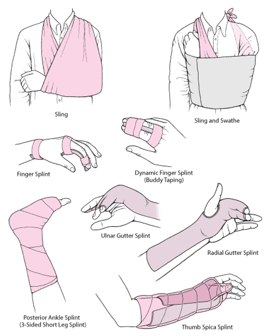
Splints, slings, or swathes are usually used after closed reduction of a dislocation to immobilize the joint. After a successful reduction of a dislocation, a cast is not usually needed.
Immobilization reduces pain and helps with healing by preventing further injury to surrounding tissues. Immobilization is helpful for most moderate or severe dislocations. Joints on both sides of the injury are immobilized.
If immobilization lasts too long (for example, for more than a few weeks in young adults), the joint may become stiff, sometimes permanently, and muscles may shorten (causing contractures) or shrink (waste away, or atrophy). Blood clots may develop. Such problems can develop quickly, and contractures can become permanent, usually in older adults. Consequently, doctors typically encourage movement as soon as possible.
A splint can be used to immobilize some stable dislocations, particularly if they need to be kept immobile for only a few days or less. During initial treatment, splints are also used to immediately immobilize moderate and severe dislocations, particularly very unstable ones, until the person can be evaluated thoroughly. Splints allow people to apply ice and to move more than a cast does.
A splint is a long, narrow slab of plaster, fiberglass, or aluminum applied with elastic wrap or tape. Because the slab does not completely encircle the limb, there is room for some expansion due to swelling. Thus, a splint typically does not increase the risk of developing compartment syndrome. Some injuries that eventually require a cast are first immobilized with a splint until most of the swelling resolves.
A sling by itself can provide some support. Slings can be useful when complete immobilization has undesirable effects. For example, if a shoulder is completely immobilized, the tissues around the joint may become stiff, sometimes within days, preventing the shoulder from moving (called frozen shoulder). Slings limit movement of the shoulder and elbow but allow movement of the hand.
A swathe, which is a piece of cloth or a strap, may be used with a sling to prevent the arm from swinging outward, especially at night. The swathe is wrapped around the back and over the injured part.
Commonly Used Techniques for Immobilizing a Joint

Surgery
Occasionally, dislocations cannot be reduced using closed reduction, and surgery is required to realign the joint in its normal position. Once the joint has been realigned, additional surgery is often not necessary.
Sometimes surgery is required to treat fractures that accompany dislocations, to stabilize the joint, or to remove debris from the joint.
Rehabilitation and Prognosis for Dislocations
Many dislocations and related injuries heal well and result in few problems. However, some do not completely heal even though they are diagnosed and treated appropriately.
How long an injury takes to heal varies from weeks to months depending on:
Type of injury
Location of the injury
The person's age
Other disorders present
For example, children heal much faster than adults, and certain disorders (including those that cause problems with circulation, such as diabetes and peripheral vascular disease) slow healing.
People usually feel some discomfort during activities even after injuries have healed enough to allow them to put their full weight on the injured part. Some people also notice that the injured part is more painful and stiffer when the weather is cold.
Being immobilized makes joints stiff, and muscles weaken and shrink because they are not used. If a limb is immobilized in a cast, the affected joint becomes stiffer each week, and eventually people become unable to fully extend and flex their limb. Such problems can develop quickly and become permanent, usually in older adults.
To prevent or minimize stiffness and to help people maintain muscle strength, doctors or physical therapists may recommend daily exercise, including range-of-motion exercises and muscle-strengthening exercises. While the injury is healing, people can exercise the rest of their body, as instructed by their doctor or physical therapist.
After the injury has healed sufficiently and the joint is no longer immobilized, people can start exercising the injured limb. When exercising, they should pay attention to how the injured limb feels and avoid exercising too forcefully. If the muscles are too weak for people to exercise them, a therapist moves their limbs for them (called passive exercise). However, ultimately, to regain full strength of an injured limb, people must move their own muscles (called active exercise).
Exercises to improve range of motion and muscle strength and to strengthen and stabilize the injured joint can help prevent dislocations from recurring and help prevent long-term problems.
Spotlight on Aging: Dislocations
People who are over 65 are more likely to dislocate a joint, partly because they are more likely to fall—a common cause of dislocations. Older adults are more likely to fall for the following reasons:
Other injuries tend to accompany a dislocation more often in older adults. For example, a shoulder dislocation is more likely to cause a torn rotator cuff in older adults than in younger people. In older adults, recovery is often more complicated and slower than it is in younger people because:
Even minor injuries can greatly interfere with older adults' ability to do normal daily activities, such as eating, dressing, bathing, and even walking, especially if they used a walker before the injury. Immobilization: Being immobilized (for example, when bed rest is necessary) is a particular problem in older adults. In older adults, being immobilized is more likely to cause:
Pressure sores develop when blood flow to an area is cut off or greatly reduced. In older adults, blood flow to a limb may already be reduced. When the weight of an injured limb rests on a cast, blood flow is reduced even more, and pressure sores may form. If bed rest is required, pressure sores can develop on areas of skin that touch the bed. These areas should be diligently inspected for any sign that the skin is breaking down. Because immobilization is more likely to cause problems in older adults, treatment of dislocations and other musculoskeletal injuries focuses on helping older adults return to daily activities as quickly as possible. |
Drug Information for the Topic


