


Bladder cancer is usually transitional cell (urothelial) carcinoma. Patients usually present with hematuria (most commonly) or irritative voiding symptoms such as frequency and/or urgency; later, urinary obstruction can cause pain. Diagnosis is by cystoscopy and biopsy. Treatment is with fulguration, transurethral resection, intravesical instillations, radical surgery, chemotherapy, external beam radiation, or a combination.
In the United States, about 83,190 new cases of bladder cancer and about 16,840 deaths (2024 estimates) occur each year (1). Bladder cancer is the fourth most common cancer among men and is less common among women; male:female incidence is about 3:1. Bladder cancer is more common among White people than Black people, and incidence increases with age.
Risk factors include the following:
Smoking (the most common risk factor, causing ≥ 50% of new cases)
Long-term cyclophosphamide use
Chronic irritation (eg, in schistosomiasis, by chronic catheterization, or by bladder calculi)
Exposure to hydrocarbons, tryptophan metabolites, or industrial chemicals, notably aromatic amines (aniline dyes, such as naphthylamine used in the dye industry) and chemicals used in the rubber, electric, cable, paint, and textile industries
Types of bladder cancer include
Transitional cell carcinomas (urothelial carcinoma), which account for > 90% of bladder cancers. Most are papillary carcinomas, which tend to be superficial and well-differentiated and to grow outward; sessile tumors are more insidious, tending to invade early and metastasize.
Squamous cell carcinomas, which are less common and usually occur in patients with parasitic bladder infestation or chronic mucosal irritation.
Adenocarcinomas, which may occur as primary tumors or rarely reflect metastasis from intestinal carcinoma. Metastasis to the bladder should be ruled out.
In > 40% of patients, tumors recur at the same or another site in the bladder, particularly if tumors are large or poorly differentiated or if several tumors are present. Bladder cancer tends to metastasize to the lymph nodes, lungs, liver, and bone.
In the bladder, carcinoma in situ is high grade but noninvasive and usually multifocal; it tends to recur.
Reference
1. Siegel RL, Giaquinto AN, Jemal A. Cancer statistics, 2024 [published correction appears in CA Cancer J Clin. 2024 Mar-Apr;74(2):203. doi: 10.3322/caac.21830]. CA Cancer J Clin. 2024;74(1):12-49. doi:10.3322/caac.21820
Symptoms and Signs of Bladder Cancer
Most patients present with unexplained hematuria (gross or microscopic). Some patients present with anemia, and hematuria is detected during evaluation. Irritative voiding symptoms (dysuria, burning, frequency) and pyuria are also common at presentation. Pelvic pain occurs with advanced cancer, when a pelvic mass may be palpable.
Diagnosis of Bladder Cancer
Cystoscopy with biopsy
Urine cytology
Bladder cancer is suspected clinically. If patients present with hematuria, workup is risk-stratified and involves a combination of diagnostic cystoscopy and imaging (CT urogram or renal ultrasound [1]). Urine cytology, which can detect malignant cells, should also be done. Cystoscopy and biopsy of abnormal areas or resection of tumors are required for diagnosis and clinical staging. Urinary antigen tests are available but are not routinely recommended for use in diagnosis. They are used sometimes if cancer is suspected but cytology results are negative.
Cystoscopy with blue light after intravesical instillation of hexyl-aminolevulinate can improve initial detection of bladder cancer as well as recurrence-free survival. Higher detection rates are expected to improve clinical outcomes by reducing future recurrences and by facilitating earlier recognition that certain tumors are unresponsive to therapy (thus, sparing some patients unnecessary treatments).
For nonmuscle-invasive bladder cancer (carcinoma in situ, Ta, T1) tumors, which comprise 70 to 80% of bladder cancers, cystoscopy with biopsy (with simultaneous complete resection) is sufficient for staging. However, if biopsy shows the tumor is more invasive than a superficial or flat tumor, then repeat resection should be considered within 4 to 8 weeks, taking care to include muscle tissue. If a tumor is found to invade the detrusor muscle (≥ stage T2), blood tests, abdominal and pelvic CT, and chest x-ray are done to determine tumor extent and evaluate for metastases. MRI can be considered for local staging. Patients with invasive tumors undergo bimanual examination (rectal examination in men, rectovaginal examination in women) while under anesthesia for cystoscopy and biopsy. The standard TNM (tumor, node, metastasis) staging system is used (see table and table ).

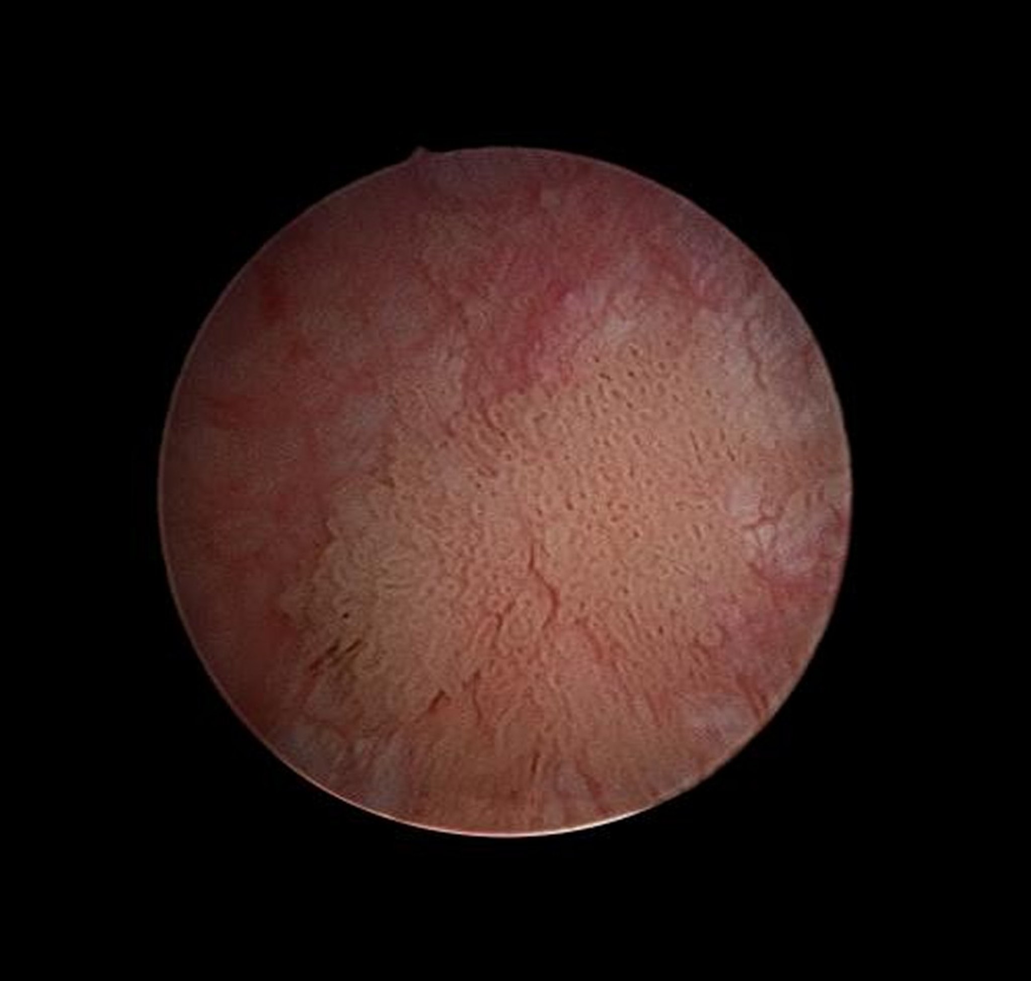
This cystoscopic view inside the bladder of a male with a transitional cell carcinoma shows a tumor (center) in the bladder wall.
DR P. MARAZZI/SCIENCE PHOTO LIBRARY

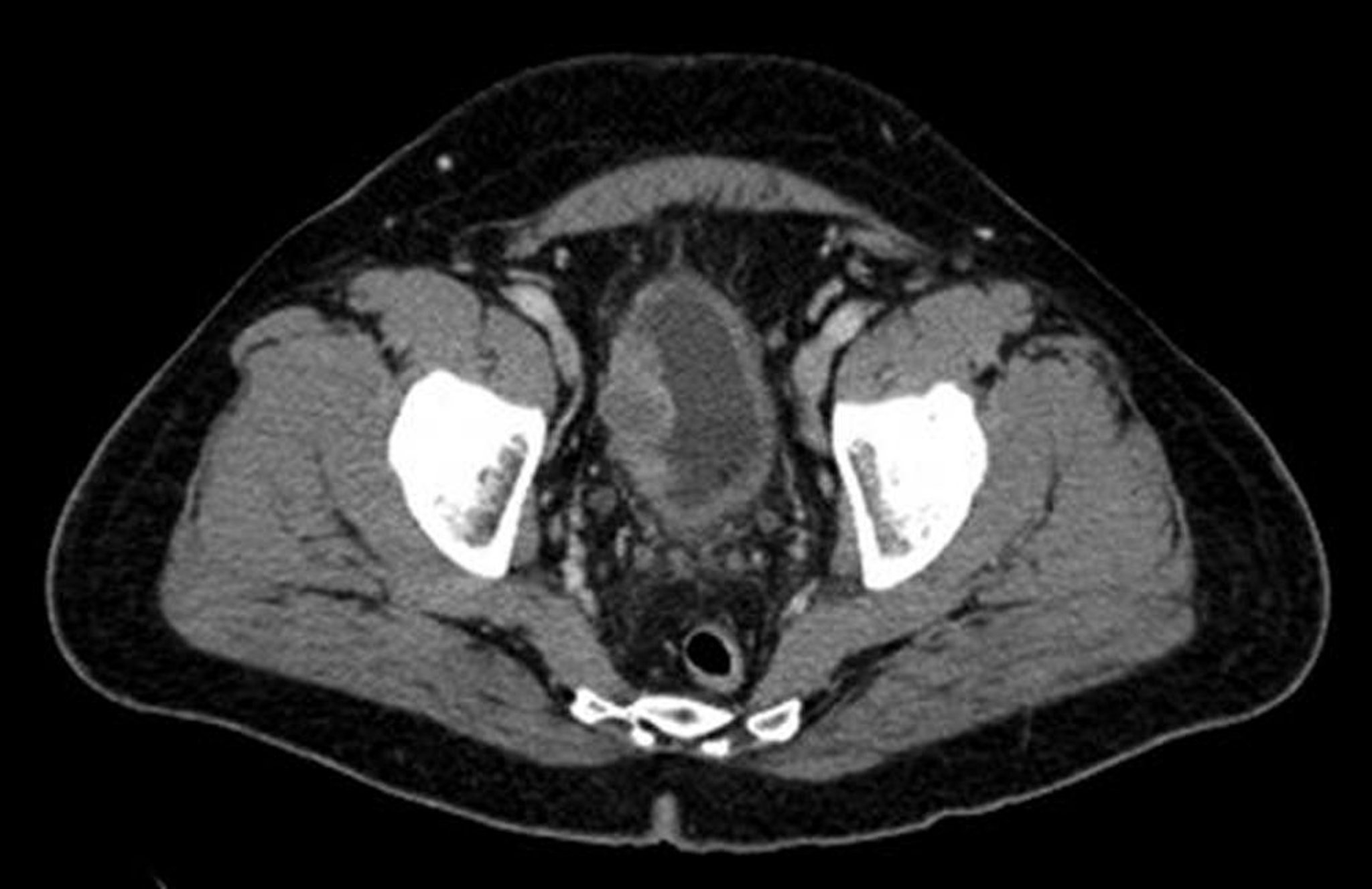
This CT scan shows a right-sided bladder cancer.
© Springer Science+Business Media
AJCC/TNM* Staging of Bladder Cancer
Stage | Tumor | Regional Lymph Node Metastasis | Distant Metastasis |
|---|---|---|---|
0a | Ta | N0 | M0 |
0is | Tis | N0 | M0 |
I | T1 | N0 | M0 |
II | T2a, T2b | N0 | M0 |
IIIA | T3a, T3b, T4a | N0 | M0 |
OR | |||
T1, T2, T3, T4a | N1 | M0 | |
IIIB | T1, T2, T3, T4a | N2, N3 | M0 |
IVA | T4b | Any N | M0 |
OR | |||
Any T | Any N | M1a | |
IVB | Any T | Any N | M1b |
* For AJCC/TNM definitions, see table TNM Definitions for Bladder Cancer. Data adapted from American Cancer Society, Bladder Cancer Stages. Accessed February 3, 2025. | |||
AJCC = American Joint Commission on Cancer; T = primary tumor; N = regional lymph node metastasis; M = distant metastasis. | |||
TNM Definitions for Bladder Cancer*
Feature | Definition |
|---|---|
Primary tumor | |
Ta | Noninvasive papillary carcinoma |
Tis | Flat tumors (carcinoma in situ [CIS]) |
T1 | Invades subepithelial connective tissue |
T2 | Invades muscle |
T2a | Invades superficial muscle (inner half) |
T2b | Invades deep muscle (outer half) |
T3 | Invades perivesical tissue |
T3a | Invades perivesical tissue microscopically |
T3b | Invades perivesical tissue macroscopically (extravesical mass) |
T4 | Invades adjacent organs |
T4a | Invades prostate, seminal vesicles, uterus, or vagina |
T4b | Invades pelvic or abdominal wall |
Regional lymph node metastasis | |
NX | Not assessable |
N0 | No lymph node metastases |
N1 | Single node in true pelvis |
N2 | ≥ 2 nodes in true pelvis |
N3 | ≥ 1 common iliac node |
Distant metastasis | |
M0 | No distant metastases |
M1a | Present in nodes distant to common iliacs |
M1b | Non-nodal metastases |
* Data adapted from American Cancer Society, Bladder Cancer Stages. Accessed February 3, 2025. | |
TNM = tumor, node, metastasis. | |
Diagnosis reference
1. Barocas DA, Boorjian SA, Alvarez RD, et al: AUA/SUFU Guideline. J Urol. 204(4):778-786, 2020. doi: 10.1097/JU.0000000000001297
Treatment of Bladder Cancer
Transurethral resection and intravesical immunotherapy or chemotherapy (for nonmuscle-invasive bladder cancers)
Cystectomy or radiation with chemotherapy (for cancers that invade muscle)
Superficial cancers
Nonmuscle invasive bladder cancers (NMIBC) should be completely removed by transurethral resection or fulguration. Postoperative instillation of chemotherapeutic agents (mitomycin-C and gemcitabine) within 24 hours has been shown to reduce recurrences. Repeated outpatient bladder instillations may also reduce recurrences in low-risk NMIBC. Carcinoma in situ and other high-grade nonmuscle-invasive urothelial carcinomas are treated with Bacille Calmette-Guérin (BCG) intravesical instillations or radical cystectomy (1). BCG instillations can be done at intervals from weekly to monthly over 1 to 3 years. In patients for whom BCG causes symptoms of significant discomfort, such as bladder irritation or dysuria, or in whom the bladder cancers recur or progress, second-line options include intravesical chemotherapy (gemcitabine/docetaxel), intravesical therapy with nadofaragene firadenovec-vncg (a nonreplicating adenoviral vector-based gene therapy), intravesical therapy with nogapendekin alfa inbakicept (an IL-15 agonist) given in conjunction with BCG, intravenous pembrolizumab, early cystectomy, and clinical trial enrollment.
Invasive cancers
Tumors that penetrate the muscle (ie, ≥ stage T2) usually require radical cystectomy (removal of bladder and adjacent structures) with concomitant urinary diversion; partial cystectomy is possible for < 5% of patients. Neoadjuvant chemotherapy with a cisplatin-containing regimen prior to cystectomy is considered standard of care in eligible patients. Lymphadenectomy at the time of surgery is required for staging and potential therapeutic benefit; however, the extent required is debatable.
Urinary diversion following cystectomy traditionally involves routing urine through an ileal conduit to an abdominal stoma and collecting it in an external drainage bag. Alternatives such as orthotopic neobladder or continent cutaneous diversion are becoming common and are appropriate for select patients. For both procedures, an internal reservoir is constructed from the intestine. For the orthotopic neobladder, the reservoir is connected to the urethra. Patients empty the reservoir by relaxing the pelvic floor muscles and increasing abdominal pressure, so that urine passes through the urethra almost naturally. Most patients maintain urinary control during the day, but some incontinence may occur at night. For continent cutaneous urinary diversion, the reservoir is connected to a continent abdominal stoma. Patients empty the reservoir by self-catheterization at regular intervals throughout the day.
Bladder-preservation protocols that combine aggressive transurethral resection, chemotherapy, and radiation therapy may be appropriate for a subset of patients, including those who are older or those who refuse more aggressive surgery. However, select patients with favorable disease characteristics (no hydronephrosis, solitary lesion, completely resected, no carcinoma in situ lesions) may achieve outcomes comparable to cystectomy with bladder-preservation techniques (2). These protocols may provide 5-year survival rates of 36 to 74% with 10 to 20% of patients requiring salvage cystectomy (3, 4).
Following radical cystectomy, patients should be considered for adjuvant cisplatin-based chemotherapy (if they did not receive neoadjuvant therapy) or for adjuvant nivolumab if they have significant residual disease despite neoadjuvant chemotherapy or cannot tolerate cisplatin-based chemotherapy.
Patients should be monitored every 3 to 6 months for progression or recurrence.
Metastatic and recurrent cancers
Metastases require systemic therapy, generally either cisplatin-based chemotherapy (gemcitabine/docetaxel or MVAC [methotrexate, vinblastine sulfate, doxorubicin hydrochloride (Adriamycin), and cisplatin]) or combination immunotherapy/antibody-drug conjugate (enfortumab vedotin/pembrolizumab), which is frequently effective but rarely curative unless metastases are confined to lymph nodes. If treated with chemotherapy upfront, patients can be followed by maintenance immunotherapy with avelumab. Systemic therapy may prolong life in patients with metastatic disease. The first targeted therapy, erdafitinib, is now available for use in patients with FGFR3 and FGFR2 mutations who have failed treatment with chemotherapy.
Treatment of recurrent cancer depends on clinical stage and site of recurrence and previous treatment. Local recurrence after transurethral resection of superficial tumors is usually treated with a second resection or fulguration. Early cystectomy is recommended for recurrent high-grade superficial bladder cancers. Distant recurrences are treated with systemic therapy.
Treatment references
1. Lenis AT, Lec PM, Chamie K, et al: Bladder cancer: A review. JAMA. 324(19):1980-1991, 2020. doi:10.1001/jama.2020.17598
2. Ditonno F, Veccia A, Montanaro F, et al. Trimodal therapy vs radical cystectomy in patients with muscle-invasive bladder cancer: a systematic review and meta-analysis of comparative studies. BJU Int. 2024;134(5):684-695. doi:10.1111/bju.16366
3. Kulkarni GS, Hermanns T, Wei Y, Bhindi B, Satkunasivam R, Athanasopoulos P, Bostrom PJ, Kuk C, Li K, Templeton AJ, Sridhar SS, van der Kwast TH, Chung P, Bristow RG, Milosevic M, Warde P, Fleshner NE, Jewett MAS, Bashir S, Zlotta AR. Propensity Score Analysis of Radical Cystectomy Versus Bladder-Sparing Trimodal Therapy in the Setting of a Multidisciplinary Bladder Cancer Clinic. J Clin Oncol. 2017 Jul 10;35(20):2299-2305. doi: 10.1200/JCO.2016.69.2327. Epub 2017 Apr 14. PMID: 2841001
4. Giacalone NJ, Shipley WU, Clayman RH, et al. Long-term Outcomes After Bladder-preserving Tri-modality Therapy for Patients with Muscle-invasive Bladder Cancer: An Updated Analysis of the Massachusetts General Hospital Experience. Eur Urol. 2017;71(6):952-960. doi:10.1016/j.eururo.2016.12.020
Prognosis for Bladder Cancer
Nonmuscle-invasive bladder cancers (stage Ta, Tis, or T1) have a high rate of local recurrence, but a subset of patients progress to more advanced cancer. Low-grade and stage Ta tumors rarely cause death. High-grade and stage T1 tumors may progress to muscle-invasive bladder cancer. Carcinoma in situ (stage Tis) may be more aggressive than comparable papillary tumors and should be treated as high-grade tumors. For patients with invasion of the bladder musculature, the 5-year survival rate is about 50%, but neoadjuvant chemotherapy improves these results in chemosensitive patients.
Generally, prognosis for patients with progressive or metastatic invasive bladder cancer is poor. Prognosis for patients with squamous cell carcinoma or adenocarcinoma of the bladder is also poor because these cancers are usually highly infiltrative and often detected at an advanced stage.
Key Points
Transitional (urothelial) cell carcinoma is > 90% of bladder cancers.
Suspect bladder cancer in patients with unexplained hematuria or other urinary symptoms (particularly in middle-aged or older men).
Diagnose bladder cancer via cystoscopic biopsy and, if there is muscle invasion, do imaging studies for staging.
Remove superficial cancers by transurethral resection or fulguration, followed by repeated bladder instillations of medications.
If cancer penetrates the muscle, treat with neoadjuvant cisplatin-based chemotherapy followed typically by radical cystectomy with urinary diversion or, less frequently, radiation plus chemotherapy.
Drug Information for the Topic


