


Lymphatic filariasis is infection with any of 3 species of Filarioidea. Acute symptoms include fever, lymphadenitis, lymphangitis, epididymitis, and funiculitis (inflammation of the spermatic cord). Chronic symptoms include abscesses, hyperkeratosis, polyarthritis, hydroceles, lymphedema, and elephantiasis. Tropical pulmonary eosinophilia with bronchospasm, fever, and pulmonary infiltrates is another manifestation of infection. Diagnosis is by detection of microfilariae in blood or lymphatic tissue biopsy specimens, ultrasound visualization of adult worms in lymphatics, or serologic testing. Treatment is with diethylcarbamazine; antibiotics are used for complicating bacterial cellulitis.
(See also Approach to Parasitic Infections and Overview of Filarial Nematode Infections.)
Bancroftian filariasis is present in tropical and subtropical areas of Africa, Asia, the Pacific, and the Americas, including Haiti. Brugian filariasis is endemic in South and Southeast Asia.
About 51 million people were infected as of 2018, and 40 million have been disfigured by the disease. In 2000 the World Health Organization launched its Global Program to Eliminate Lymphatic Filariasis. As a result, substantial progress has been made in stopping the spread of infection through large-scale, annual treatment of eligible people in areas where infection is present. In 2020, more than 860 million people lived in areas where enough infection was present to require such annual treatment.
Lymphatic filariasis is caused by Wuchereria bancrofti (about 90% of cases), Brugia malayi, or B. timori. Transmission is by mosquitoes. Infective larvae from the mosquito migrate to the lymphatics, where they develop into threadlike adult worms within 6 to 12 months. Females are 80 to 100 mm long; males are about 40 mm long. Gravid adult females produce microfilariae that circulate in blood.
Symptoms and Signs
Infection can result in microfilaremia without overt clinical manifestations. Symptoms and signs are caused primarily by adult worms. Microfilaremia gradually disappears after people leave the endemic area.
Acute inflammatory filariasis consists of 4- to 7-day episodes (often recurrent) of fever and inflammation of lymph nodes with lymphangitis (termed acute adenolymphangitis) or acute epididymitis and spermatic cord inflammation; secondary bacterial infections are common. Localized involvement of a limb may cause an abscess that drains externally and leaves a scar. Adenolymphangitis episodes usually precede onset of chronic disease by ≥ 2 decades. Acute filariasis is more severe in previously unexposed immigrants to endemic areas than in native residents.
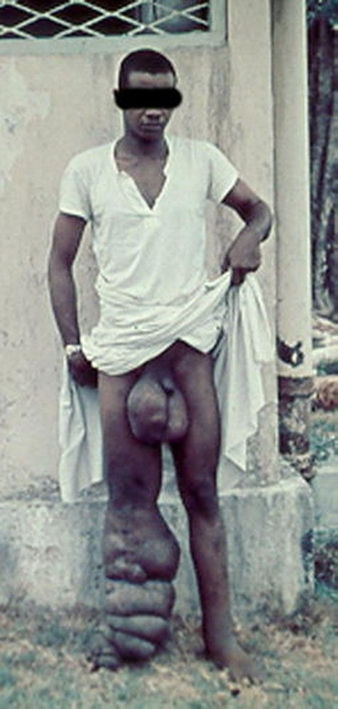
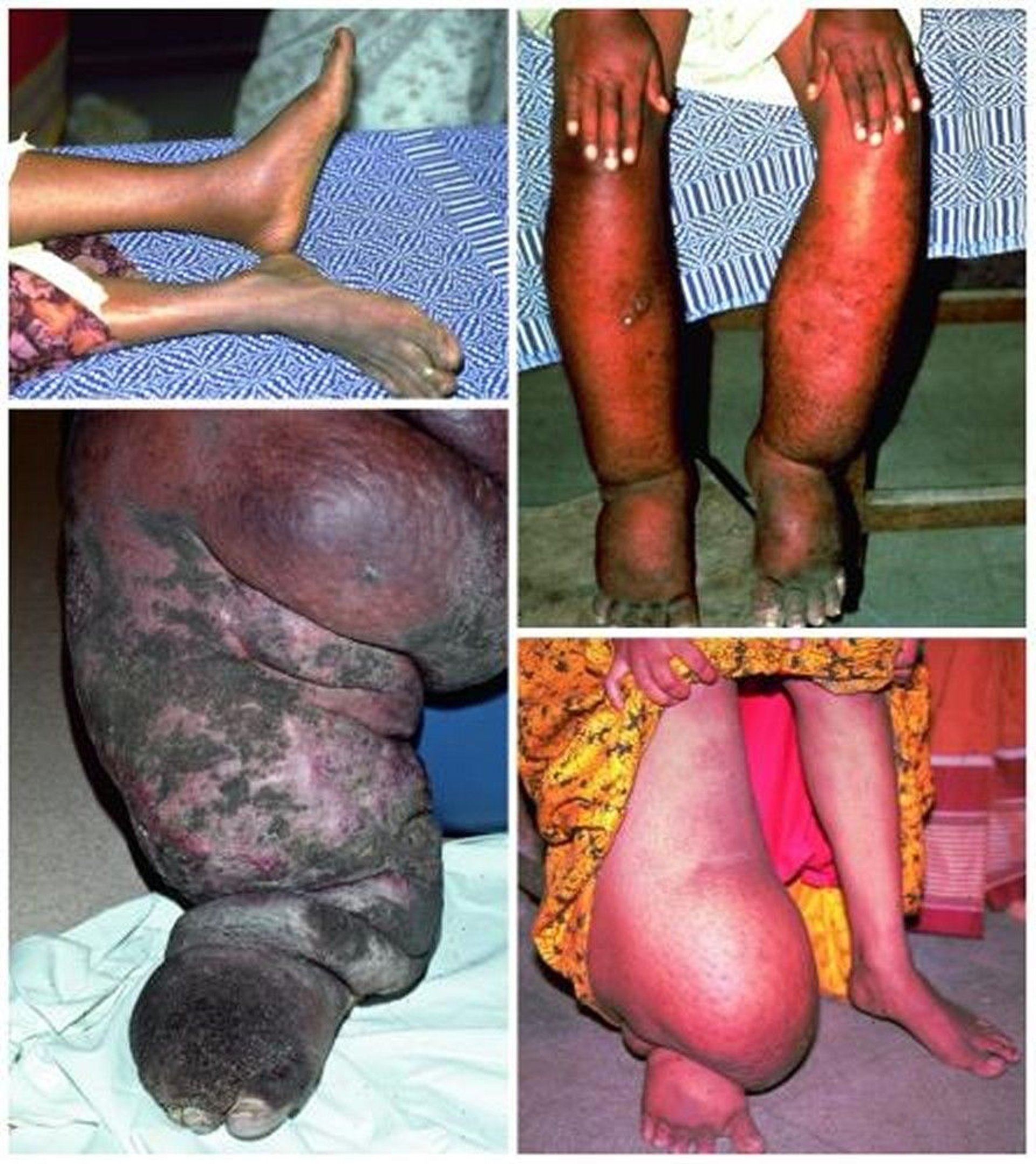
Chronic filarial disease develops insidiously after many years. In most patients, asymptomatic lymphatic dilation occurs, but chronic inflammatory responses to adult worms and secondary bacterial infections may result in chronic lymphedema of the affected body area. Increased local susceptibility to bacterial and fungal infections further contributes to its development. Chronic pitting lymphedema of a lower extremity can progress to elephantiasis (chronic lymphatic obstruction). W. bancrofti can cause hydrocele and scrotal elephantiasis. Other forms of chronic filarial disease are caused by disruption of lymphatic vessels or aberrant drainage of lymph fluid, leading to chyluria and chyloceles.

© Springer Science+Business Media

© Springer Science+Business Media
Extralymphatic signs include chronic microscopic hematuria and proteinuria and mild polyarthritis, all presumed to result from immune complex deposition.
Tropical pulmonary eosinophilia is an uncommon manifestation with recurrent bronchospasm, transitory lung infiltrates, low-grade fever, and marked eosinophilia. It is most likely due to hypersensitivity reactions to microfilariae. Chronic lung involvement can lead to pulmonary fibrosis.
Diagnosis
Microscopic examination of blood samples or lymphatic tissue biopsy
Antigen test for W. bancrofti (available internationally, but not in the United States)
Antibody tests
Microscopic detection of microfilariae in blood establishes the diagnosis of lymphatic filariasis. Filtered or centrifuged concentrates of blood are more sensitive than thick blood films. Blood samples must be obtained when microfilaremia peaks—at night in most endemic areas, but during the day in many Pacific islands. Viable adult worms can be visualized in dilated lymphatics by ultrasonography; their movement has been called the filarial dance.
Several blood tests are available:
Antibody detection: Enzyme immunoassay tests for antifilarial IgG1 and IgG4
Antigen detection: A rapid-format immunochromatographic test for W. bancrofti antigen
Patients with active filarial infection typically have elevated levels of antifilarial IgG4 in the blood. However, there is substantial antigenic cross-reactivity between filariae and other helminths, and a positive serologic test does not distinguish between past and current filarial infection. A rapid diagnostic test for W. bancrofti antigen is used internationally in filariasis elimination programs, but it is not licensed in the United States. Polymerase chain reaction (PCR) assays for W. bancrofti and B. malayi are available in research laboratories. Adults of both species may be identified in biopsy specimens of lymphatic tissue.
Treatment
Diethylcarbamazine
Diethylcarbamazine (DEC) kills microfilariae and a variable proportion of adult worms. In the United States, DEC is available from the Centers for Disease Control and Prevention (CDC) after laboratory confirmation of filariasis.
Pearls & Pitfalls
|
Treatment of acute lymphatic filariasis
DEC 2 mg/kg orally 3 times a day for 12 days has traditionally been used; 6 mg/kg orally once is an alternative. Generally, the 1-day regimen seems to be as effective as the 12-day regimen.
Adverse effects with DEC are usually limited and depend on the number of microfilariae in the blood. The most common are dizziness, nausea, fever, headache, and pain in muscles or joints, which are thought to be related to release of filarial antigens.
Before treatment with DEC, patients should be assessed for coinfection with Loa loa (loiasis) or Onchocerca volvulus (onchocerciasisLoa loa microfilarial levels due to risk of life-threatening side effects including encephalopathy.
A number of drug combinations and regimens have been used in mass treatment programs.
Wolbachia endosymbiont bacteria within filaria, leading to death of adult filarial worms. It can be given with DEC or used alone.
Attacks of acute adenolymphangitis usually resolve spontaneously, although antibiotics may be required to control secondary bacterial infections.
Treatment of chronic lymphedema
Chronic lymphedema requires meticulous skin care, including use of systemic antibiotics to treat secondary bacterial infections; these antibiotics may slow or prevent progression to elephantiasis.
Whether DEC therapy prevents or lessens chronic lymphedema remains controversial.
Conservative measures such as elastic bandaging of the affected limb reduce swelling.
Surgical decompression using nodal-venous shunts to improve lymphatic drainage offers some long-term benefit in extreme cases of elephantiasis. Massive hydroceles can also be managed surgically, but recurrence is common.
Treatment of tropical pulmonary eosinophilia
Tropical pulmonary eosinophilia responds to DEC 2 mg/kg orally 3 times a day for 14 to 21 days, but relapses occur in up to 25% of patients and require additional courses of therapy.
Prevention
The World Health Organization launched the Global Program to Eliminate Lymphatic Filariasisloiasisonchocerciasis
Key Points
Lymphatic filariasis is transmitted by mosquitoes; infective larvae migrate to the lymphatics, where they develop into adult worms.
Adult worms inside the lymphatics can cause inflammation resulting in acute adenolymphangitis or epididymitis or in chronic lymphatic obstruction, which, in some patients, leads to elephantiasis or hydrocele.
Diagnose based on microscopic detection of microfilariae in filtered or centrifuged concentrates of blood that is drawn at the time of day when microfilaremia peaks (varies by species).
Tests for antigen, antibodies, and parasite DNA are alternatives to diagnosis by microscopy.
Treat with diethylcarbamazine after excluding coinfection with Loa loa and Onchocerca volvulus.
The Global Program to Eliminate Lymphatic Filariasis has reduced transmission in many endemic areas.


