

Radial head fractures frequently result from a fall on an outstretched arm and may be difficult to see on radiographs.
(See also Overview of Fractures.)
The radial head is palpated on the lateral elbow as a structure that rotates during pronation and supination and that articulates with the lateral epicondyle. The lateral epicondyle and radial head typically form an isosceles triangle with the olecranon. Joint effusions (common with radial head fractures) may be palpable over this triangle.
Radial head fractures are more common among adults than children.
Symptoms and Signs of Radial Head Fractures
When the radial head is fractured, pain at the radial head is worse during supination, and the radial head is tender. Swelling due to hemarthrosis is usually present. Passive motion of the elbow may be limited. Capitellum fracture may occur simultaneously.
Diagnosis of Radial Head Fractures
Physical examination
Radiographs
Anteroposterior, lateral, and oblique views are taken. But because radiographs may show only indirect evidence of fracture, diagnosis relies heavily on physical examination.

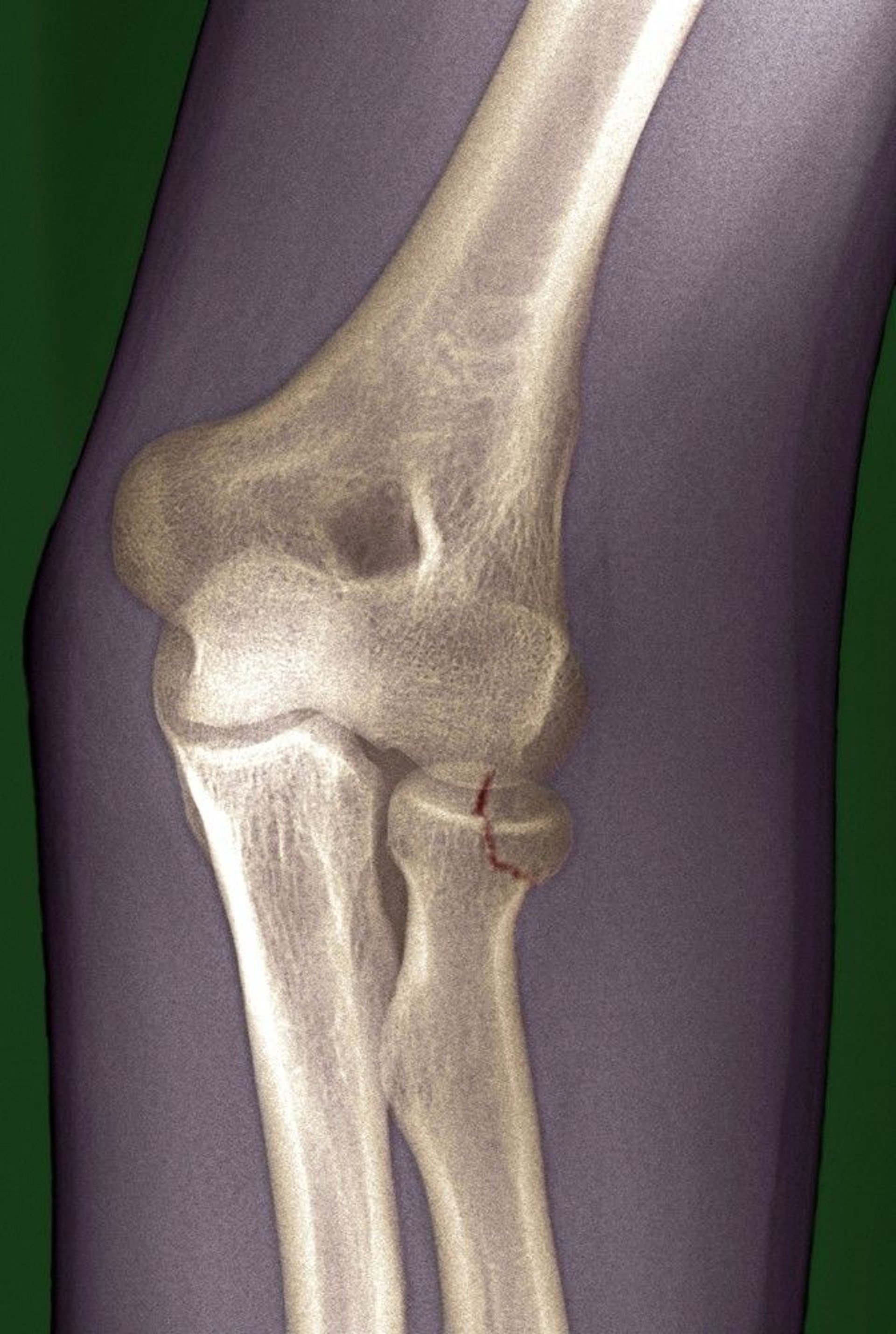
The fracture line (marked in red here) is subtle and could be easily missed.
DU CANE MEDICAL IMAGING LTD/SCIENCE PHOTO LIBRARY

The anterior humeral line and radiocapitellar line are normal. However, an anterior fat pad is present, suggesting a joint effusion.
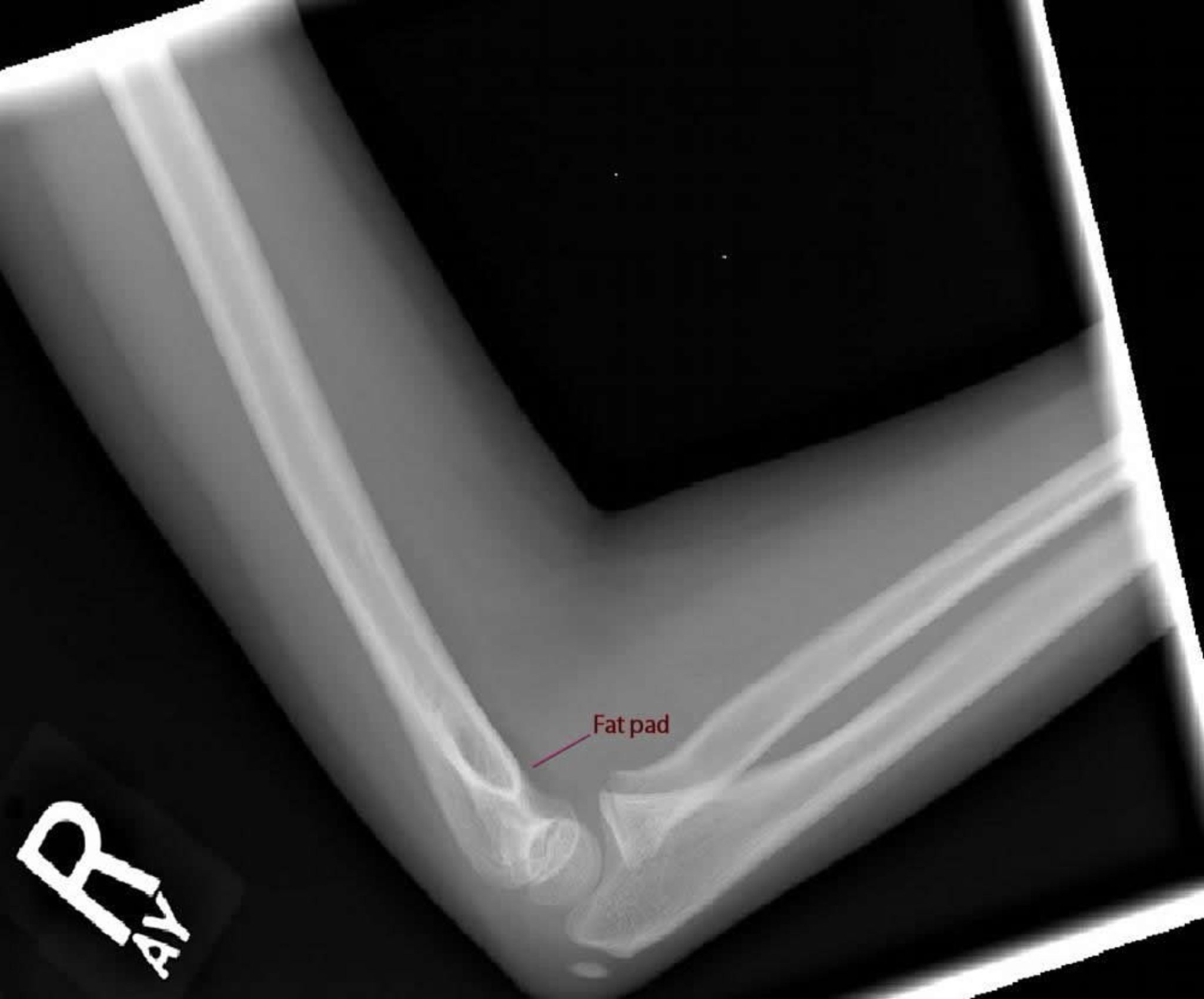
Image courtesy of Danielle Campagne, MD.
Routine anteroposterior and lateral radiographs often do not show the fracture but usually show a joint effusion, which is indicated by the presence of abnormal fat pads on radiographs. Displacement of the anterior fat pad may indicate joint effusion but is not specific; visibility of the posterior fat pad on a true lateral view is specific for joint effusion but not highly sensitive. Patients with localized radial head tenderness and effusion require oblique views (which are more sensitive for fracture) or presumptive treatment of a fracture.
The radiocapitellar line is a line through the midshaft of the radius on a lateral radiograph of the elbow. Normally, this line transects the middle of the capitellum. Sometimes in children, the only sign of fracture on radiographs is displacement of this line.
Pearls & Pitfalls
|
Arthrocentesis may be done to remove blood from the joint to help differentiate mechanical blockage of passive joint motion from restriction due to pain and muscle spasm. Then, a local anesthetic is injected to relieve pain.
Stability is tested by applying stress to the elbow medially and laterally and checking for laxity or increased motion. If the joint does not move when stress is applied, the fracture is stable and associated ligaments are probably uninjured.
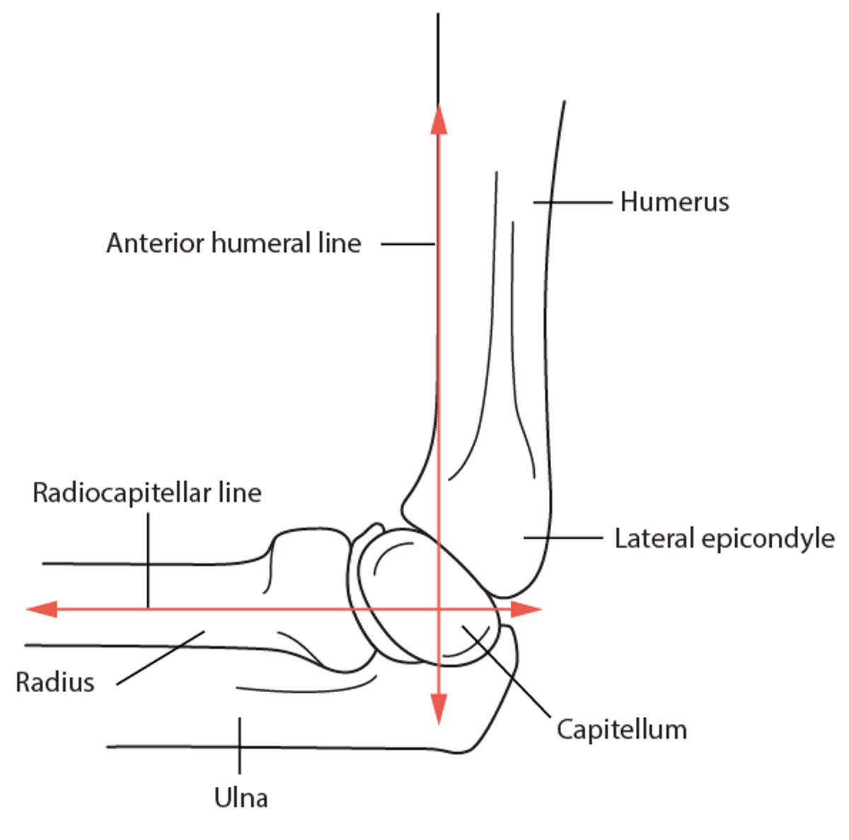
Anterior Humeral Line and Radiocapitellar Line
Normally, the anterior humeral line, which is drawn along the anterior border of the humerus on a lateral radiograph, transects the middle of the capitellum. If the line transects none or only the anterior part of the capitellum, a distal humeral fracture with posterior displacement may be present. The radiocapitellar line, which is drawn through the midshaft of the radius, normally bisects the capitellum. If it does not, an occult fracture should be suspected. |

Treatment of Radial Head Fractures
Usually a sling and range-of-motion exercises
Rarely surgical repair
Fractures with minimal displacement and no restriction of passive elbow motion or instability can be treated with a sling, which can be applied for comfort with the elbow flexed 90°. Elbow range-of-motion exercises should be started as soon as patients can tolerate them.
If the elbow is unstable or motion is mechanically blocked, fractures are treated surgically.
Key Points
The radial head is tender, and pain is worse during supination.
Take anteroposterior, lateral, and oblique radiographs, but because radiographs may show only indirect evidence of fracture, physical examination is very important in diagnosis.
If the radial head is tender and clinical or radiographic evidence suggests an elbow effusion, treat presumptively for radial head fracture even if the radiograph shows no fracture.


