

Most vertebral compression fractures are a consequence of osteoporosis, are asymptomatic or minimally symptomatic, and occur with no or minimal trauma.
(See also Overview of Fractures.)
Vertebral compression fractures due to osteoporosis are common in the thoracic spine (usually below T6) and lumbar spine, particularly near the T12-L1 junction. There may be no preceding trauma or only minimal trauma (eg, a minor fall, sudden bending, lifting, coughing). Patients who have had an osteoporotic vertebral fracture are at higher risk of other vertebral and nonvertebral fractures.
Occasionally, compression or other vertebral fractures result from significant force (eg, a motor vehicle crash, a fall from a height, a gunshot wound). In such cases, a spinal cord injury is often also present, and the spine may be fractured in > 1 place. If the cause was a fall or jump from a height, one or both heels may also be fractured; 10% of all patients with a calcaneal fracture also have a thoracolumbar fracture (because of the axial load to the skeleton when landing on the heels) (1).
Reference
1. Germann CA, Perron AD, Miller MD, Powell SM, Brady WJ. Orthopedic pitfalls in the ED: calcaneal fractures. Am J Emerg Med. 2004;22(7):607-611. doi:10.1016/j.ajem.2004.09.008
Symptoms and Signs of Vertebral Compression Fractures
Osteoporotic vertebral fractures are asymptomatic or cause only loss of height or kyphosis in approximately two thirds of patients. In other patients, pain may develop immediately or later. The pain may radiate into the abdomen. Radicular pain, weakness, and reflex or sphincter abnormalities are uncommon. The pain typically decreases after about 4 weeks and resolves after about 12 weeks.
Nonosteoporotic vertebral compression fractures cause acute pain, bone tenderness at the fracture site, and usually muscle spasm.
Diagnosis of Vertebral Compression Fractures
Radiograph
Osteoporotic fractures are usually diagnosed by radiograph. Findings are usually
Loss of vertebral height (particularly > 6 cm or more than half the height of the vertebral body)
Decreased radiodensity
Loss of trabecular structure
Anterior wedging
Vertebral osteoporotic fractures are commonly diagnosed as incidental findings. If patients do not have risk factors for osteoporosis (eg, older age), these fractures are unlikely.
Solitary fractures above T4 suggest cancer rather than osteoporosis. If patients are not known to have osteoporosis, dual-energy x-ray absorptiometry (DXA) should be done. If osteoporosis is newly diagnosed, patients should be evaluated for causes of secondary osteoporosis.
If significant trauma has occurred, CT is done to evaluate the entire spinal column, and if neurologic deficits or symptoms are present, MRI of the appropriate section of the spinal cord is done.

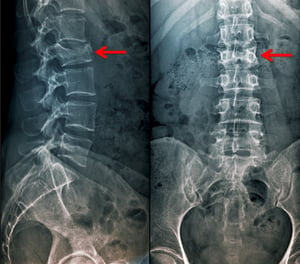
This compression fracture of the 2nd lumbar vertebra is visible as a loss of vertebral body height in the anteroposterior as well as the lateral view.
This compression fracture of the 2nd lumbar vertebra is visible as a loss of vertebral body height in the anteroposteri
Scott Camazine/SCIENCE PHOTO LIBRARY

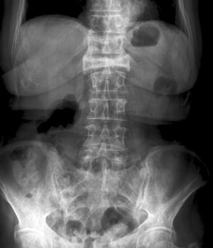
This compression fracture of the 1st lumbar vertebra is visible on the anteroposterior view as a severe loss of height and increased radiodensity.
This compression fracture of the 1st lumbar vertebra is visible on the anteroposterior view as a severe loss of height
ZEPHYR/SCIENCE PHOTO LIBRARY

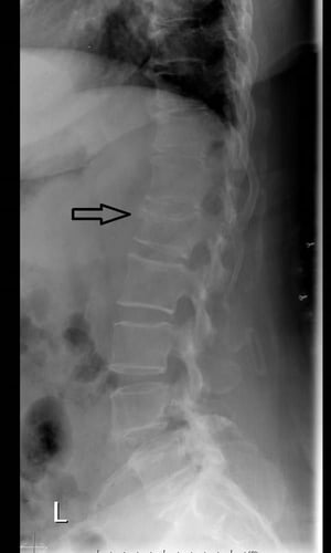
This lateral spine radiograph shows loss of height and anterior wedging due to a vertebral compression fracture.
This lateral spine radiograph shows loss of height and anterior wedging due to a vertebral compression fracture.
Image courtesy of Danielle Campagne, MD.

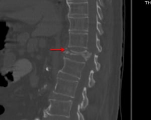
This CT scan (sagittal view) shows marked compression fracture of T12 with mild degenerative disease (eg, anterior vertebral body osteophytes) elsewhere.
This CT scan (sagittal view) shows marked compression fracture of T12 with mild degenerative disease (eg, anterior vert
© Springer Science+Business Media
This compression fracture of the 2nd lumbar vertebra is visible as a loss of vertebral body height in the anteroposterior as well as the lateral view.
This compression fracture of the 2nd lumbar vertebra is visible as a loss of vertebral body height in the anteroposteri
Scott Camazine/SCIENCE PHOTO LIBRARY
This compression fracture of the 1st lumbar vertebra is visible on the anteroposterior view as a severe loss of height and increased radiodensity.
This compression fracture of the 1st lumbar vertebra is visible on the anteroposterior view as a severe loss of height
ZEPHYR/SCIENCE PHOTO LIBRARY
This lateral spine radiograph shows loss of height and anterior wedging due to a vertebral compression fracture.
This lateral spine radiograph shows loss of height and anterior wedging due to a vertebral compression fracture.
Image courtesy of Danielle Campagne, MD.
This CT scan (sagittal view) shows marked compression fracture of T12 with mild degenerative disease (eg, anterior vertebral body osteophytes) elsewhere.
This CT scan (sagittal view) shows marked compression fracture of T12 with mild degenerative disease (eg, anterior vert
© Springer Science+Business Media
If the cause was a fall or jump from a height, clinicians should check for calcaneus fractures and for additional vertebral fractures. If suicidal ideation is suspected or expressed, a psychiatrist should be consulted.
Treatment of Vertebral Compression Fractures
Analgesics
Early mobilization and physical therapy
Treatment of vertebral fractures focuses on pain relief and early mobilization. Analgesics are given. Early resumption of normal activity helps limit further bone loss and disability.
Physical therapists can help by teaching correct lifting techniques and prescribing exercises to strengthen paravertebral muscles, but therapy may need to be delayed until pain is controlled.
Osteoporosis, if present, should be treated (eg, with a bisphosphonate). Calcitonin may also be used and can help relieve pain and increase bone density. , if present, should be treated (eg, with a bisphosphonate). Calcitonin may also be used and can help relieve pain and increase bone density.
Bracing is commonly prescribed, but its efficacy is unclear.
In some cases, vertebroplasty, sometimes preceded by kyphoplasty, can relieve severe pain. In vertebroplasty, methyl methacrylate is injected into the vertebral body. In kyphoplasty, the vertebral body is expanded with a balloon.
These procedures may reduce deformity in the injected vertebrae but do not reduce and may even increase the risk of fractures in adjacent vertebrae. Other risks may include rib fractures, cement leakage, and pulmonary edema or myocardial infarction (MI).
If fractures result from significant trauma, the spine is immobilized immediately, and CT or MRI is done to evaluate the stability of the fractures. Spinal cord injuries, if present, are treated promptly, and supportive care (eg, analgesics, early mobilization) is provided.
Key Points
Most vertebral fractures result from osteoporosis.
Approximately two thirds of vertebral osteoporotic fractures are asymptomatic or cause only loss of height or kyphosis.
Suspect cancer if patients have a solitary fracture above T4.
If patients are not known to have osteoporosis, schedule dual-energy x-ray absorptiometry.
Encourage early mobilization.
Drugs Mentioned In This Article


