Search
SectionsIndexSymptoms
  • Cardiovascular Disorders
  • Clinical Pharmacology
  • Critical Care Medicine
  • Dental Disorders
  • Dermatologic Disorders
  • Ear, Nose, and Throat Disorders
  • Endocrine and Metabolic Disorders
  • Eye Disorders
  • Gastrointestinal Disorders
  • Genitourinary Disorders
  • Geriatrics
  • Gynecology and Obstetrics
  • Hematology and Oncology
  • Hepatic and Biliary Disorders
  • Immunology; Allergic Disorders
  • Infectious Diseases
  • Injuries; Poisoning
  • Musculoskeletal and Connective Tissue Disorders
  • Neurologic Disorders
  • Nutritional Disorders
  • Pediatrics
  • Psychiatric Disorders
  • Pulmonary Disorders
  • Special Subjects
ABCDEFGHI
JKLMNOPQR
STUVWXYZ
  • Abdominal Pain, Acute
  • Abdominal pain, Chronic
  • Alopecia
  • Amenorrhea
  • Amnesia
  • Anosmia
  • Bleeding, Excessive
  • Breast Lumps
  • Chest Pain
  • Constipation in Adults
  • Constipation in Children
  • Cough in Adults
  • Cough in Children
  • Crying
  • Diarrhea in Adults
  • Diarrhea in Children
  • Diplopia
  • Dizziness
  • Dry Mouth
  • Dysmenorrhea
  • Dyspepsia
  • Dysphagia
  • Dyspnea
  • Dysuria
  • Earache
  • Ear Discharge
  • Edema
  • Edema During Late Pregnancy
  • Epistaxis
  • Erectile dysfunction
  • Eyelid Swelling
  • Eye Pain
  • Fever
  • Fever, Acute, in Adults
  • Fever, Chronic (FUO)
  • Fever in Infants and Children
  • Floaters
  • Gas
  • Gastrointestinal Bleeding
  • Halitosis
  • Headache
  • Hearing Loss
  • Hearing Loss: Sudden Deafness
  • Hematospermia
  • Hematuria
  • Hemoptysis
  • Hiccups
  • Hirsutism
  • Insomnia and Excessive Daytime Sleepiness
  • Itching
  • Itching, Anal
  • Jaundice in Adults
  • Jaundice in Neonates
  • Joint Pain, Monarticular
  • Joint Pain, Polyarticular
  • Knee pain
  • Lump in Throat
  • Nasal Congestion and Rhinorrhea
  • Nausea and Vomiting During Early pPregnancy
  • Nausea and Vomiting in Adults
  • Nausea and Vomiting in Infants and Children
  • Neck and Back Pain
  • Neck Mass
  • Nipple Discharge
  • Orthostatis Hypotension
  • Pain
  • Pain, Chronic
  • Palpitations
  • Pelvic Pain
  • Pelvic Pain During Early Pregnancy
  • Polyuria
  • Priapism
  • Red Eye
  • Scrotal Pain
  • Sore Throat
  • Stomatitis
  • Stridor
  • Syncope
  • Tearing
  • Tinnitus
  • Toothache
  • Tremor
  • Urinary Frequency
  • Urinary Incontinence in Adults
  • Urinary Incontinence in Children
  • Urinary Retention
  • Urticaria
  • Vaginal Bleeding
  • Vaginal Bleeding During Early Pregnancy
  • Vaginal Bleeding During Late Pregnancy
  • Vaginal Itching and Discharge
  • Vision, Blurred
  • Vision Loss, Acute
  • Weakness, Generalized
  • Wheezing
In This Topic
Neurologic Disorders
Spinal Cord Disorders
Overview of Spinal Cord Disorders
Anatomy
Symptoms and Signs
Diagnosis
Back to Top
Resources
  • About The Merck Manual
  • Ready Reference Guides
  • Trade Names of Some Commonly Used Drugs
  • Normal Laboratory Values
  • Clinical Calculators
  • Multimedia
  • Selected Links
Manuals available online
'/home/index.html' + bookPageLink
 
'/professional/index.html'
These and other Manuals available
in print, online, and as mobile applications.

See more at MerckManuals.com
Sections in Health Care Professionals
  • Cardiovascular Disorders
  • Clinical Pharmacology
  • Critical Care Medicine
  • Dental Disorders
  • Dermatologic Disorders
  • Ear, Nose, and Throat Disorders
  • Endocrine and Metabolic Disorders
  • Eye Disorders
  • Gastrointestinal Disorders
  • Genitourinary Disorders
  • Geriatrics
  • Gynecology and Obstetrics
  • Hematology and Oncology
  • Hepatic and Biliary Disorders
  • Immunology; Allergic Disorders
  • Infectious Diseases
  • Injuries; Poisoning
  • Musculoskeletal and Connective Tissue Disorders
  • Neurologic Disorders
  • Nutritional Disorders
  • Pediatrics
  • Psychiatric Disorders
  • Pulmonary Disorders
  • Special Subjects
Chapters in Neurologic Disorders
  • Approach to the Neurologic Patient
  • Neurotransmission
  • Autonomic Nervous System
  • Pain
  • Function and Dysfunction of the Cerebral Lobes
  • Stroke (CVA)
  • Coma and Impaired Consciousness
  • Delirium and Dementia
  • Seizure Disorders
  • Sleep and Wakefulness Disorders
  • Headache
  • Brain Infections
  • Prion Diseases
  • Meningitis
  • Neuro-ophthalmologic and Cranial Nerve Disorders
  • Craniocervical Junction Abnormalities
  • Movement and Cerebellar Disorders
  • Demyelinating Disorders
  • Peripheral Nervous System and Motor Unit Disorders
  • Spinal Cord Disorders
  • Intracranial and Spinal Tumors
Topics in Spinal Cord Disorders
  • Overview of Spinal Cord Disorders
  • Acute Transverse Myelitis
  • Spinal Cord Arteriovenous Malformations
  • Cervical Spondylosis and Spondylotic Cervical Myelopathy
  • Hereditary Spastic Paraparesis
  • Spinal Cord Infarction
  • Spinal Cord Compression
  • Spinal Epidural Abscess
  • Spinal Subdural or Epidural Hematoma
  • Syrinx
  • Tropical Spastic Paraparesis/HTLV-1–Associated Myelopathy (TSP/HAM)
 
  • Merck Manual
  • >
  • Health Care Professionals
  • >
  • Neurologic Disorders
  • >
  • Spinal Cord Disorders
  • 4
 
Overview of Spinal Cord Disorders

Share This

Spinal cord disorders can cause permanent severe neurologic disability. For some patients, such disability can be avoided or minimized if evaluation and treatment are rapid. Spinal cord disorders usually result from conditions extrinsic to the cord—eg, compression due to spinal stenosis, herniated disk, tumor, abscess, or hematoma. Less commonly, disorders are intrinsic to the cord. Intrinsic disorders include infarction, hemorrhage, transverse myelitis, HIV infection, poliovirus infection (see Enteroviruses: Poliomyelitis), syphilis (which can cause tabes dorsalis—see Sexually Transmitted Diseases (STDs): Late or tertiary syphilis), trauma (see Spinal Trauma), vitamin B12 deficiency (which causes subacute combined degeneration—see Vitamin Deficiency, Dependency, and Toxicity: Symptoms and Signs), decompression sickness (see Injury During Diving or Work in Compressed Air: Decompression Sickness), lightning injury (which can cause keraunoparalysis—see Electrical and Lightning Injuries: Lightning Injuries), radiation therapy (which can cause myelopathy), syrinx, and spinal cord tumor (see Intracranial and Spinal Tumors: Spinal Cord Tumors). Arteriovenous malformations may be extrinsic or intrinsic. Copper deficiency may result in myelopathy similar to that caused by vitamin B12 deficiency. Spinal nerve roots outside of the spinal cord may also be damaged (see Peripheral Nervous System and Motor Unit Disorders: Nerve Root Disorders).

Anatomy

The spinal cord extends caudally from the medulla at the foramen magnum and terminates at the upper lumbar vertebrae, usually between L1 and L2, where it forms the conus medullaris. In the lumbosacral region, nerve roots from lower cord segments descend within the spinal column in a nearly vertical sheaf, forming the cauda equina.
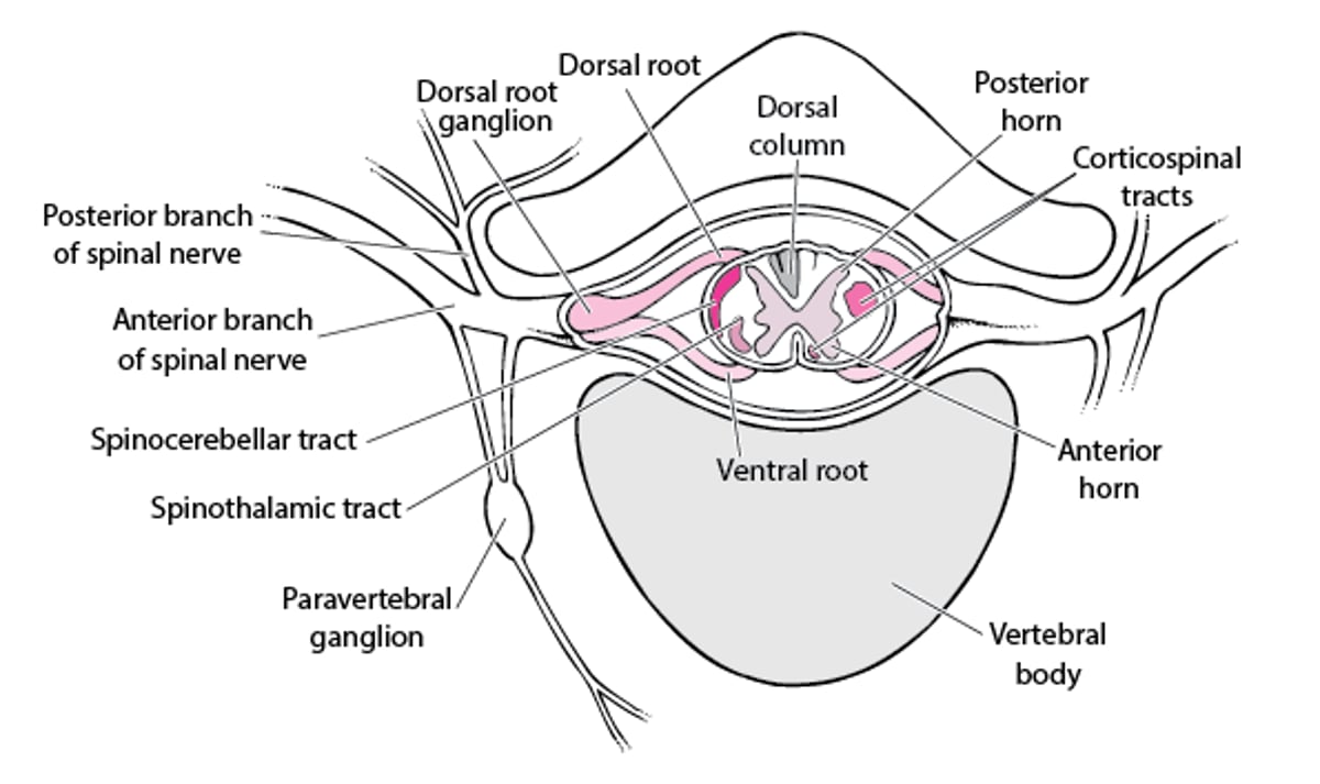
The white matter at the cord's periphery contains ascending and descending tracts of myelinated sensory and motor nerve fibers. The central H-shaped gray matter is composed of cell bodies and nonmyelinated fibers (see Fig. 1: Spinal Cord Disorders: Spinal nerve.Figures). The anterior (ventral) horns of the “H” contain lower motor neurons, which receive impulses from the motor cortex via the descending corticospinal tracts and, at the local level, from internuncial neurons and afferent fibers from muscle spindles. The axons of the lower motor neurons are the efferent fibers of the spinal nerves. The posterior (dorsal) horns contain sensory fibers that originate in cell bodies in the dorsal root ganglia. The gray matter also contains many internuncial neurons that carry motor, sensory, or reflex impulses from dorsal to ventral nerve roots, from one side of the cord to the other, or from one level of the cord to another. The spinothalamic tract transmits pain and temperature sensation contralaterally in the spinal cord; most other tracts transmit information ipsilaterally. The cord is divided into functional segments (levels) corresponding approximately to the attachments of the 31 pairs of spinal nerve roots.

Fig. 1

Spinal nerve.

Symptoms and Signs

Neurologic dysfunction due to spinal cord disorders occurs at the involved spinal cord segment (see Table 1: Spinal Cord Disorders: Effects of Spinal Cord Dysfunction by Segmental LevelTables) and at all segments below it. The exception is the central cord syndrome (see Table 2: Spinal Cord Disorders: Spinal Cord SyndromesTables), which may spare segments below.

Table 1

PrintOpen table Open table in new window
Effects of Spinal Cord Dysfunction by Segmental Level

Location of Lesion*

Possible Effects

At or above C5

Respiratory paralysis

Quadriplegia

Between C5 and C6

Paralysis of legs, wrists, and hands

Weakness of shoulder abduction and elbow flexion

Loss of biceps jerk reflex

Loss of brachioradialis deep tendon reflex

Between C6 and C7

Paralysis of legs, wrists, and hands, but shoulder movement and elbow flexion usually possible

Between C7 and C8

Loss of triceps jerk reflex

Paralysis of legs and hands

At C8 to T1

Horner syndrome (constricted pupil, ptosis, facial anhidrosis)

Paralysis of legs

Between T1 and conus medullaris

Paralysis of legs

*Abbreviations refer to vertebrae; the cord is shorter than the spine, so that moving down the spine, cord segments and vertebral levels are increasingly out of alignment. At all levels of cord injury, deep tendon reflexes are altered (initially decreased and later becoming brisk) below the level of the lesion, bowel and bladder control is lost, and sensation is lost below the level of injury.

Spinal cord disorders cause various patterns of deficits depending on which nerve tracts within the cord or which spinal roots outside the cord are damaged. Disorders affecting spinal nerves, but not directly affecting the cord, cause sensory or motor abnormalities or both only in the areas supplied by the affected spinal nerves.

Table 2

PrintOpen table in new window Open table in new window
Spinal Cord Syndromes

Syndrome

Cause

Symptoms and Signs

Anterior cord syndrome

Lesions disproportionately affecting the anterior spinal cord, commonly due to infarction (eg, caused by occlusion of the anterior spinal artery)

Malfunction of all tracts except the posterior columns, thus sparing position and vibratory sensation

Brown-Séquard syndrome (rare)

Unilateral spinal cord lesions, typically due to penetrating trauma

Ipsilateral paresis

Ipsilateral loss of touch, position, and vibratory sensation

Contralateral loss of pain and temperature sensation*

Central cord syndrome affecting the cervical spinal cord

Lesions affecting the center of the cervical spinal cord, mainly central gray matter (including spinothalamic tracts, which cross), commonly due to trauma, syrinx, or tumors in the central spinal cord

Paresis tending to be more severe in the upper extremities than in the lower extremities and sacral regions

Tendency to lose pain and temperature sensation in a capelike distribution over the upper neck, shoulders, and upper trunk, with light touch, position, and vibratory sensation relatively preserved (dissociated sensory loss)

Conus medullaris syndrome

Lesions around L1

Distal leg paresis

Perianal and perineal loss of sensation (saddle anesthesia)

Erectile dysfunction

Urinary retention, frequency, or incontinence

Fecal incontinence

Hypotonic anal sphincter

Abnormal bulbocavernosus and anal wink reflexes

Transverse myelopathy

Lesions affecting all or most tracts of the spinal cord at ≥ 1 segmental levels

Deficits in all functions mediated by the spinal cord (because all tracts are affected to some degree)

*Occasionally, only part of one side of the spinal cord malfunctions (partial Brown-Séquard syndrome).

Spinal cord dysfunction causes paresis, loss of sensation, reflex changes, and autonomic dysfunction (eg, bowel, bladder, and erectile dysfunction; loss of sweating). Dysfunction may be partial (incomplete). Autonomic and reflex abnormalities are usually the most objective signs of cord dysfunction; sensory abnormalities are the least objective.

Corticospinal tract lesions cause upper motor neuron dysfunction. Acute, severe lesions (eg, infarction, traumatic lesions) cause spinal shock with flaccid paresis (decreased muscle tone, hyporeflexia, and no extensor plantar responses). After days or weeks, upper motor neuron dysfunction evolves into spastic paresis (increased muscle tone, hyperreflexia, and clonus). Extensor plantar responses and autonomic dysfunction are present. Flaccid paresis that lasts more than a few weeks suggests lower motor neuron dysfunction (eg, due to Guillain-Barré syndrome).

Specific cord syndromes include transverse sensorimotor myelopathy, Brown-Séquard syndrome, central cord syndrome, anterior cord syndrome, and conus medullaris syndrome (see Table 2: Spinal Cord Disorders: Spinal Cord SyndromesTables).

Cauda equina syndrome, which involves damage to nerve roots at the caudal end of the cord, is not a spinal cord syndrome. However, it mimics conus medullaris syndrome, causing distal leg paresis and sensory loss in and around the perineum and anus (saddle anesthesia), as well as bladder, bowel, and pudendal dysfunction (eg, urinary retention, urinary frequency, urinary or fecal incontinence, erectile dysfunction, loss of rectal tone, abnormal bulbocavernosus and anal wink reflexes). In cauda equina syndrome (unlike in spinal cord injury), muscle tone and deep tendon reflexes are decreased in the legs.

Diagnosis

  • MRI

Neurologic deficits at segmental levels suggest a spinal cord disorder. Similar deficits, especially if unilateral, may result from nerve root or peripheral nerve disorders, which can usually be differentiated clinically. Level and pattern of spinal cord dysfunction help determine presence and location of a spinal cord lesion but not always type of lesion.

MRI is the most accurate imaging test for spinal cord disorders; MRI shows spinal cord parenchyma, soft-tissue lesions (eg, abscesses, hematomas, tumors, abnormalities involving intervertebral disks), and bone lesions (eg, erosion, severe hypertrophic changes, collapse, fracture, subluxation, tumors). Myelography with a radiopaque dye followed by CT is used less often. It is not as accurate as MRI and is more invasive but may be more readily available. Plain x-rays may help detect bone lesions.

Last full review/revision November 2012 by Michael Rubin, MDCM

Content last modified January 2013

Buy the Book

Mobile Versions

Back to Top

Previous: Overview of Peripheral Nervous System Disorders

Next: Acute Transverse Myelitis

Audio
Figures
Photographs
Sidebars
Tables
Videos

Copyright     © 2010-2013 Merck Sharp & Dohme Corp., a subsidiary of Merck & Co., Inc., Whitehouse Station, N.J., U.S.A.    Privacy    Terms of Use