


Trisomy 18 is a chromosomal disorder caused by an extra chromosome 18 that results in intellectual disability and physical abnormalities.
Trisomy 18 caused by an extra chromosome 18.
Infants are typically small and have many physical abnormalities and problems with internal organs.
Tests can be done before or after birth to confirm the diagnosis.
There is no cure for trisomy 18, but some specific symptoms and complications caused by the syndrome can be treated.
(See also Overview of Chromosome and Gene Disorders.)
Chromosomes are structures within cells that contain DNA and many genes. Genes are segments of deoxyribonucleic acid (DNA) and contain the code for a specific protein that functions in one or more types of cells in the body. Genes contain instructions that determine how the body is supposed to look and function.
An extra chromosome, making 3 of a kind (instead of the usual 2), is called trisomy. Children who have trisomy 18 have a third chromosome 18.
Trisomy 18 occurs in about 4 out of 10,000 pregnancies. The extra chromosome almost always comes from the mother. Mothers who are over age 35 are at increased risk of having a child with trisomy 18. There are more girls than boys with trisomy 18.
Fewer than 10% of affected infants survive beyond the first year of life.
Symptoms of Trisomy 18
In the womb, affected fetuses are not typically very active, and there is often excess amniotic fluid, a small placenta, and restricted growth.
Physical abnormalities
At birth, newborns often have a low birth weight and their muscles and body fat are underdeveloped. Newborns are typically limp and have a weak cry. The mouth and jaw may be small, which gives the newborn's face a pinched appearance.
The hands are clenched in fists, and the index fingers often overlap the middle and ring fingers. The fingernails are underdeveloped. Skinfolds, especially over the back of the neck, are common. The big toes are shortened and frequently bend upward. Clubfeet and rocker-bottom feet are common.
Other visible deformities are common, including a small head, low-set and malformed ears, a narrow pelvis, and a short breastbone (sternum).
Physical abnormalities may be obvious at birth. However, some newborns have abnormalities that are not as obvious.
Common Types of Clubfoot

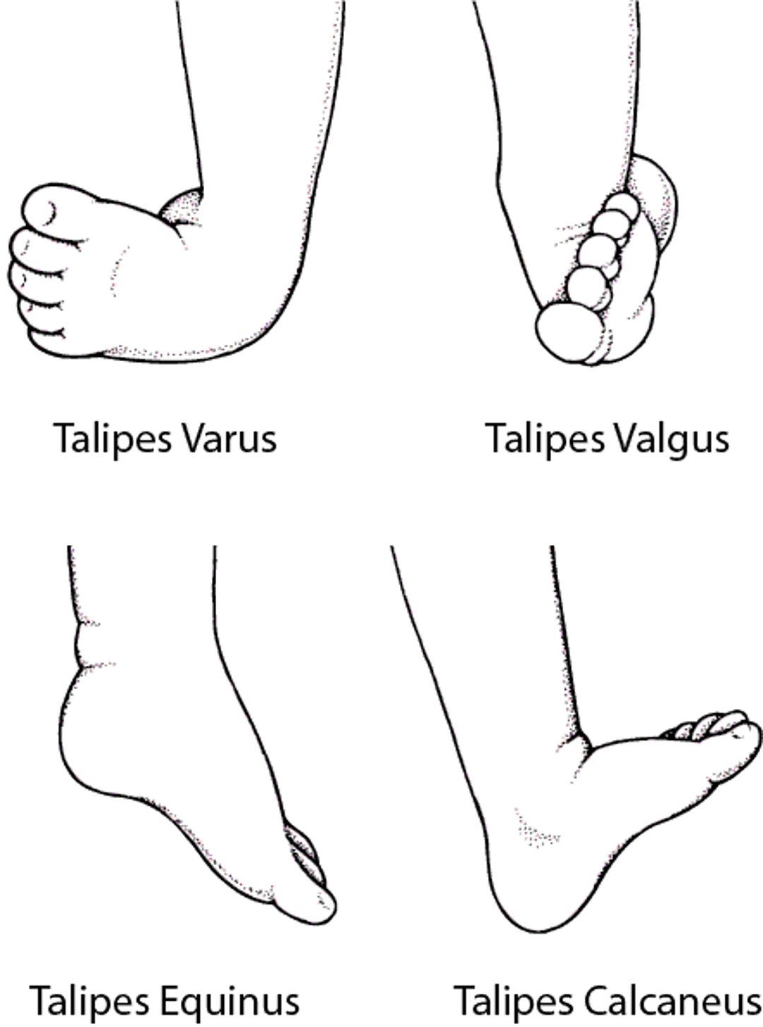

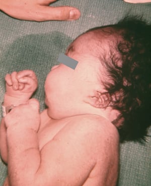
Image courtesy of the Centers for Disease Control and Prevention Public Health Image Library.


Image courtesy of the Centers for Disease Control and Prevention Public Health Image Library.

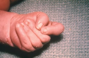
Image courtesy of the Centers for Disease Control and Prevention Public Health Image Library.
Image courtesy of the Centers for Disease Control and Prevention Public Health Image Library.
Image courtesy of the Centers for Disease Control and Prevention Public Health Image Library.
Image courtesy of the Centers for Disease Control and Prevention Public Health Image Library.
Internal abnormalities
Internal organs also have defects. Severe abnormalities may be present in the heart, lungs, digestive tract, and kidneys. Newborns may also have hernias, muscles that have separated from the abdominal wall, or both. Boys may have undescended testes.
Diagnosis of Trisomy 18
Before birth, ultrasound of the fetus or blood tests of the mother
Before birth, chorionic villus sampling, amniocentesis, or both
After birth, the appearance of the infant and blood tests of the infant
(See also Next-generation sequencing technologies.)
Before birth, trisomy 18 may be suspected based on findings detected during an ultrasound of the fetus. Doctors also can do a test to find deoxyribonucleic acid (DNA) from the fetus in the mother's blood and use this DNA to detect an increased risk of trisomy 18. This test is called noninvasive prenatal screening (NIPS) or cell-free fetal DNA analysis.
If doctors suspect trisomy 18 based on the results of these tests, they often confirm the diagnosis by doing chorionic villus sampling to obtain and test a small sample of the placenta, amniocentesis to obtain and test a sample of the fluid that surrounds the fetus (amniotic fluid), or both.

After birth, the infant's physical appearance may suggest the diagnosis of trisomy 18. To confirm the diagnosis, the infant's chromosomes typically are analyzed using a blood test.
Treatment of Trisomy 18
Support for the family
There is no cure for trisomy 18.
Children with trisomy 18 have severe developmental delay and disability. They should receive physical and speech therapies starting early. It is recommended that family members seek support.
Treatment of some of the abnormalities caused by trisomy 18 has resulted in some people living longer. These people are at increased risk of developing certain tumors, such as in the liver (hepatoblastoma) and kidneys (Wilms tumor). To help detect these tumors and other complications, doctors may recommend that children have periodic blood tests and imaging studies such as ultrasound of the abdomen and kidneys.
Prognosis for Trisomy 18
Trisomy 18 is fatal for more than 50% of children within the first week of life, and only about 10% are still alive at 1 year of age. However, people who have trisomy 18 are living longer, and there are adults with trisomy 18.
More Information
The following English-language resources may be useful. Please note that The Manual is not responsible for the content of these resources.


