


A corneal ulcer is an open sore on the cornea (the clear layer in front of the iris and pupil) that is usually caused by an eye infection.
Contact lenses, injuries, disorders, medications, and nutritional deficiencies can also cause open sores (ulcers) to form on the cornea.
Ache, foreign body sensation (the ache and foreign body sensation can be severe), redness, tearing, and light sensitivity are common symptoms.
Doctors diagnose an ulcer based on the appearance of the person's cornea.
Antibiotic, antiviral, or antifungal eye drops are usually given as soon as possible.
(See also Herpes Simplex Keratitis.)
Causes of Corneal Ulcer

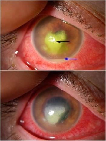
The top image shows an opaque bacterial corneal ulcer (usually gray-white, but yellowish in this photo because it was stained with fluorescein dye—black arrow) and reddening of the conjunctiva. A layer of pus (blue arrow) lies inside the eye at the very bottom of the iris. The bottom image shows improvement after 1 week of antibiotic eye drops.
Corneal ulcers may be caused by infections with bacteria, fungi, viruses, or parasites such as Acanthamoeba (which lives in contaminated water). Ulcers may begin with a corneal injury, such as with severe dry eyes, if a foreign object scratches, penetrates, or lodges in the eye or, more often, if the eye is irritated by a contact lens, especially when contact lenses are worn during sleep or are not adequately disinfected (see Care and complications). Viral corneal ulcers (often due to herpesvirus) can be triggered to recur by physical stress or may recur spontaneously. A deficiency of vitamin A and protein may lead to the formation of a corneal ulcer. However, such ulcers are rare in the United States.
When the eyelids do not close properly, the cornea may become dry and irritated. This kind of irritation can lead to injury and the development of a corneal ulcer. Corneal ulcers may also result from in-growing eyelashes (trichiasis), an in-turned eyelid (entropion), or eyelid inflammation (blepharitis).
Symptoms of Corneal Ulcer
Corneal ulcers cause redness, pain, usually a feeling like a foreign object is in the eye (foreign body sensation), aching, sensitivity to bright light, and increased tear production. The ulcer often appears as a white or dull and grayish spot on the cornea. Sometimes, ulcers develop over the entire cornea and may penetrate deeply. Pus may accumulate behind the cornea, sometimes forming a white layer at the bottom of the cornea (called a hypopyon). The conjunctiva usually is bloodshot. The deeper the ulcer, the more severe the symptoms and complications.


Complications of corneal ulcer
Corneal ulcers may heal with treatment, but they may leave a cloudy scar that impairs vision.
Other complications may include deep-seated infection, perforation of the cornea, displacement of the iris, and destruction of most or all of the tissue in the eye socket.
Diagnosis of Corneal Ulcer
An eye examination
Sometimes cultures
Doctors evaluate the ulcer by using a slit lamp (an instrument that enables doctors to examine the eye under high magnification). To see an ulcer clearly, a doctor may apply eye drops that contain a yellow-green dye called fluorescein. The fluorescein temporarily stains damaged areas of the cornea, making it possible for the doctor to see damaged areas of the cornea that are not otherwise visible.
In some situations, doctors scrape the surface of large ulcers for a sample. The sample is then grown in a laboratory (cultured) to identify the bacterium, fungus, virus, or protozoon causing the infection. Once the organism is identified, doctors choose the best medication to fight the infection.
Treatment of Corneal Ulcer
Antibiotic, antiviral, or antifungal eye drops
Drops that dilate the eye
Sometimes transplantation of the cornea
A corneal ulcer is an emergency that should be treated immediately.
Antibiotic, antiviral, or antifungal eye drops are usually needed immediately and must be given frequently, sometimes every hour around the clock for several days.
Drops that dilate the eye, such as atropine or scopolamine, can help with pain and reduce the chance of complications.
Corneal transplantation (keratoplasty) is sometimes needed.
Drug Information for the Topic


