


Urticaria consists of migratory, well-circumscribed, erythematous, pruritic plaques on the skin.
Urticaria also may be accompanied by angioedema, which results from mast cell and basophil activation in the deeper dermis and subcutaneous tissues and manifests as edema of the face and lips, extremities, or genitals. Angioedema can occur in the bowel and manifest as colicky abdominal pain. Angioedema can be life-threatening if airway obstruction occurs because of laryngeal edema or tongue swelling.
(See also Evaluation of the Dermatologic Patient.)
Pathophysiology of Urticaria
Urticaria results from the release of histamine, bradykinin, kallikrein, and other vasoactive substances from mast cells and basophils in the superficial dermis, resulting in intradermal edema caused by capillary and venous vasodilation and occasionally caused by leukocyte infiltration.
The process can be immune mediated or nonimmune mediated.
Immune-mediated mast cell activation includes
Type I hypersensitivity reactions, in which allergen-bound IgE antibodies bind to high-affinity cell surface receptors on mast cells and basophils
Autoimmune disorders, in which antibodies to an IgE receptor functionally cross-link IgE receptors and cause mast cell degranulation
Nonimmune-mediated mast cell activation includes
Direct nonallergic activation of mast cells by certain medications or substances
Drug-induced cyclooxygenase inhibition that activates mast cells by poorly understood mechanisms
Activation by physical or emotional stimuli; mechanism is poorly understood but possibly involves the release of neuropeptides that interact with mast cells
Etiology of Urticaria
Urticaria is classified as acute (< 6 weeks) or chronic (> 6 weeks); acute cases (70%) are more common than chronic (30%).
Acute urticaria (see table ) most often results from
Type I hypersensitivity reactions
A presumptive trigger (eg, medication, food ingestion, insect bite or sting, infection) occasionally can be identified.
Chronic urticaria most often results from
Idiopathic causes
Autoimmune disorders
Chronic urticaria often lasts months to years, eventually resolving without a cause being found.
Some Causes of Urticaria
Cause | Suggestive Findings | Diagnostic Approach |
|---|---|---|
Acute urticaria | ||
Contact or inhaled allergens (eg, latex, animal saliva, dust, pollen, molds, dander) | Onset within minutes or hours after contact with offending agent | Clinical examination alone Sometimes allergy testing |
Medication/substance effects
| Urticaria within 48 hours of drug exposure Angioedema common with ACE inhibitors | Clinical examination alone Sometimes allergy testing |
Emotional or physical stimuli
| Onset typically within seconds or minutes of offending stimulus | Clinical examination, including reproducible response to suspected stimulus |
Infections | Symptoms of systemic infection* | Testing for specific suspected underlying infection Resolution of urticaria after eradication of the infection |
Ingested allergens (eg, peanuts, tree nuts, fish, shellfish, wheat, eggs, milk, soybeans) | Urticaria within minutes or hours after ingestion of offending agent | Clinical examination Sometimes allergy testing |
Insect bites or stings (Hymenoptera venom) | Urticaria within seconds or minutes after insect bite or sting | Clinical examination alone |
Urticaria with or without fever, polyarthralgias, polyarthritis, lymphadenopathy, proteinuria, edema, and abdominal pain within 7–10 days after parenteral administration of a biologic-based medication or substance | Clinical examination alone | |
Transfusion reactions | Urticaria usually within a few minutes after initiating blood product transfusion (or switching to a new unit of blood product) | Clinical examination alone |
Chronic urticaria | ||
Autoimmune disorders (eg, SLE, Sjögren syndrome, autoimmune thyroid disease, cryoglobulinemia, urticarial vasculitis) | Evidence of systemic autoimmune disease, including hypothyroidism or hyperthyroidism (autoimmune thyroiditis); hepatitis, renal failure, and polyarthritis (cryoglobulinemia); malar rash, serositis, and polyarthritis (SLE); dry eyes and dry mouth (Sjögren syndrome); cutaneous ulcers or hypopigmented lesions after resolution of urticaria (urticarial vasculitis) | Specific to type of autoimmune disease under consideration TSH measurement Thyroid autoantibodies (eg, thyroid peroxidase antibodies, antimicrosomal antibodies) Cryoglobulin titers Serum complement levels (C3, C4, C1q) Rheumatologic serologies (eg, ANA, RF, anti-CCP, anti-SS-A, anti-SS-B, anti-Sm, anti-RNP, anti-Jo-1) Skin biopsy (cryoglobulinemia, urticarial vasculitis) |
Signs of underlying cancer (eg, weight loss, night sweats, abdominal pain, cough, hemoptysis, jaundice, lymphadenopathy, melena) | Specific to the type of suspected underlying cancer | |
Chronic idiopathic urticaria | Occurrence of daily (or almost daily) wheals, and itching for at least 6 weeks, with no obvious cause | Diagnosis of exclusion |
Medications/substance (same as those causing acute urticaria) | Unexplained urticaria in a patient chronically taking prescription, over-the-counter, or herbal drugs | Clinical examination Sometimes allergy testing Resolution with stoppage of offending medication or substance |
Endocrine abnormalities (eg, thyroid dysfunction, elevated progesterone level) | Heat or cold intolerance, bradycardia or tachycardia, hyporeflexia or hyperreflexia Patients taking progesterone-containing oral contraceptives or hormone replacement therapy or those with cyclic urticaria that appears during the second half of the menstrual cycle and resolves with menstruation | Clinical examination Usually TSH measurement |
Physical stimuli (same as those causing acute urticaria) and sometimes emotional exacerbation of urticaria | Urticaria typically within seconds or minutes of offending stimulus | Clinical examination, including reproducible response to suspected stimulus |
Systemic mastocytosis (urticaria pigmentosa) | Presence of small pigmented papules that turn into wheals with mild trauma (eg, gentle stroking) Possible concomitant anemia, abdominal pain, easy flushing, and recurrent headaches | Skin biopsy Serum tryptase level |
* Patients should be asked about recent travel to a developing country. | ||
ANA = antinuclear antibodies; CCP = anticyclic citrullinated peptide; CMV = cytomegalovirus; EBV = Epstein-Barr virus; RF = rheumatoid factor; SLE = systemic lupus erythematosus; TSH = thyroid-stimulating hormone. | ||


Urticarial lesions (wheals or hives) are migratory, elevated, pruritic, reddish plaques caused by local dermal edema.
Urticarial lesions (wheals or hives) are migratory, elevated, pruritic, reddish plaques caused by local dermal edema.
Photo provided by Thomas Habif, MD.

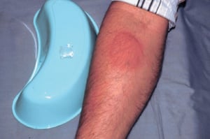
This photo shows a positive ice cube test in a patient with idiopathic cold-induced urticaria. This photo was taken 5 minutes after the ice cube was removed.
This photo shows a positive ice cube test in a patient with idiopathic cold-induced urticaria. This photo was taken 5 m
© Springer Science+Business Media

Urticaria pigmentosa may manifest as erythematous plaque-like lesions on the skin.
Urticaria pigmentosa may manifest as erythematous plaque-like lesions on the skin.
© Springer Science+Business Media

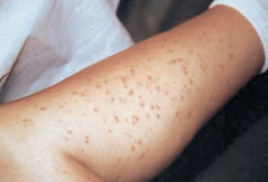
Systemic mastocytosis can cause yellowish tan to reddish brown macules and papules.
Systemic mastocytosis can cause yellowish tan to reddish brown macules and papules.
© Springer Science+Business Media

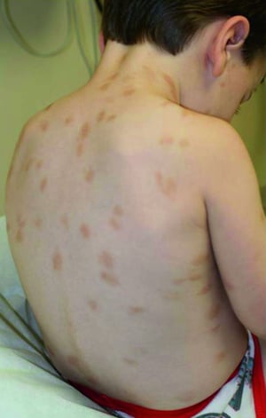
This photo shows reddish brown macules on the back of a school-aged child.
This photo shows reddish brown macules on the back of a school-aged child.
© Springer Science+Business Media

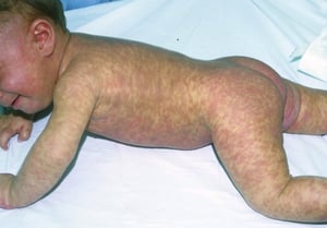
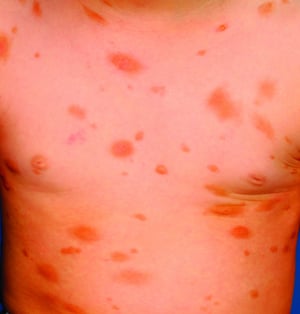
The infant shown here has profuse papulonodular and plaque lesions of urticaria pigmentosa.
The infant shown here has profuse papulonodular and plaque lesions of urticaria pigmentosa.
© Springer Science+Business Media

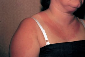
This photo shows solar urticaria in a woman who had been wearing a tank top. These hives appear within minutes of sun exposure.
This photo shows solar urticaria in a woman who had been wearing a tank top. These hives appear within minutes of sun e
© Springer Science+Business Media

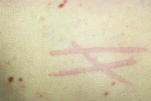
Dermatographism, or skin-writing, may occur when the skin is lightly scratched and results in raised red lines.
Dermatographism, or skin-writing, may occur when the skin is lightly scratched and results in raised red lines.
© Springer Science+Business Media
Urticarial lesions (wheals or hives) are migratory, elevated, pruritic, reddish plaques caused by local dermal edema.
Urticarial lesions (wheals or hives) are migratory, elevated, pruritic, reddish plaques caused by local dermal edema.
Photo provided by Thomas Habif, MD.
This photo shows a positive ice cube test in a patient with idiopathic cold-induced urticaria. This photo was taken 5 minutes after the ice cube was removed.
This photo shows a positive ice cube test in a patient with idiopathic cold-induced urticaria. This photo was taken 5 m
© Springer Science+Business Media
Urticaria pigmentosa may manifest as erythematous plaque-like lesions on the skin.
Urticaria pigmentosa may manifest as erythematous plaque-like lesions on the skin.
© Springer Science+Business Media
Systemic mastocytosis can cause yellowish tan to reddish brown macules and papules.
Systemic mastocytosis can cause yellowish tan to reddish brown macules and papules.
© Springer Science+Business Media
This photo shows reddish brown macules on the back of a school-aged child.
This photo shows reddish brown macules on the back of a school-aged child.
© Springer Science+Business Media
The infant shown here has profuse papulonodular and plaque lesions of urticaria pigmentosa.
The infant shown here has profuse papulonodular and plaque lesions of urticaria pigmentosa.
© Springer Science+Business Media
This photo shows solar urticaria in a woman who had been wearing a tank top. These hives appear within minutes of sun exposure.
This photo shows solar urticaria in a woman who had been wearing a tank top. These hives appear within minutes of sun e
© Springer Science+Business Media
Dermatographism, or skin-writing, may occur when the skin is lightly scratched and results in raised red lines.
Dermatographism, or skin-writing, may occur when the skin is lightly scratched and results in raised red lines.
© Springer Science+Business Media
Evaluation of Urticaria
Because there are no definitive diagnostic tests for urticaria, evaluation largely relies on history and physical examination.
History
History of present illness should include a detailed account of the individual episodes of urticaria, including distribution, size, and appearance of lesions; frequency of occurrence; duration of individual lesions; and any prior episodes. Activities and exposures during, immediately before, and within the past 24 hours of the appearance of urticaria should be noted. Clinicians specifically should ask about recent exercise; exposure to potential allergens (see table ), insects, or animals; new laundry detergent or soaps; new foods; recent infections; or recent stressful life events. The patient should be asked about the duration between any suspected trigger and the appearance of urticaria and which particular triggers are suspected. Important associated symptoms include pruritus, rhinorrhea, swelling of the face and tongue, and dyspnea.
Review of systems should seek symptoms of causative disorders, including fever, fatigue, abdominal pain, and diarrhea (infection); heat or cold intolerance, tremor, or weight change (autoimmune thyroiditis); joint pain (cryoglobulinemia, systemic lupus erythematosus [SLE]); malar rash (SLE); dry eyes and dry mouth (Sjögren syndrome); cutaneous ulcers and hyperpigmented lesions after resolution of urticaria (urticarial vasculitis); small pigmented papules (mastocytosis); lymphadenopathy (viral illness, cancer, serum sickness); acute or chronic diarrhea (viral or parasitic enterocolitis); and fevers, night sweats, or weight loss (cancer).
Past medical history should include a detailed allergy history, including known atopic conditions (eg, allergies, asthma, eczema) and known possible causes (eg, autoimmune disorders, cancer). All medication use should be reviewed, including over-the-counter medications and herbal products, specifically any agents particularly associated with urticaria (see table ). Family history should elicit any history of systemic rheumatic disease, other autoimmune disorders, or cancer. Social history should cover any recent travel and any risk factors for transmission of infectious disease (eg, hepatitis, HIV).
Physical examination
Vital signs should note the presence of bradycardia or tachycardia and tachypnea. General examination should immediately seek any signs of respiratory distress and also note cachexia, jaundice, or agitation.
Examination of the head should note any swelling of the face, lips, or tongue; scleral icterus; malar rash; tender and enlarged thyroid; lymphadenopathy; or dry eyes and dry mouth. The oropharynx should be inspected and the sinuses should be palpated and transilluminated for signs of occult infection (eg, sinus infection, tooth abscess).
Abdominal examination should note any masses, hepatomegaly, splenomegaly, or tenderness. Neurologic examination should note any tremor or hyperreflexia or hyporeflexia. Musculoskeletal examination should note the presence of any inflamed or deformed joints.
Skin examination should note the presence and distribution of urticarial lesions as well as any cutaneous ulceration, hyperpigmentation, small papules, or jaundice. Urticarial lesions usually appear as well-demarcated transient swellings involving the dermis. These swellings are typically erythematous and vary in size from pinprick to covering wide areas. Some lesions can be very large. In other cases, smaller urticarial lesions may become confluent. However, skin lesions also may be absent at the time of the visit. Maneuvers to evoke physical urticaria can be done during the examination, including exposure to vibration (tuning fork), warmth (tuning fork held under warm water), cold (stethoscope, chilled tuning fork, or ice cube), water, or pressure (lightly scratching an unaffected area with a fingernail).
Red flags
The following findings are of particular concern:
Angioedema (swelling of the face, lips, or tongue)
Stridor, wheezing, or other respiratory distress
Hyperpigmented lesions, ulcers, or urticaria that persist > 48 hours
Signs of systemic illness (eg, fever, lymphadenopathy, jaundice, cachexia)
Interpretation of findings
Acute urticaria is nearly always due to some defined exposure to a medication, substance, or physical stimulus or an acute infectious illness. However, the trigger is not always clear from the history, particularly because allergy may develop without warning to a previously tolerated substance.
Most chronic urticaria is idiopathic. The next most common cause is an autoimmune disorder. The causative autoimmune disease is sometimes clinically apparent. Urticarial vasculitis sometimes is associated with systemic rheumatic diseases (particularly SLE or Sjögren syndrome). In urticarial vasculitis, urticaria is accompanied by findings of leukocytoclastic vasculitis on histopathology; it should be considered when the urticaria is painful rather than pruritic, lasts > 48 hours, does not blanch, or is accompanied by vesicles or purpura.
Testing
Usually, no testing is needed for an isolated episode of urticaria unless symptoms and signs suggest a specific disorder (eg, infection).
Unusual, recurrent, or persistent cases warrant further evaluation. Referral for allergy skin testing should be done, and routine laboratory tests should consist of complete blood count, blood chemistries, liver tests, and thyroid-stimulating hormone (TSH). Further testing should be guided by symptoms and signs (eg, of autoimmune disorders) and any abnormalities on the screening tests (eg, hepatitis serologies and ultrasonography for abnormal liver tests; ova and parasites for eosinophilia; cryoglobulin titer for elevated liver tests or elevated creatinine; thyroid autoantibodies for abnormal TSH).
Skin biopsy should be done if there is any uncertainty as to the diagnosis or if wheals persist > 48 hours (to rule out urticarial vasculitis).
Clinicians should be cautious when recommending the patient do an empiric challenge (eg, “Try such and such again and see whether you get a reaction”) because subsequent reactions may be more severe.
Treatment of Urticaria
Any identified causes are treated or remedied. Implicated medications or foods should be stopped.
Nonspecific symptomatic treatment (eg, taking cool baths, avoiding hot water and scratching, wearing loose clothing) may be helpful.
Systemic treatment
Antihistamines remain the mainstay of treatment. They must be taken on a regular basis, rather than as needed. Newer oral antihistamines often are preferred because of once-daily dosing and because some are less sedating. Appropriate choices include
Cetirizine 10 mg once/day
Fexofenadine 180 mg once/day
Desloratadine 5 mg once/day
Levocetirizine 5 mg once/day
Older oral antihistamines (eg, hydroxyzine 10 to 25 mg every 4 to 6 hours; diphenhydramine 25 to 50 mg every 6 hours) are sedating but inexpensive and sometimes quite effective.
Systemic corticosteroids (eg, prednisone 30 to 40 mg orally once/day) are given for severe symptoms but should not be used long term. Topical corticosteroids or topical antihistamines are not beneficial.
Patients with chronic idiopathic urticaria often do not respond to antihistamines or other medications commonly used. Omalizumab, a monoclonal antibody that targets circulating immunoglobulin E (IgE), can suppress certain allergic reactions and may help relieve symptoms (1). Dupilumab, a monoclonal antibody that targets interleukins (IL)-4 and IL-13, is another agent that may also be helpful in reducing itch and severity of urticaria in patients aged 12 years and older who remain symptomatic despite antihistamine treatment (2).
Angioedema
Patients who have angioedema involving the oropharynx or any involvement of the airway should receive subcutaneous epinephrine 0.3 mL of 1:1000 solution and be admitted to the hospital. On discharge, patients should be supplied with and trained in the use of an auto-injectable epinephrine pen.
Treatment references
1. Zhao ZT, Ji CM, Yu WJ, et al: Omalizumab for the treatment of chronic spontaneous urticaria: A meta-analysis of randomized clinical trials. J Allergy Clin Immunol 137(6):1742-1750.e4, 2016. doi: 10.1016/j.jaci.2015.12.1342
2. Maurer M, Casale TB, Saini SS, et al: Dupilumab in patients with chronic spontaneous urticaria (LIBERTY-CSU CUPID): Two randomized, double-blind, placebo-controlled, phase 3 trials. J Allergy Clin Immunol 154(1):184-194, 2024. doi:10.1016/j.jaci.2024.01.028
Geriatrics Essentials: Urticaria
The older oral antihistamines (eg, hydroxyzine, diphenhydramine) are sedating and can cause confusion, urinary retention, and delirium. They should be used cautiously to treat urticaria in older adults.
Key Points
Urticaria can be caused by allergic or nonallergic mechanisms.
Most acute cases are caused by an allergic reaction to a specific substance.
Most chronic cases are idiopathic or result from autoimmune disease.
Concomitant systemic symptoms require a thorough evaluation for the etiology.
Treatment is based on severity; nonsedating antihistamines and avoidance of triggers are first-line options.
Topical corticosteroids and topical antihistamines are not beneficial.
Drug Information for the Topic


