


Linear immunoglobulin A (IgA) disease is an uncommon bullous disease distinguished from bullous pemphigoid and dermatitis herpetiformis by linear deposits of IgA in the basement membrane zone. Diagnosis is by skin biopsy and direct immunofluorescence. Treatment is with topical corticosteroids.
Bullae are elevated, fluid-filled blisters ≥ 10 mm in diameter.
Linear IgA disease has 2 main clinical variants—bullous disease of childhood and adult linear IgA disease. Although they vary clinically in minor ways, their immunofluorescence patterns are identical. The IgA autoantibodies target several antigens within the dermal–epidermal junction.
Infections and penicillins trigger many childhood and adult cases (1). Vancomycin, diclofenac, nonsteroidal anti-inflammatory drugs (NSAIDs), captopril, and lithium also have been suggested as causes. Risk of linear IgA disease is increased in patients who have inflammatory bowel disease (possibly with a related pathophysiology that involves a generation of autoantibodies) or lymphoproliferative cancers (in adults) but not other autoimmune disorders.
General reference
1. Khan M, Park L, Skopit S: Management options for linear immunoglobulin A (IgA) bullous dermatosis: A literature review. Cureus 15(3):e36481, 2023. doi: 10.7759/cureus.36481
Symptoms and Signs of Linear IgA Disease
In linear IgA disease, vesicular or bullous skin lesions occur frequently in a clustered (herpetiform) arrangement. In younger children, the face and perineum are often involved, and spread to the limbs, trunk, hands, feet, and scalp is common. In adults, the trunk is almost always involved, and the scalp, face, and limbs are often involved. Lesions are often pruritic and may burn. Mucosal involvement is common in both age groups; milia (superficial epidermal inclusion cysts) are not characteristic.

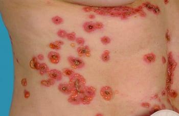
This photo shows a vesicular rash on the trunk of an adult with IgA disease.
Diagnosis of Linear IgA Disease
Skin biopsy and direct immunofluorescence
Diagnosis of linear immunoglobulin A disease is by skin biopsy and direct immunofluorescence. The histologic features are not specific, but direct immunofluorescence shows IgA deposited along the basement membrane zone in a linear fashion.
Treatment of Linear IgA Disease
Withdrawal of causative medications
For mild disease, topical corticosteroids
For children, penicillin or erythromycin
Drug-induced disease may be treated solely with withdrawal of the causative medication.
Mild disease can be treated with topical corticosteroids. Oral penicillin (or erythromycin) can be used in children (1). Dapsone and sulfonamides (using doses and precautions similar to those for dermatitis herpetiformis) and colchicine are alternatives for all ages. Often the cutaneous lesions respond before the mucosal lesions. Spontaneous remission occurs in most patients after 3 to 6 years.
Treatment reference
1. Khan M, Park L, Skopit S: Management options for linear immunoglobulin A (IgA) bullous dermatosis: A literature review. Cureus 15(3):e36481, 2023. doi: 10.7759/cureus.36481
Key Points
Linear immunoglobulin A (IgA) disease is an uncommon bullous disease with manifestations similar to those of bullous pemphigoid and dermatitis herpetiformis.
The trigger is often an infection or medication.
Skin biopsy and direct immunofluorescence can help confirm the diagnosis.
Treat by withdrawing any causative medication and sometimes using topical corticosteroids and, in children, erythromycin.
Drug Information for the Topic


