


Tinea pedis is a dermatophyte infection of the feet. Diagnosis is based on clinical appearance and sometimes by potassium hydroxide (KOH) wet mount, particularly if the infection manifests as hyperkeratotic, ulcerative, or vesiculobullous or is not interdigital. Treatment is with topical antifungals, occasionally oral antifungals, moisture reduction, and drying agents.
Tinea pedis is the most common dermatophytosis because moisture resulting from foot sweating facilitates fungal growth. It affects up to 25% of adults (1) worldwide, and has a lower, but still significant prevalence in children (2).
Tinea pedis may occur as any of 4 clinical forms or in combination:
Chronic hyperkeratotic
Chronic intertriginous
Acute ulcerative
Vesiculobullous
Chronic hyperkeratotic tinea pedis due to Trichophyton rubrum causes a distinctive pattern of lesion, manifesting clinically as scaling and thickening of the soles, which often extends beyond the plantar surface in a moccasin distribution.
Patients who are not responding as expected to antifungal therapy may have another less common cause of plantar rash. Differential diagnosis is sterile maceration (due to hyperhidrosis and occlusive footgear), contact dermatitis (due to type IV delayed hypersensitivity to various materials in shoes, particularly adhesive cement, thiuram compounds in footwear that contains rubber, and chromate tanning agents used in leather footwear), irritant contact dermatitis, and psoriasis.
Dermatophytid (id) reactions may occur as an immune response to the fungal infection, causing a rash elsewhere on the body, often on the hands or fingers. This secondary rash can be pruritic and may present as vesicles, papules, or eczematous patches.

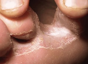
In this photo, macerated, scaling lesions are visible between the 3rd and 4th interdigital spaces and extend to the subdigital skin of the foot.
In this photo, macerated, scaling lesions are visible between the 3rd and 4th interdigital spaces and extend to the sub
Image provided by Thomas Habif, MD.

In this photo, scaling and maceration are visible in the 4th interdigital space.
In this photo, scaling and maceration are visible in the 4th interdigital space.
© Springer Science+Business Media

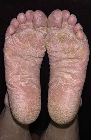
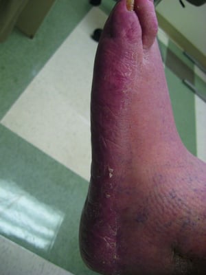
In this photo, chronic hyperkeratotic tinea pedis manifests as scaling and thickening of the soles that extends beyond the plantar surface in a moccasin distribution.
In this photo, chronic hyperkeratotic tinea pedis manifests as scaling and thickening of the soles that extends beyond
Image provided by Thomas Habif, MD.

© Springer Science+Business Media
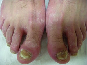
In this photo, onychomycosis is also visible.
In this photo, onychomycosis is also visible.
© Springer Science+Business Media

© Springer Science+Business Media

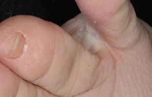
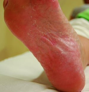
In this photo, scaling is visible in the interdigital space on close inspection.
In this photo, scaling is visible in the interdigital space on close inspection.
© Springer Science+Business Media
In this photo, macerated, scaling lesions are visible between the 3rd and 4th interdigital spaces and extend to the subdigital skin of the foot.
In this photo, macerated, scaling lesions are visible between the 3rd and 4th interdigital spaces and extend to the sub
Image provided by Thomas Habif, MD.
In this photo, scaling and maceration are visible in the 4th interdigital space.
In this photo, scaling and maceration are visible in the 4th interdigital space.
© Springer Science+Business Media
In this photo, chronic hyperkeratotic tinea pedis manifests as scaling and thickening of the soles that extends beyond the plantar surface in a moccasin distribution.
In this photo, chronic hyperkeratotic tinea pedis manifests as scaling and thickening of the soles that extends beyond
Image provided by Thomas Habif, MD.
© Springer Science+Business Media
In this photo, onychomycosis is also visible.
In this photo, onychomycosis is also visible.
© Springer Science+Business Media
© Springer Science+Business Media
In this photo, scaling is visible in the interdigital space on close inspection.
In this photo, scaling is visible in the interdigital space on close inspection.
© Springer Science+Business Media
Chronic intertriginous tinea pedis is characterized by scaling, erythema, and erosion of the interdigital and subdigital skin of the feet, most commonly affecting the lateral 3 toes.
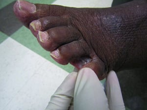
Acute ulcerative tinea pedis (most often caused by Trichophyton mentagrophytes var. interdigitale) typically begins in the 3rd and 4th interdigital spaces and extends to the lateral dorsum and/or the plantar surface of the arch. These toe web lesions are usually macerated and have scaling borders.
Secondary bacterial infection, cellulitis, and lymphangitis are common complications.

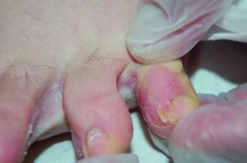
In this photo, maceration and scaling are visible in the 4th interdigital space. Erythema and edema on the 5th toe suggest secondary bacterial infection.
Vesiculobullous tinea pedis, in which vesicles develop on the soles and coalesce into bullae, is the less common result of an exacerbation of interdigital tinea pedis. Risk factors include occlusive shoes and excessive environmental heat and humidity.
General references
1. Gupta AK, Wang T, Lincoln SA, Bakotic WL. Interdigital and Plantar Foot Infections: A Retrospective Analysis of Molecularly Diagnosed Specimens in the United States and a Literature Review. Microorganisms. 2025;13(1):184. Published 2025 Jan 16. doi:10.3390/microorganisms13010184
2. Stenderup JEB, Goandal NF, Saunte DML. Systematic Review of the Prevalence of Tinea Pedis in Children. Pediatr Dermatol. 2025;42(3):539-551. doi:10.1111/pde.15947
Diagnosis of Tinea Pedis
Primarily physical examination
Potassium hydroxide (KOH) wet mount
Diagnosis of tinea pedis is usually evident based on clinical examination and review of risk factors.
If the appearance is not diagnostic or if the infection manifests as hyperkeratotic, ulcerative, or vesiculobullous, a potassium hydroxide wet mount is helpful.
The differential diagnosis of tinea pedis includes:
Hand and foot dermatitis (dyshidrotic dermatitis)
Palmoplantar psoriasis (see table )
Treatment of Tinea Pedis
Topical and occasionally oral antifungals
Moisture reduction and drying agents
The safest tinea pedis treatment is topical antifungals, but recurrence is common and treatment must often be prolonged. (See table .)
A more durable response may be achieved by oral medications including itraconazole and terbinafine (1). Concomitant topical antifungal use may further reduce recurrence.
Moisture reduction on the feet and in footwear is necessary for preventing recurrence. Permeable or open-toe footwear and sock changes are important especially during warm weather. Interdigital spaces should be manually dried after bathing. Drying agents are also recommended; options include antifungal powders (eg, miconazole), gentian violet, Burow solution (5% aluminum acetate) soaks, and 20 to 25% aluminum chloride solution.
Treatment reference
1. Bell-Syer SE, Khan SM, Torgerson DJ. Oral treatments for fungal infections of the skin of the foot. Cochrane Database Syst Rev. 2012;10(10):CD003584. Published 2012 Oct 17. doi:10.1002/14651858.CD003584.pub2
Key Points
Tinea pedis is the most common dermatophytosis because moisture resulting from foot sweating facilitates fungal growth.
Consider the diagnosis if patients have lesions of the toes and/or feet that are intertriginous, ulcerative, hyperkeratotic, or vesicobullous.
Also consider hand and feet dermatitis (dyshidrotic dermatitis), palmoplantar psoriasis, and allergic contact dermatitis.
Treat using topical and occasionally oral antifungals as well as drying measures and agents.
Drug Information for the Topic


