

Lichen planus is a recurrent, pruritic, inflammatory eruption characterized by small, discrete, polygonal, flat-topped, violaceous papules that may coalesce into rough scaly plaques, often accompanied by oral and/or genital lesions. Diagnosis is usually clinical and supported by skin biopsy. Treatment generally requires topical or intralesional glucocorticoids. Severe cases may require phototherapy or systemic glucocorticoids, retinoids, or immunosuppressants.
Lichen planus (LP) is a chronic, immune-mediated inflammatory disease affecting the skin, mucous membranes (especially oral and/or genital), scalp, and nails. Oral LP is the most common subtype. It is more prevalent in women, with prevalence increases progressively after age 40 (1).
General reference
1. González-Moles MÁ, Warnakulasuriya S, González-Ruiz I, et al. Worldwide prevalence of oral lichen planus: A systematic review and meta-analysis. Oral Dis. 2021;27(4):813-828. doi:10.1111/odi.13323
Etiology of Lichen Planus
Lichen planus (LP) is thought to be caused by a T cell–mediated autoimmune reaction against basal epithelial keratinocytes in people with genetic predisposition (1). Medications (especially beta-blockers, nonsteroidal anti-inflammatory drugs [NSAIDs], angiotensin-converting enzyme inhibitors, sulfonylureas, gold, antimalarials, penicillamine, and thiazides) can cause LP; drug-induced LP (sometimes called lichenoid drug eruption) may be indistinguishable from nondrug-induced LP or may have a pattern that is more eczematous. ). Medications (especially beta-blockers, nonsteroidal anti-inflammatory drugs [NSAIDs], angiotensin-converting enzyme inhibitors, sulfonylureas, gold, antimalarials, penicillamine, and thiazides) can cause LP; drug-induced LP (sometimes called lichenoid drug eruption) may be indistinguishable from nondrug-induced LP or may have a pattern that is more eczematous.
Associations of oral LP with hepatitis (hepatitis B infection, hepatitis B vaccine, and, particularly, hepatitis C–induced liver insufficiency) and primary biliary cholangitis (formerly known as primary biliary cirrhosis) have been reported.
Etiology reference
1. Lukács J, Schliemann S, Elsner P. Lichen planus and lichenoid reactions as a systemic disease. Clin Dermatol. 2015;33(5):512-519. doi:10.1016/j.clindermatol.2015.05.001
Symptoms and Signs of Lichen Planus
Typical lesions are pruritic, violaceous, polygonal, flat-topped papules and plaques. Erythema may look more violaceous or brown on dark skin than on light skin. Lesions initially are 2 to 4 mm in diameter, with angular borders and a distinct sheen in cross-lighting.
They are usually symmetrically distributed, most commonly on the flexor surfaces of the wrists, legs, trunk, glans penis, and oral and vaginal mucosae but can be widespread. The face is rarely involved. Onset may be abrupt or gradual.
Children are affected infrequently.

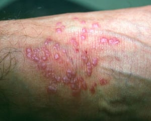
This image shows small papules with a sheen in cross-lighting typical of lichen planus.
This image shows small papules with a sheen in cross-lighting typical of lichen planus.
Image courtesy of Karen McKoy, MD.

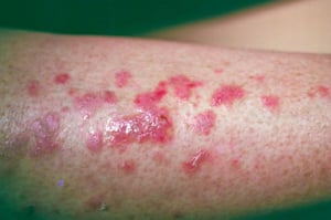
This image shows vesiculobullous lesions (some ruptured) resulting from lichen planus.
This image shows vesiculobullous lesions (some ruptured) resulting from lichen planus.
Image courtesy of Karen McKoy, MD.

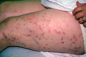
This image shows papules and plaques resulting from lichen planus.
This image shows papules and plaques resulting from lichen planus.
Image courtesy of Karen McKoy, MD.
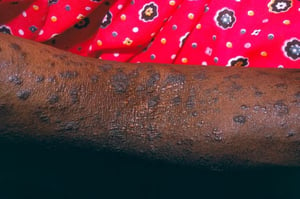
This photo shows papules and plaques of lichen planus. Violaceous flat-topped papules are visible on the dorsal arm. Erythema may look more violaceous or brown on dark skin.
This photo shows papules and plaques of lichen planus. Violaceous flat-topped papules are visible on the dorsal arm. Er
Image courtesy of Karen McKoy, MD.

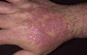
This image shows violaceous flat-topped papules coalescing into plaques on the dorsal hand of a patient with lichen planus.
This image shows violaceous flat-topped papules coalescing into plaques on the dorsal hand of a patient with lichen pla
Image provided by Thomas Habif, MD.
This image shows small papules with a sheen in cross-lighting typical of lichen planus.
This image shows small papules with a sheen in cross-lighting typical of lichen planus.
Image courtesy of Karen McKoy, MD.
This image shows vesiculobullous lesions (some ruptured) resulting from lichen planus.
This image shows vesiculobullous lesions (some ruptured) resulting from lichen planus.
Image courtesy of Karen McKoy, MD.
This image shows papules and plaques resulting from lichen planus.
This image shows papules and plaques resulting from lichen planus.
Image courtesy of Karen McKoy, MD.
This photo shows papules and plaques of lichen planus. Violaceous flat-topped papules are visible on the dorsal arm. Erythema may look more violaceous or brown on dark skin.
This photo shows papules and plaques of lichen planus. Violaceous flat-topped papules are visible on the dorsal arm. Er
Image courtesy of Karen McKoy, MD.
This image shows violaceous flat-topped papules coalescing into plaques on the dorsal hand of a patient with lichen planus.
This image shows violaceous flat-topped papules coalescing into plaques on the dorsal hand of a patient with lichen pla
Image provided by Thomas Habif, MD.
During the acute phase, new papules may appear at sites of minor skin injury (Koebner phenomenon), such as a superficial scratch. Lesions may coalesce or change over time, becoming hyperpigmented, atrophic, hyperkeratotic (hypertrophic lichen planus [LP]), or vesiculobullous. Although pruritic, lesions are rarely excoriated or crusted. If the scalp is affected, patchy scarring alopecia (lichen planopilaris) may occur.
Oral LP may occur without cutaneous lesions. Reticulated, lacy, bluish white, linear lesions (Wickham striae) are a hallmark of oral LP, especially on the buccal mucosae. Tongue margins and gingival mucosae in edentulous areas may also be affected. An erosive form of LP may occur in which the patient develops shallow, often painful, recurrent oral ulcers, which, if long-standing, rarely become cancerous. Chronic exacerbations and remissions are common.

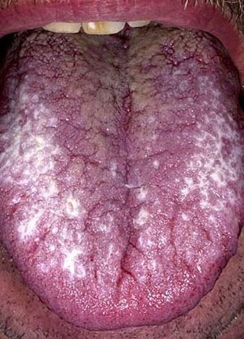
Lichen planus lesions can occur in the oral cavity. Reticulated, lacy, bluish white, linear lesions (Wickham striae, seen here on the sides of the tongue) are a hallmark of oral lichen planus, particularly on the buccal mucosa.
Vulvar and vaginal mucosae are often involved. Up to approximately 60% of women with oral mucosal findings also have vulvar LP (1). In men, genital involvement is common, especially of the glans penis.
Nails are involved in up to 10% of cases (2). Findings vary in intensity with nail bed discoloration, longitudinal ridging and lateral thinning, and complete loss of the nail matrix and nail, with scarring of the proximal nail fold onto the nail bed (pterygium formation).

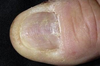
This photo shows longitudinal ridging of the thumbnail of a patient with lichen planus of the nail.
Symptoms and signs references
1. Belfiore P, Di Fede O, Cabibi D, et al. Prevalence of vulval lichen planus in a cohort of women with oral lichen planus: an interdisciplinary study. Br J Dermatol. 2006;155(5):994-998. doi:10.1111/j.1365-2133.2006.07480.x
2. Kharghoria G, Grover C, Bhattacharya SN, Sharma S. Histopathological evaluation of nail lichen planus: A cross-sectional study. J Cutan Pathol. 2021;48(1):11-17. doi:10.1111/cup.13783
Diagnosis of Lichen Planus
Physical examination
Biopsy with immunohistochemistry
Although the diagnosis of lichen planus (LP) is suggested by appearance of the lesions, the differential diagnosis may include any of the papulosquamous disorders, cutaneous lupus erythematosus, and secondary syphilis, among others.
Oral or vaginal LP may resemble leukoplakia, and the oral lesions must also be distinguished from candidiasis, carcinoma, aphthous ulcers, pemphigus, mucous membrane (cicatricial) pemphigoid, and chronic erythema multiforme.
Typically, biopsy is performed for diagnostic confirmation. Direct immunofluorescence may be used to distinguish LP from other lichenoid dermatoses, especially for oral lesions (1).
If LP is diagnosed, laboratory testing for liver dysfunction, including hepatitis B and C infections, should be considered.
Diagnosis reference
1. Nukaly HY, Halawani IR, Alghamdi SMS, et al. Oral Lichen Planus: A Narrative Review Navigating Etiologies, Clinical Manifestations, Diagnostics, and Therapeutic Approaches. J Clin Med. 2024;13(17):5280. Published 2024 Sep 5. doi:10.3390/jcm13175280
Treatment of Lichen Planus
Local treatments
Systemic treatments
Sometimes light therapy
Asymptomatic lichen planus (LP) does not require treatment. Medications suspected of triggering LP should be stopped; it can takes weeks to months after the offending medication has been stopped for the lesions to resolve.
Local treatments
Few controlled studies have evaluated treatments. Options differ based on the location and extent of disease.
Most cases of LP on the trunk or extremities can be treated with topical treatments (1). Topical glucocorticoids are first-line treatment for most cases of localized disease. High-potency ointments or creams (eg, clobetasol, fluocinonide, betamethasone) may be used on the thicker lesions on the extremities; lower-potency glucocorticoids (eg, hydrocortisone, desonide) may be used on the face, groin, and axillae. As always, courses should be limited to reduce risk of glucocorticoid–related skin atrophy.). Topical glucocorticoids are first-line treatment for most cases of localized disease. High-potency ointments or creams (eg, clobetasol, fluocinonide, betamethasone) may be used on the thicker lesions on the extremities; lower-potency glucocorticoids (eg, hydrocortisone, desonide) may be used on the face, groin, and axillae. As always, courses should be limited to reduce risk of glucocorticoid–related skin atrophy.
Intralesional glucocorticoids (eg, triamcinolone acetonide solution diluted with saline to 5 to 10 mg/mL) can be used every 4 weeks for hyperkeratotic plaques, scalp lesions, and lesions resistant to other therapies.Intralesional glucocorticoids (eg, triamcinolone acetonide solution diluted with saline to 5 to 10 mg/mL) can be used every 4 weeks for hyperkeratotic plaques, scalp lesions, and lesions resistant to other therapies.
Systemic treatments and phototherapy
Local therapy is impractical for generalized LP; thus, an oral medication or phototherapy is used. Oral glucocorticoids (eg, prednisone 20 mg once a day for 2 to 6 weeks followed by a taper) may be used for severe cases. The disease may rebound when therapy ceases; however, long-term systemic glucocorticoids should not be used.Local therapy is impractical for generalized LP; thus, an oral medication or phototherapy is used. Oral glucocorticoids (eg, prednisone 20 mg once a day for 2 to 6 weeks followed by a taper) may be used for severe cases. The disease may rebound when therapy ceases; however, long-term systemic glucocorticoids should not be used.
Oral retinoids (eg, acitretin) are indicated for otherwise recalcitrant cases. Light therapy using narrowband ultraviolet B (NBUVB) is an alternative to oral therapies, especially if they have failed or are contraindicated.Oral retinoids (eg, acitretin) are indicated for otherwise recalcitrant cases. Light therapy using narrowband ultraviolet B (NBUVB) is an alternative to oral therapies, especially if they have failed or are contraindicated.
Based on case reports and case series, other systemic options may include methotrexate, cyclosporine, mycophenolate mofetil (MMF), hydroxychloroquine, and azathioprine. Janus kinase (JAK) inhibitors have been effective in the management of cutaneous LP in limited studies but continue to be under evaluation. One randomized trial of oral baricitinib in adults with cutaneous LP has suggested rapid clearance (Based on case reports and case series, other systemic options may include methotrexate, cyclosporine, mycophenolate mofetil (MMF), hydroxychloroquine, and azathioprine. Janus kinase (JAK) inhibitors have been effective in the management of cutaneous LP in limited studies but continue to be under evaluation. One randomized trial of oral baricitinib in adults with cutaneous LP has suggested rapid clearance (2). TYK2 inhibition (deucravacitinib) is also being explored for cutaneous LP (). TYK2 inhibition (deucravacitinib) is also being explored for cutaneous LP (3). Observational data also support the use of apremilast (a phosphodiesterase-4 inhibitor used to treat psoriasis) (). Observational data also support the use of apremilast (a phosphodiesterase-4 inhibitor used to treat psoriasis) (4).
There are also reports of favorable outcomes with off-label use of IL-17, IL-23, and tumor necrosis factor inhibitors in the management of LP (5).
Oral lichen planus
Treatment of oral LP differs slightly from the treatment of other affected areas. Viscous lidocaine may help relieve symptoms of erosive ulcers; because inflamed mucous membranes can absorb high amounts, dose should not exceed 200 mg (eg, 10 mL of a 2% solution) or 4 mg/kg (in children) 4 times a day. Tacrolimus 0.1% ointment applied twice daily may induce lasting remission, but the data are limited. Treatment of oral LP differs slightly from the treatment of other affected areas. Viscous lidocaine may help relieve symptoms of erosive ulcers; because inflamed mucous membranes can absorb high amounts, dose should not exceed 200 mg (eg, 10 mL of a 2% solution) or 4 mg/kg (in children) 4 times a day. Tacrolimus 0.1% ointment applied twice daily may induce lasting remission, but the data are limited.
Other treatment options include topical (in an adhesive base), intralesional, and systemic glucocorticoids.
Erosive oral LP may respond to hydroxychloroquine, mycophenolate mofetil, or Erosive oral LP may respond to hydroxychloroquine, mycophenolate mofetil, orcyclosporine rinses. Emerging strategies include JAK and TYK2 inhibitors, which are still under evaluation.
Treatment references
1. Husein-ElAhmed H, Gieler U, Steinhoff M. Lichen planus: a comprehensive evidence-based analysis of medical treatment. J Eur Acad Dermatol Venereol. 2019;33(10):1847-1862. doi:10.1111/jdv.15771
2. Hwang A, Kechter J, Do T, et al. Oral Baricitinib in the Treatment of Cutaneous Lichen Planus. Preprint.. Oral Baricitinib in the Treatment of Cutaneous Lichen Planus. Preprint. medRxiv. 2024;2024.01.09.24300946. Published 2024 Jan 11. doi:10.1101/2024.01.09.24300946
3. Stolte KN, Mesas-Fernández A, Meier K, et al. TYK2 inhibition with deucravacitinib ameliorates erosive oral lichen planus. Exp Dermatol. 2024;33(4):e15080. doi:10.1111/exd.15080. TYK2 inhibition with deucravacitinib ameliorates erosive oral lichen planus. Exp Dermatol. 2024;33(4):e15080. doi:10.1111/exd.15080
4. Viswanath V, Joshi P, Dhakne M, et al: Evaluation of the efficacy and safety of apremilast in the management of lichen planus. : Evaluation of the efficacy and safety of apremilast in the management of lichen planus.Clin Cosmet Investig Dermatol 15:2593-2600, 2022. doi: 10.2147/CCID.S390591
5. Mital R, Gray A, Minta A, et al: Novel and off-label biologic use in the management of hidradenitis suppurativa, pyoderma gangrenosum, lichen planus, and seborrheic dermatitis: A narrative review. Dermatol Ther (Heidelb) 13(1):77–94, 2023. doi: 10.1007/s13555-022-00860-5
Prognosis for Lichen Planus
Many cases resolve without intervention, presumably because the inciting agent is no longer present. Recurrence after years may be due to reexposure to the trigger or some change in the triggering mechanism.
Vulvovaginal lichen planus may be chronic and refractory to therapy, causing decreased quality of life and vaginal or vulvar scarring.
Oral mucosal lesions usually persist for life.
Key Points
Lichen planus (LP) is thought to be an autoimmune disorder in patients with a genetic predisposition but may be caused by medications or be associated with disorders such as hepatitis C.
LP is characterized by recurrent, pruritic papules that are polygonal, flat-topped, and violaceous and can coalesce into plaques.
Oral and genital lesions can develop, become chronic, and cause morbidity.
Diagnose LP by clinical appearance and, if necessary, biopsy.
Treat localized LP with topical or injected glucocorticoids.
Treat generalized LP with oral medications or phototherapy.
Drug Information for the Topic


