


Perichondritis of the ear can be an inflammatory, but not necessarily infectious, process, resulting in diffuse swelling, redness, and pain of the pinna or an abscess between the cartilage and the perichondrium.
Perichondritis of the ear is an inflammation of the overlying skin and tissue (perichondrium) covering the cartilage of the ear. Infections are only one cause of perichondritis.
Causes of perichondritis include:
Trauma (including burns)
Insect bites
Ear piercings through the cartilage
Systemic inflammatory conditions (eg, vasculitides such as granulomatosis with polyangiitis, relapsing polychondritis)
Spread of superficial infections of the pinna
Because the cartilage’s blood supply is provided by the perichondrium, an injury that separates the perichondrium from both sides of the cartilage may lead to avascular necrosis and a deformed pinna (called cauliflower ear) in a matter of weeks. Abscesses may occur, sometimes due to gram-positive bacteria (eg, Staphylococcus aureus) (1). Septic necrosis may also ensue, often with infection by gram-negative bacilli.

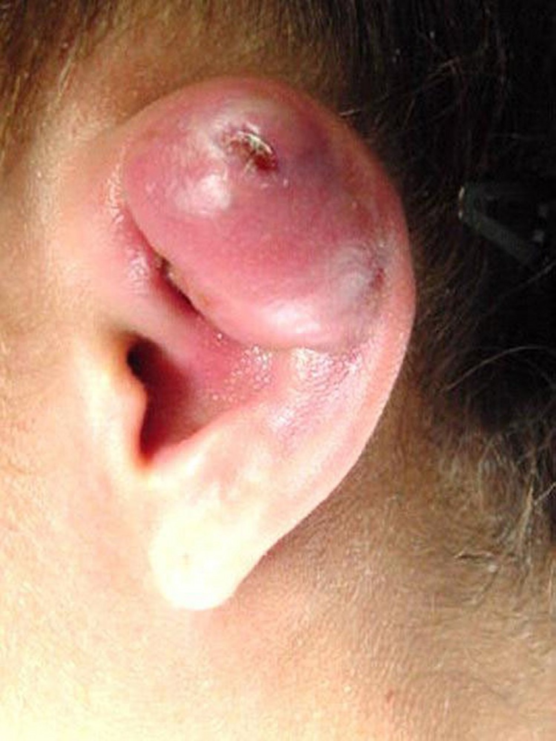
Severe perichondritis with abscess formation is seen in this image. The infection was caused by an ear piercing through the cartilaginous portion of the pinna.
Image provided by Bechara Ghorayeb, MD.
Symptoms of perichondritis include erythema, pain, and swelling. The course of perichondritis can be indolent, recurrent, long-term, and destructive.
The diagnosis of perichondritis is primarily made clinically.
General reference
1. Klug TE, Holm N, Greve T, et al. Perichondritis of the auricle: bacterial findings and clinical evaluation of different antibiotic regimens. Eur Arch Otorhinolaryngol. 2019;276(8):2199-2203. doi:10.1007/s00405-019-05463-z
Treatment of Perichondritis of the Ear
Prompt oral antibiotic therapy, typically a fluoroquinolone, sometimes with an aminoglycoside plus a semisynthetic penicillin
Often a systemic glucocorticoid
For an abscess, prompt incision and drainage with through-and-through tacking sutures (also called quilting sutures) to reattach the perichondrium to the cartilage
Patients with diffuse inflammation of the entire pinna are given empiric antibiotics (eg, fluoroquinolones, which have good cartilage penetration) and often a systemic glucocorticoid for its anti-inflammatory effects. Any foreign material (eg, ring, splinter) should be removed. If the etiology is not clearly infectious (eg, an infected piercing), patients should be evaluated for an inflammatory disorder (see Overview of Vasculitis and Relapsing Polychondritis).
Perichondrial abscesses are incised, and a small drain is left in place for 24 to 72 hours. Systemic antibiotics are initiated with a fluoroquinolone or an aminoglycoside plus a semisynthetic penicillin. Subsequent antibiotic choice is guided by culture and sensitivity. Warm compresses may help. It is important to ensure that the perichondrium is reapproximated to the cartilage to maintain the blood supply to the cartilage and to prevent necrosis. Reapproximation is ensured by inserting 1 or 2 mattress sutures through the entire thickness of the pinna, preferably through dental rolls or a non-stick dressing on both sides of the pinna.


