


Vocal fold paralysis has numerous causes and can affect speaking, breathing, and swallowing. The left vocal fold is affected twice as often as the right, and females are affected more often than males (3:2). Diagnosis is based on direct visualization. An extensive assessment may be necessary to determine the cause. Several direct surgical approaches are available if treating the cause is not curative.
Etiology of Vocal Fold Paralysis
Vocal fold paralysis may result from lesions or dysfunction at the level of the nucleus ambiguus, its supranuclear tracts, the main trunk of the vagus, or the recurrent laryngeal nerves. The left vocal fold is paralyzed more often than the right because the left recurrent nerve takes a longer course from the brain stem to the larynx, providing more opportunity for compression, traction, or surgical injuries (1).
Paralysis may be
Unilateral (most common)
Bilateral
Unilateral vocal fold paralysis is most common. About one-third of unilateral paralyses are neoplastic in origin, one-third are iatrogenic, and the remainder are idiopathic, infectious, or due to a central cause (2). Central causes include intracranial tumors, vascular insults, and demyelinating diseases that cause nucleus ambiguus paralysis. Tumors at the base of the skull and trauma to the neck cause vagus paralysis. Recurrent laryngeal nerve paralysis is caused by neck or thoracic lesions (eg, aortic aneurysm; mitral stenosis; mediastinal tuberculous adenitis; tumors of the thyroid gland, esophagus, lung, or mediastinal structures), trauma, thyroidectomy, neurotoxins (eg, lead, arsenic, mercury), neurotoxic infections (eg, diphtheria), cervical spine injury or surgery, Lyme disease, and viral illness. Neuronitis probably accounts for most idiopathic cases (3). Females appear to be affected more commonly than males, with a ratio of approximately 3:2 (4).
Bilateral vocal fold paralysis is a life-threatening disorder caused by thyroid and cervical surgery, tracheal intubation, trauma, and neurodegenerative and neuromuscular diseases.
Paradoxical vocal fold motion (also called paradoxical vocal fold dysfunction) is discussed elsewhere in The Manual.
Etiology references
1. Williams MJ, Ayylasomayajula A, Behkam R, et al. A computational study of the role of the aortic arch in idiopathic unilateral vocal-fold paralysis. J Appl Physiol (1985). 2015;118(4):465-474. doi:10.1152/japplphysiol.00638.2014
2. Ramadan HH, Wax MK, Avery S. Outcome and changing cause of unilateral vocal cord paralysis. Otolaryngol Head Neck Surg. 1998;118(2):199-202. doi:10.1016/S0194-5998(98)80014-4
3. Mau T, Husain S, Sulica L. Pathophysiology of iatrogenic and idiopathic vocal fold paralysis may be distinct. Laryngoscope. 2020;130(6):1520-1524. doi:10.1002/lary.28281
4. Cantarella G, Dejonckere P, Galli A, et al. A retrospective evaluation of the etiology of unilateral vocal fold paralysis over the last 25 years. Eur Arch Otorhinolaryngol. 2017;274(1):347-353. doi:10.1007/s00405-016-4225-9
Symptoms and Signs of Vocal Fold Paralysis
Vocal fold paralysis results in the loss of ability to perform vocal fold abduction and adduction. Paralysis may affect phonation, respiration, and deglutition, and food and fluids may be aspirated into the trachea. The paralyzed vocal fold generally lies 2 to 3 mm lateral to the midline.

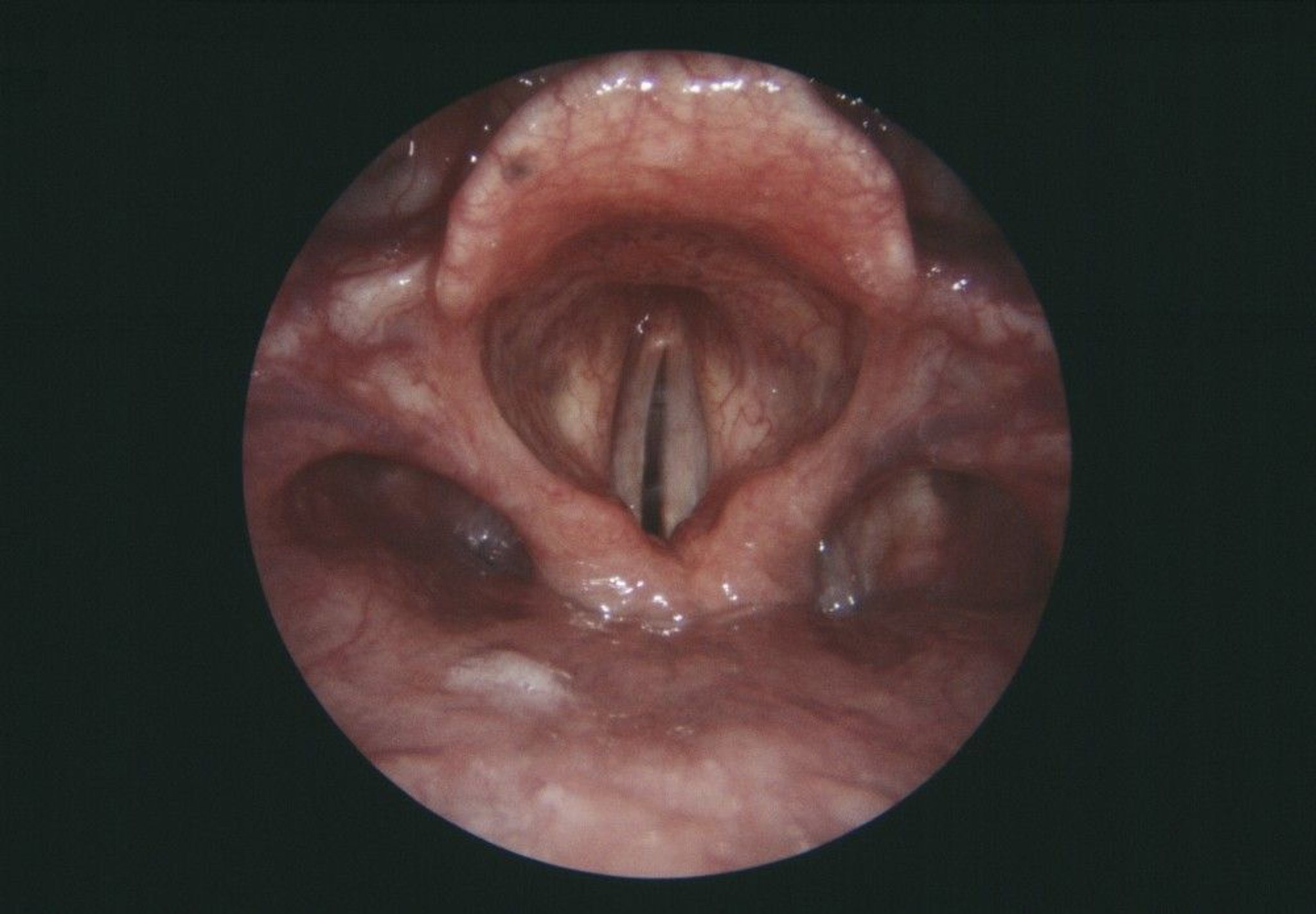
This endoscope view of the larynx (voice box) shows a paralyzed right vocal fold (gray, center left).
CNRI/SCIENCE PHOTO LIBRARY
In recurrent laryngeal nerve paralysis, the vocal fold may move with phonation but not with inspiration.
In unilateral paralysis, the voice may be hoarse and breathy, but the airway is usually not obstructed because the normal fold abducts sufficiently.
In bilateral paralysis, both vocal folds generally lie within 2 to 3 mm of the midline, and the voice is of good quality but of limited intensity and pitch modulation. The airway, however, is inadequate, resulting in stridor and dyspnea with moderate exertion as each vocal fold is drawn to the midline glottis by an inspiratory Bernoulli effect. There is also a danger of aspiration.
Diagnosis of Vocal Fold Paralysis
Laryngoscopy (g specifically strobovideolaryngoscopy)
Various tests for possible causes (CT, MRI, thyroid scan, barium swallow, upper GI endoscopy)
The diagnosis of vocal fold paralysis is based on laryngoscopy, the gold standard for diagnosing vocal fold paralysis. Strobovideolaryngoscopy, a highly specialized version of this diagnostic procedure, is preferred by many laryngologists because it provides detailed information on vocal fold vibration and mucosal wave characteristics (1).
The underlying cause must always be sought. Evaluation is guided by abnormalities identified on history and physical examination. During the history, the physician asks about all possible causes of peripheral neuropathy, including chronic heavy metal exposure (arsenic, lead, mercury), adverse drug effects of phenytoin and vincristine use, and history of systemic rheumatic diseases (eg, systemic lupus erythematosus, rheumatoid arthritis), Lyme disease, sarcoidosis, diabetes, and alcohol use disorder. Further evaluation may include enhanced CT or MRI of the head, neck, and chest to identify tumors, vascular anomalies, or inflammatory processes. Thyroid scan, barium swallow or bronchoscopy, and esophagoscopy may also help confirm the underlying cause.
Cricoarytenoid joint fixation must be differentiated from a neuromuscular etiology. Fixation is best documented by absence of passive mobility during rigid laryngoscopy under general anesthesia. Cricoarytenoid fixation may result from conditions such as rheumatoid arthritis, external blunt trauma, and prolonged endotracheal intubation.
Diagnosis reference
1. Wu AP, Sulica L. Diagnosis of vocal fold paresis: current opinion and practice. Laryngoscope. 2015;125(4):904-908. doi:10.1002/lary.25004
Treatment of Vocal Fold Paralysis
For unilateral paralysis, surgical procedures to position vocal folds closer together
For bilateral paralysis, surgical procedures and measures to maintain airway
In unilateral paralysis, treatment is directed at improving voice quality through augmentation, medialization, or reinnervation. Voice therapy is a helpful adjunct if enough closure can be achieved (1).
Augmentation involves injecting a paste of plasticized particles, collagen, hyaluronic acid, micronized dermis, or autologous fat into the paralyzed vocal fold; this helps bring the 2 folds closer together during adduction to improve the voice and prevent aspiration.
Medialization is shifting the vocal fold toward the midline by inserting an adjustable implant laterally to the affected fold. This can be performed with a local anesthetic in the operating room, allowing the size and position of the implant to be “tuned” to the patient’s voice.
Reinnervation, which restores tone and positioning, but not motion, to the paralyzed vocal fold, is increasingly being used in pediatric and adult (< 55 years) patients with a nonrecoverable paralysis (2, 3).
In bilateral paralysis, an adequate airway must be reestablished. Tracheotomy may be needed permanently or temporarily during an upper respiratory infection because of airway obstruction. An arytenoidectomy with lateralization of the true vocal fold opens the glottis and improves the airway but may adversely affect voice quality. Posterior laser cordectomy opens the posterior glottis and may be preferred to arytenoidectomy. Successful laser establishment of a posterior glottic airway usually obviates the need for long-term tracheotomy while preserving a serviceable voice quality. Selective reinnervation procedures are increasingly successful in very select patients (4).
Treatment references
1. Chen X, Wan P, Yu Y, et al. Types and timing of therapy for vocal fold paresis/paralysis after thyroidectomy: a systematic review and meta-analysis. J Voice. 2014;28(6):799-808. doi:10.1016/j.jvoice.2014.02.003
2. Espinosa MC, Ongkasuwan J. Recurrent laryngeal nerve reinnervation: is this the standard of care for pediatric unilateral vocal cord paralysis? Curr Opin Otolaryngol Head Neck Surg. 26(6):431-436, 2018. doi:10.1097/MOO.0000000000000499
3. Anthony, B., Parker, N., Patel, R. et al. Surgical considerations for laryngeal reinnervation and future research directions. Curr Otorhinolaryngol Rep. 8, 224–229, 2020. https://doi.org/10.1007/s40136-020-00294-7
4. Dunya, G., Orb, Q.T., Smith, M.E. et al. A review of treatment of bilateral vocal fold movement impairment. Curr Otorhinolaryngol Rep. 9, 7–15, 2021. https://doi.org/10.1007/s40136-020-00320-8
Key Points
Vocal fold paralysis can be caused by a lesion or dysfunction anywhere in the neural pathway to the larynx (the nucleus ambiguus, its supranuclear tracts, the main trunk of the vagus, the recurrent laryngeal nerves).
Most paralyses are unilateral and affect mainly the voice, but bilateral paralysis can occur and obstruct the airway.
Paralysis is diagnosed by laryngoscopy, but identification of the cause typically requires imaging (eg, MRI) and other tests.
Patients with bilateral paralysis often require tracheal intubation/tracheotomy initially, before corrective surgical procedures are attempted.
Various surgical procedures are available to improve voice quality in unilateral paralysis or to improve airway patency in long-term bilateral paralysis.
Drug Information for the Topic


